8.11 Urolithiasis
Urolithiasis, commonly referred to as kidney stones or renal calculi, refers to stones that develop within the urinary tract. Nephrolithiasis refers to stones in the kidney, and ureterolithiasis refers to stones in the ureter(s).
Pathophysiology and Risk Factors
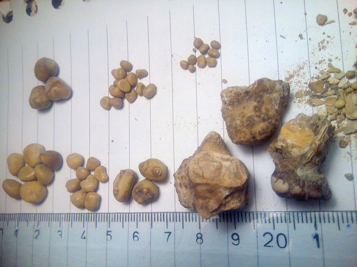
Urolithiasis typically occurs due to the crystallization of substances present in urine, such as calcium, oxalate, uric acid, cystine, or phosphate. Different types of stones form based on the predominant mineral content. See Figure 8.15[1] for an image of renal calculi.

Risk factors for stone development include insufficient fluid intake, excessive dietary intake of high mineral content, and hypercalcemia related to hyperparathyroidism or other medical conditions. Risk factors also include gout, repeated urinary tract infections, anatomical abnormalities within the urinary tract, and family history of urolithiasis.[2],[3]
Assessment
Signs and symptoms of urolithiasis can vary depending on the size and location of the stone, associated complications of the blockage, and an individual’s tolerance of the pain associated with urolithiasis. See Table 8.11a for clinical manifestations of urolithiasis across body systems.
Table 8.11a. Clinical Manifestations of Urolithiasis[4],[5]
| Body System | Physical Assessment Findings |
|---|---|
| Urinary | Flank or abdominal pain (often described as severe and intermittent that may radiate to the groin or lower abdomen), hematuria, dysuria, urgency, and presence of crystals in urine |
| Integumentary | Pallor or diaphoresis due to severe pain |
| Cardiovascular | Tachycardia due to severe pain and associated anxiety |
| Gastrointestinal | Nausea and vomiting due to severe pain |
| Musculoskeletal | Guarding of the abdomen or affected side due to severe pain, restlessness, or inability to find a comfortable position |
Diagnostic Testing
Diagnostic testing for urolithiasis involves various examinations and imaging studies to confirm the presence of urinary stones; determine their location, size, composition; and assess any associated complications. A urinalysis is performed to detect the presence of blood, crystals, or signs of infection in the urine. A 24-hour urine collection can help to measure various components in the urine such as high levels of calcium, oxalate, uric acid, or citrate. Imaging studies are ordered to visualize the presence of stones. A CT scan performed without contrast can provide detailed images of the urinary tract, allowing for localization, measurement, and characterization of the stones. X-rays can be useful in identifying certain types of stones, especially calcium stones.[6],[7]
Nursing Diagnoses
Nursing diagnoses for clients with urolithiasis can help guide nursing care and address the specific needs of these individuals.
Common nursing diagnoses include the following:
- Acute Pain
- Impaired Urinary Elimination
- Risk for Infection
- Readiness for Enhanced Knowledge
Outcome Identification
Outcome identification includes setting short- and long-term goals and creating expected outcome statements customized for the client’s specific needs. Expected outcomes are statements of measurable action for the client within a specific time frame that are responsive to nursing interventions. Examples of expected outcomes for clients with urolithiasis are as follows:
- The client will report a reduction in pain intensity to 3/10 or less on a 0-10 pain scale, within one hour following analgesic administration and nonpharmacological pain management interventions.
- The client will void without pain, indicating the resolution of impaired urinary elimination, by discharge.
- The client will verbalize three lifestyle modifications such as dietary changes to prevent recurrent stone formation by the end of the teaching session.
Interventions
Medical Interventions
Medical interventions for urolithiasis aim to manage symptoms, facilitate the passage of stones, and prevent stone recurrence. In some cases, if the stone is small, the client may be advised to let it pass on its own.
Medication Therapy
Several types of medications may be prescribed to manage the severe pain associated with urolithiasis:
- Analgesics: Analgesics like nonsteroidal anti-inflammatory drugs (NSAIDs) or opioids may be prescribed to manage severe pain associated with kidney stones.
- Alpha-blockers: Alpha-blockers like tamsulosin help relax the muscles in the ureter, facilitating the passage of stones and reducing pain.
- Calcium Channel Blockers: Specific calcium-channel medications can help relax the ureter and assist in stone passage.
- Intravenous (IV) Fluids: IV fluids maintain hydration and help flush out the stone(s).
Lithotripsy & Ureteroscopy
Shock wave lithotripsy (SWL) utilizes shock waves to break larger stones into smaller fragments, making them easier to pass through the urinary tract.
If an individual is unable to pass the stone, ureteroscopy may be utilized. Ureteroscopy refers to the insertion of a thin scope through the urethra and bladder and into the ureter to remove stones or break them up with laser therapy. Ureteral stenting may be performed to relieve obstruction caused by kidney stones. Stones lodged in the ureter can cause partial or complete blockage, resulting in severe pain, damage to the kidneys from urine backflow, and urinary stasis. The stent bypasses the obstruction, allowing urine to flow from the kidney to the bladder, reducing pressure, and minimizing associated symptoms.
Surgical Intervention
Percutaneous nephrolithotomy refers to the removal of large kidney stones using a nephroscope inserted through a small incision in the client’s back.[8],[9]
Nursing Interventions
Nursing interventions for urolithiasis aim to alleviate symptoms, facilitate stone passage, prevent complications, and provide support and education to clients. The client’s urine is often strained, and the stone(s) is collected and sent to the lab for analysis of its makeup. This information can help guide interventions to prevent future stones.
Health Teaching and Health Promotion
Nurses encourage adequate hydration by drinking adequate amounts of fluids to help dilute urine and prevent the concentration of minerals that can form stones. Other dietary changes to reduce the risk of stone formation are encouraged, such as the intake of oxalate-rich foods, sodium, and animal proteins. See Table 8.11b for a summary of suggested dietary modifications based on the type of stone present.
Table 8.11b. Dietary Modifications Based on Stone Type[10],[11]
| Stone Type | Dietary Modifications |
|---|---|
| Calcium Oxalate Stones | Consume adequate calcium from sources like low-fat dairy and reduce intake of high-oxalate foods like spinach, nuts, tea, and chocolate. Limit salt intake to prevent excessive calcium in urine. |
| Uric Acid Stones | Maintain adequate fluid intake to increase urine volume and dilute uric acid. Reduce intake of purine-rich foods like red meat, shellfish, and organ meats. Limit sodium intake to minimize uric acid excretion. |
| Struvite Stones | Avoid excess consumption of foods high in phosphates like dairy products, organ meats, and some processed foods. Acidifying urine through diet or medications may be recommended. |
| Cystine Stones | Increase fluid intake to maintain high urine output and reduce cystine concentration. Limit sodium intake to help decrease cystine excretion. Reduce intake of animal proteins because they contain cystine precursors. |
Symptom Management
Nurses teach about prescribed analgesics and nonpharmacological interventions to manage severe pain. Nonpharmacological interventions include application of heat, repositioning, and ambulation to help facilitate the passage of the stone. Comfortable positions that may alleviate pain include lying on the affected side or assuming a position that eases the passage of stones. Stress management techniques like relaxation breathing, guided imagery, and distractions can also help manage discomfort. Clients may feel nauseated from the sensations of severe pain, so antiemetic medications may also be required.
Evaluation
During the evaluation stage, nurses determine the effectiveness of nursing interventions for a specific client. The previously identified expected outcomes are reviewed to determine if they were met, partially met, or not met by the time frames indicated. If outcomes are not met or only partially met by the time frame indicated, the nursing care plan is revised. Evaluation should occur every time the nurse implements interventions with a client, reviews updated laboratory or diagnostic test results, or discusses the care plan with other members of the interprofessional team.
![]() RN Recap: Urolithiasis
RN Recap: Urolithiasis
View a brief YouTube video overview of urolithiasis[12]:
- “Kidney_stones_(_renal_calculi_),_Бубрежни_камења_15.jpg” by Jakupica is licensed under CC BY-SA 4.0 ↵
- Curhan, G. C., Aronson, M., & Preminger, G. M. (2023). Kidney stones in adults: Diagnoses and acute management of suspected nephrolithiasis. UpToDate. https://www.uptodate.com/ ↵
- Preminger, G. M., & Curhan, G. C. (2023). Kidney stones in adults: Evaluation of the patient with established stone disease. UpToDate. https://www.uptodate.com/ ↵
- Preminger, G. M., & Curhan, G. C. (2023). Kidney stones in adults: Evaluation of the patient with established stone disease. UpToDate. https://www.uptodate.com/ ↵
- National Institute of Diabetes and Digestive and Kidney Diseases. (2017). Kidney stones. National Institutes of Health. https://www.niddk.nih.gov/health-information/urologic-diseases/kidney-stones ↵
- Open RN Project. (2024, June 23). Health Alterations - Chapter 8 - Urolithiasis [Video]. You Tube. CC BY-NC 4.0 https://youtu.be/3Xl76JjQgpA?si=54N-fTU9LpMsc6YY ↵
Stones that develop within the urinary tract.
Stones in the kidney.
Stones in the ureter(s).
The utilization of shock waves to break larger stones into smaller fragments, making them easier to pass through teh urinary tract.
The insertion of a thin scope through the urethra and bladder and into the ureter to remove stones or break them up with laser therapy.
The removal of large kidney stones using a nephroscope inserted through a small incision in the client’s back.

