4.2 Review of Anatomy and Physiology of the Immune System
Anatomy of the Lymphatic and Immune Systems
The lymphatic system is the system of vessels, cells, and organs that transports fluid called lymph to the bloodstream and also filters pathogens from the blood. The immune system is the complex collection of cells and organs that destroys or neutralizes pathogens that would otherwise cause infection, disease, or death. The lymphatic system, for most people, is associated with the immune system to such a degree that the two systems are virtually indistinguishable.
Lymph Vessels
Lymph capillaries are interlaced in interstitial space, the space between individual cells in the tissues. Interstitial fluid enters the lymphatic capillaries and becomes lymph. Lymph is a clear-to-white fluid that transports immune system cells, as well as dietary lipids and fat-soluble vitamins absorbed in the small intestine called chyle. See Figure 4.1[1] for an image of lymph capillaries.
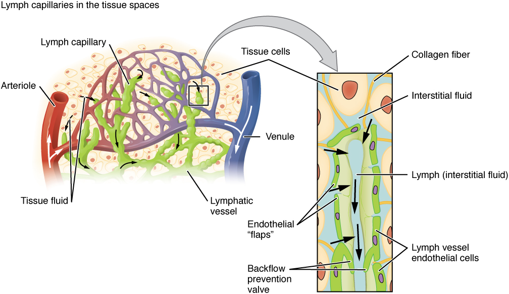
Lymph capillaries empty into larger lymphatic vessels that become larger and larger until they ultimately empty into the left and right subclavian veins in the neck. This is where lymph enters the bloodstream.
Lymph Nodes
Immune system cells use lymphatic vessels to not only make their way from the interstitial space into the circulation, but also use lymph nodes as major staging areas for the development of critical immune responses. A lymph node is one of the small, bean-shaped organs located throughout the lymphatic system. Lymph nodes store immune system cells that help the body fight infection and also filter the lymph fluid to remove foreign material such as bacteria and cancer cells.
Lymph travels through the lymph nodes near the groin, armpits, neck, chest, and abdomen. Humans have about 500–600 lymph nodes throughout the body. See Figure 4.2[2] for an illustration of lymph vessels and nodes.
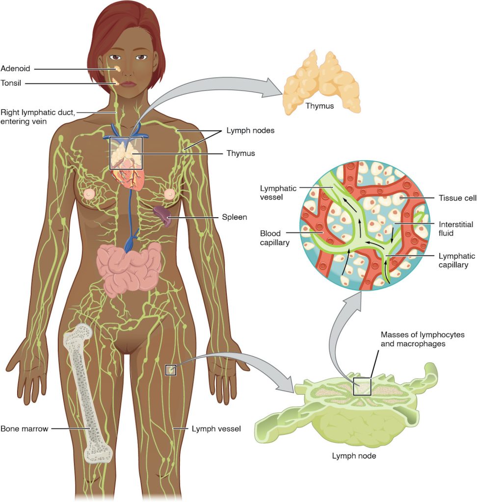
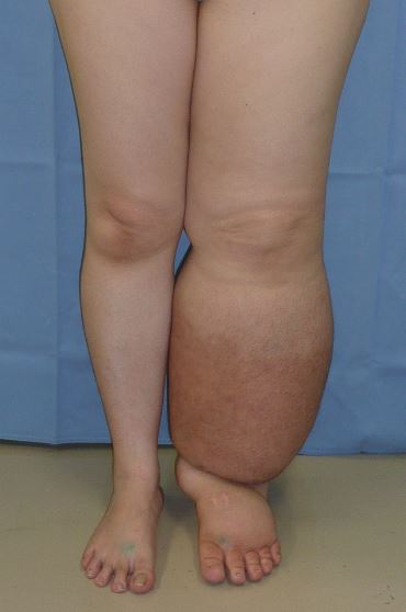
A major distinction between the lymphatic and cardiovascular systems is that lymph is not actively pumped by the heart but is forced through the vessels by the contraction of skeletal muscles during body movements and breathing. One-way valves in lymphatic vessels keep the lymph moving toward the heart. When the lymphatic system is damaged in some way, such as by being blocked by cancer cells or destroyed by injury, lymph “backs up” from the lymph vessels into interstitial spaces. This inappropriate accumulation of fluid is referred to as lymphedema. Lymphedema can also occur after lymph nodes are removed as part of cancer treatment. See Figure 4.3[3] for an image of lymphedema.
Primary Lymphoid Organs: Bone Marrow and Thymus
The primary lymphoid organs are the bone marrow and thymus gland. The lymphoid organs are where lymphocytes mature, proliferate, and are selected to attack pathogens.
A lymphocyte is a type of white blood cell that fights infection. There are two main types of lymphocytes: B cells and T cells. B cells produce antibodies that are used to attack invading bacteria, viruses, and toxins. T cells destroy the body’s own cells that have been taken over by viruses or become cancerous.[4] Recall that all blood cells, including lymphocytes, are formed in the bone marrow. The B cell undergoes nearly all of its development in the bone marrow, whereas the immature T cell, called a thymocyte, leaves the bone marrow and matures in the thymus gland. The thymus gland is an organ found in the space between the sternum and the aorta of the heart.
Secondary Lymphoid Organs
Lymphocytes develop and mature in the primary lymphoid organs, but they mount immune responses from the secondary lymphoid organs. Secondary lymphoid organs include the lymph nodes, spleen, and tonsils.
Spleen
The spleen is sometimes called the “filter of the blood.” The spleen stores and filters red blood cells and also functions as the location of immune responses to blood-borne pathogens. Therefore, clients who have had a splenectomy are at increased risk for infection.
Tonsils
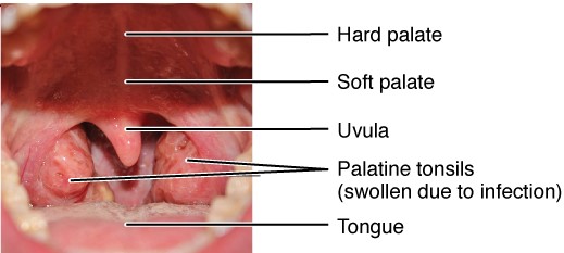
The tonsils are lymphoid nodules located along the inner surface of the pharynx and are important in developing immunity to oral pathogens. The tonsil located at the back of the throat, the pharyngeal tonsil, is sometimes referred to as the adenoid when swollen. See Figure 4.4[5] for an illustration of the tonsils. Such swelling is an indication of an active immune response to infection.
View a supplementary YouTube video[6] from Crash Course on the lymphatic system: Lymphatic System: Crash Course Anatomy & Physiology #44.
The Immune Response
There are two basic processes the body uses to defend against pathogens (i.e., microorganisms that cause infection). These processes are referred to as the innate immune response and the adaptive immune response.
Innate Immune Response
The innate immune response defends the body against pathogens in a nonspecific manner. It is called “innate” because it is present from the moment we are born. The innate immune response includes physical barriers and internal defenses.
Physical Barriers
Physical barriers are the body’s basic defenses against infection. They include skin and mucous membranes, as well as mucus and enzymes that physically destroy and/or remove pathogens and debris from areas of the body where they might cause harm or infection.
Internal Defenses
Internal defenses include phagocytosis, the inflammatory response, and fever.[7]
Phagocytosis
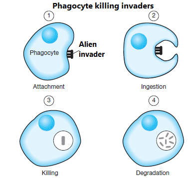
Phagocytosis refers to the process of specific white blood cells engulfing and destroying pathogens (i.e., “eating” them). Phagocytes include neutrophils and macrophages. Neutrophils are the most abundant type of white blood cells. They are the first to arrive during the innate immune response. After destroying a pathogen, neutrophils self-destruct and become pus. Macrophages are created from monocytes, a type of white blood cell. Each macrophage can destroy multiple pathogens, and they also clean up the destroyed neutrophils.[8] See Figure 4.5[9] for an image of phagocytosis.
Inflammatory Response
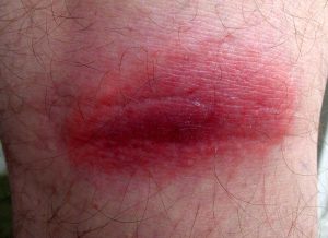
Inflammation is characterized by heat, redness, pain, and swelling. Although inflammation is often perceived as a negative consequence of injury or disease, it is an important process that recruits immune defenses to eliminate pathogens, remove damaged and dead cells, and initiate repair mechanisms.
During the inflammatory response, the damaged cells release chemicals including histamine, bradykinin, and prostaglandins. These chemicals cause redness, heat, and increased permeability of blood vessels, so fluid leaks into tissues, causing swelling. These chemicals also attract phagocytes and lymphocytes. See Figure 4.6[10] for an image of inflammation.
Fever
Fever is part of the inflammatory response that extends beyond the site of infection and affects the entire body, resulting in an overall increase in body temperature. Fever enhances the innate immune response by stimulating white blood cells to kill pathogens. The rise in body temperature also inhibits the growth of many pathogens.
View a supplementary YouTube video[11] from Crash Course on nonspecific innate immunity: Immune System, Part 1: Crash Course Anatomy & Physiology #45.
Adaptive Immune Response
The adaptive immune response is activated when the nonspecific, innate immune response is insufficient to control an infection. The adaptive immune response requires exposure to a pathogen for the body to recognize it as a threat and respond. Exposure to the pathogen can be through direct contact or via immunization. After the adaptive immune system has been exposed to a pathogen, it develops memory cells to recognize this pathogen in the future and quickly mounts a defense.[12]
Lymphocytes, a specific type of white blood cell, play a key role in the adaptive immune response. There are two main groups of lymphocytes called B cells and T cells, both of which are formed in bone marrow. B cells mature in the bone marrow, and T cells mature in the thymus, the rationale for being called “B” cells and “T” cells. B cells are involved in humoral immunity, and T cells are involved in cell-mediated defenses.
Humoral Immunity: B cells
Humoral immunity refers to the function of B cells and their production of antibodies. B cells originate and develop in the bone marrow and then migrate in body fluids through the lymph nodes, spleen, and blood to seek out and destroy pathogens in the interstitial spaces. B cells make Y-shaped proteins called antibodies that destroy pathogens based on their antigens. Antigens are markers that tell the immune system whether something in the body is harmful or not. Antigens are found on viruses, bacteria, cancer cells, and even normal cells of the body. Each antigen has a unique shape that the immune system reads like a name tag to know whether or not it belongs in that person’s body. Antibodies are very specific to the antigens they recognize and destroy. They fit onto the antigen like a key to a lock.[13]
When a B cell finds an abnormal cell with an antigen that it has antibodies for, it binds to it and responds by cloning itself into many B cells and also releases thousands of antibodies per second to attack the pathogen. Some B cells become memory B cells that are involved in immunological memory to ensure a stronger and faster antibody response if the body is exposed to the same antigen in the future.
Passive Immunity
Passive immunity refers to transfer of immunity due to antibodies that are produced in a body other than one’s own. For example, infants have passive immunity because they are born with antibodies that are transferred through the placenta from their mother. However, these antibodies can’t create memory cells, so it won’t remember an antigen if it gets infected again. These antibodies disappear between ages 6 and 12 months of age. Breast milk also contains antibodies, so babies who are breastfed have passive immunity for a longer period of time.[14]
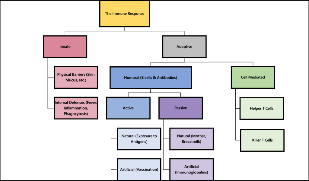
Passive immunization can also be transferred from an injection of antibodies formed by another person or animal. See Figure 4.7[15] for an illustration of the immune response.
Immunoglobulins are a specific type of antibody. There are five classes of immunoglobulins: IgG, IgM, IgA, IgD, and IgE. Immunoglobulins provide immediate protection against an antigen, but do not provide long-lasting protection. For example, immunoglobulin is administered to individuals who have been exposed to hepatitis B as passive immunization to help fight the infection.[16]
Cell-Mediated Response: T cells
When innate immunity and humoral immunity are not effective in defending against pathogens or abnormal cells, the cell-mediated response uses lymphocyte T cells to destroy the abnormal cells. There are two major types of T cells called helper T cells and killer T cells[17]:
- Helper T cells use chemical messengers to activate the adaptive immune response. They stimulate B cells to make antibodies and help develop the killer T cells.
- Killer T cells, also called cytotoxic cells, directly kill cells that have been invaded by a virus or are otherwise abnormal. They also clone themselves into many effector T cells and create memory T cells for immunological memory of the antigen.
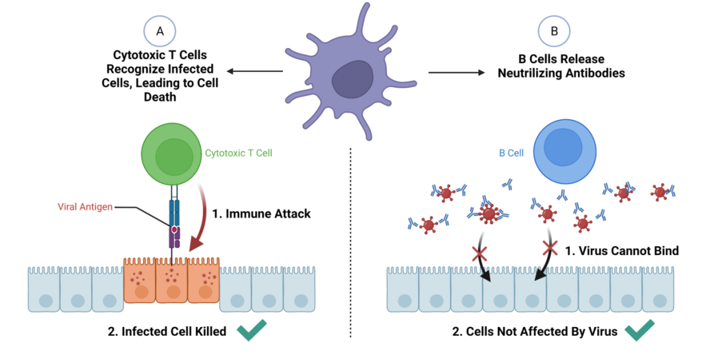
See Figure 4.8[18] for a comparison of the function of B cells and cytotoxic T cells. The purple cell is an invading virus. The B cells release antibodies to fight the virus and protect the body’s cells from being invaded by the virus. The cytotoxic T cells recognize body cells that have been infected by the virus and destroy them.
Chemical responses during the immune response including acute-phase proteins, complement proteins, and cytokines[19]:
- Acute-phase protein: An example of an acute-phase protein is C-reactive protein. High levels of C-reactive protein in blood samples indicate a serious infection or other medical condition is causing inflammation.
- Complement proteins: Complement proteins are always present in the blood and tissue fluids, allowing them to be activated quickly. They aid in the destruction of pathogens by piercing their outer membranes (cell lysis) or by making them more attractive to phagocytic cells.
- Cytokines: Cytokines are proteins secreted by cells that act as chemical messengers in immune responses. When a pathogen enters the body, the first immune cell to notice the pathogen is like the conductor of an orchestra. That cell directs all the other immune cells by creating and sending out messages (cytokines) to the rest of the organs or cells in the body to respond to and initiate inflammation.
- However, too many cytokines can have a negative effect and result in a cytokine storm. A cytokine storm is a severe immune reaction in which the body releases too many cytokines into the blood too quickly. A cytokine storm can occur as a result of an infection, autoimmune condition, or other disease. Signs and symptoms include high fever, inflammation, severe fatigue, and nausea. A cytokine storm can be severe or life-threatening and lead to multiple organ failure. For example, many COVID-19 deaths were caused by cytokine storm.
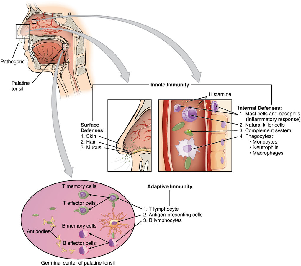
See Figure 4.9[20] for an illustration of innate immunity and specific adaptive immunity using an example of a pathogen entering the body through the nose.
View a diagram summary of immunity.
View a supplementary YouTube video[21] from Crash Course on cell-mediated immune response: Immune System, Part 3: Crash Course Anatomy & Physiology #47.
Immunological Memory
Immunological memory refers to the ability of the adaptive immune response to mount a stronger and faster immune response upon reexposure to a pathogen. Memory B and T cells for each specific pathogen are created that provide long-term protection from reinfection with that pathogen. On reexposure, these memory cells facilitate an efficient and quick immune response. For example, when an individual recovers from chicken pox, the body develops a memory of the varicella-zoster virus that will specifically protect it from reinfection if exposed to the virus again.[22]
Acquired immunity refers to immunity that develops with exposure to various antigens as an individual’s immune system builds a defense. However, pathogens can mutate and change the shape of their antigens so the immune system can no longer recognize or defend against it.[23] For example, influenza viruses mutate frequently, requiring annual influenza vaccination.
Immunization
Immunization is a method to trigger an individual’s acquired immune response and prevent future disease. A vaccine is an agent administered by injection, orally, or by nasal spray that provides active acquired immunity to a particular infectious disease. Small doses of a viral antigen are given to activate immunological memory. Memory cells allow the body to react quickly if exposed to the recognized antigen and efficiently defend against the virus.[24]
Allergies and Immune Disorders
An allergy is an inflammatory response due to a hypersensitivity to a substance that most people’s bodies perceive as harmless. Histamine, released during the immune response, is a primary cause of allergies and anaphylactic shock. Autoimmune disease is caused by the body’s inability to distinguish its own healthy cells from abnormal cells or pathogens, producing antibodies that attack its own tissues. Allergies and autoimmune diseases will be further discussed in the “Autoimmune and Hypersensitivity Reactions” section of this chapter.
Immune Response and Cancer
There are three stages of the immune response to many cancers[25]:
- Elimination occurs when the immune response first develops antibodies toward tumor-specific antigens and actively kills most cancer cells.
- Equilibrium is the period that follows elimination when remaining cancer cells are held in check.
- Escape from the immune response occurs when cancer mutates and no longer expresses specific antigens to which the immune system responds.
Immunodeficiency
Immunodeficiency results from a failure or absence of lymphocytes, phagocytes, and the complement system. Immunodeficiencies can be either primary or secondary. An example of a primary immunodeficiency caused by a genetic defect is severe combined immunodeficiency disorders (SCID). SCID is incompatible with life, and affected children typically die of infection within the first two years. Secondary immunodeficiency has multiple causes, such as medications (i.e., corticosteroids, immunosuppressive medications, and chemotherapy), nutritional deficiencies, and the human immunodeficiency virus (HIV) that causes acquired immunodeficiency syndrome (AIDS). HIV specifically impacts the helper T cells.[26]
Life Span Considerations
The following characteristics should be considered in pediatric and older adult clients.[27],[28]
Pediatric Considerations
- Children have lower levels of immunoglobulins, and humoral and cell-mediated immunity are not fully developed until age 6 or 7, increasing their susceptibility to infections.
- Newborns acquire natural immunity from their mother via breast milk.
Older Adult Considerations
- Lymphocytes can become less reactive to antigens and can lose immune function, increasing the potential for infections.
- Antibody levels are decreased.
- Immune and inflammatory responses decline due to chronic disease processes.
- With aging, thinning skin naturally occurs, making it prone to injury, susceptible to intrusion of microorganisms, and the development of infection.
- Cilia in the respiratory and gastrointestinal tracts are less active, allowing entry of pathogens.
- There is a higher incidence of cancer, infection, and autoimmune diseases in older adults.
- Many older adults live in close proximity to one another in long-term care or assisted living facilities, increasing the risk of transmission of communicable diseases.
- “2202_Lymphatic_Capillaries.jpg” by OpenStax College is licensed under CC BY 3.0 ↵
- “lymphaticimage-972x1024.jpg” by J. Gordon Betts, et al. is licensed under CC BY 4.0. Access for free at https://openstax.org/books/anatomy-and-physiology/pages/1-introduction. ↵
- “Lymphedema_limbs.JPG” by Medical doctors is licensed under CC BY-SA 4.0 ↵
- National Human Genome Research Institute. (2025). Lymphocyte. https://www.genome.gov/genetics-glossary/Lymphocyte ↵
- This work is a derivative of “2209 Location and Histology of Tonsils.jpg” by OpenStax and is licensed under CC BY 3.0. Access for free at https://openstax.org/books/anatomy-and-physiology/pages/21-1-anatomy-of-the-lymphatic-and-immune-systems ↵
- CrashCourse. (2015, November 30). Lymphatic system: Crash Course Anatomy & Physiology #44 [Video]. YouTube. All rights reserved. https://www.youtube.com/watch?v=I7orwMgTQ5I ↵
- InformedHealth.org (2023). The innate and adaptive immune systems. https://www.ncbi.nlm.nih.gov/books/NBK279396/ ↵
- A.D.A.M. Medical Encyclopedia [Internet]. (2023). Immune response. https://medlineplus.gov/ency/article/000821.htm ↵
- “33815869430_4cfda6ff3d_o.png” by zzzi zeth is in the Public Domain. ↵
- “Tickbite_Inflammation_2482a.jpg” by Túrelio is licensed under CC BY-SA 3.0 de. ↵
- CrashCourse. (2015, December 8). Immune system, Part 1: Crash Course Anatomy & Physiology #45 [Video]. YouTube. All rights reserved. https://www.youtube.com/watch?v=GIJK3dwCWCw ↵
- A.D.A.M. Medical Encyclopedia [Internet]. (2023). Immune response. https://medlineplus.gov/ency/article/000821.htm ↵
- Cleveland Clinic. (2022). Antigen. https://my.clevelandclinic.org/health/diseases/24067-antigen ↵
- A.D.A.M. Medical Encyclopedia [Internet]. A.D.A.M. Medical Encyclopedia [Internet]. (2023). Immune response. https://medlineplus.gov/ency/article/000821.htm ↵
- “Immune Response” by Open RN is licensed by CC BY-NC 4.0 ↵
- A.D.A.M. Medical Encyclopedia [Internet]. (2023). Immune response. https://medlineplus.gov/ency/article/000821.htm ↵
- A.D.A.M. Medical Encyclopedia [Internet]. (2023). Immune response. https://medlineplus.gov/ency/article/000821.htm ↵
- “General_Effector_Mechanisms_of_B_and_T_Cells.png” by סתו כסלו is licensed under CC BY-SA 4.0 ↵
- Ernstmeyer, K., & Christman, E. (Eds.). (2024). Nursing fundamentals 2E. Open RN | WisTech Open. https://wtcs.pressbooks.pub/nursingfundamentals/ ↵
- “2211_Cooperation_Between_Innate_and_Immune_Responses.jpg” by OpenStax is licensed under CC BY 3.0 ↵
- CrashCourse. (2015, December 21). Immune system, Part 3: Crash Course Anatomy & Physiology #47 [Video]. YouTube. https://www.youtube.com/watch?v=rd2cf5hValM ↵
- Ernstmeyer, K., & Christman, E. (Eds.). (2024). Nursing fundamentals 2E. Open RN | WisTech Open. https://wtcs.pressbooks.pub/nursingfundamentals/ ↵
- Cleveland Clinic. (2022). Antigen. https://my.clevelandclinic.org/health/diseases/24067-antigen ↵
- A.D.A.M. Medical Encyclopedia [Internet]. (2023). Immune response. https://medlineplus.gov/ency/article/000821.htm ↵
- Betts, J. G., Young, K. A., Wise, J. A., Johnson, E., Poe, B., Kruse, D. H., Korol, O., Johnson, J. E., Womble, M., & DeSaix, P. (2022). Anatomy and Physiology 2e. OpenStax. https://openstax.org/books/anatomy-and-physiology-2e/pages/1-introduction ↵
- Justiz, Vaillant, A. A., & Qurie, A. (2023). Immunodeficiency. StatPearls [Internet]. https://www.ncbi.nlm.nih.gov/books/NBK500027/ ↵
- Lond, M. L., Ladewig, P. W., Davidson, M. R., Ball, J. W., Bindler, R. M., & Cowen, K. J. (2017). Maternal & child nursing care (5th ed.). Pearson Education, Inc. ↵
- Stromberg, H. K. (2023). Medical-surgical nursing: Concepts & practice (5th ed.). Elsevier. ↵
The system of vessels, cells, and organs that transports fluid called lymph to the bloodstream and also filters pathogens from the blood
The complex collection of cells and organs that destroys or neutralizes pathogens that would otherwise cause infection, disease, or death.
The space between individual cells in the tissues.
A fluid that transports immune cells and waste products.
Lipids and fat-soluble vitamins absorbed in the small intestine.
Store immune system cells that help the body fight infection and also filter the lymph fluid to remove foreign material such as bacteria and cancer cells.
Inappropriate accumulation of fluid.
A type of white blood cell that fights infection.
Produce antibodies that are used to attack invading bacteria, viruses, and toxins.
Destroy the body's own cells that have been taken over by viruses or become cancerous.
An organ found in the space between the sternum and the aorta of the heart.
Stores and filters red blood cells and also functions as the location of immune responses to blood-borne pathogens.
Lymphoid nodules located along the inner surface of the pharynx and are important in developing immunity to oral pathogens.
Microorganisms that cause infection.
Defends the body against pathogens through physical defenses and internal defenses; present from the moment we are born.
The engulfment and destruction of bacteria, an important process during acute bacterial infections and inflammation.
The most abundant type of white blood cells.
Created from monocytes, a type of white blood cell, and can destroy multiple pathogens.
Characterized by heat, redness, pain, and swelling.
Activated when the nonspecific, innate immune response is insufficient to control an infection.
The function of B cells and their production of antibodies.
Y-shaped proteins that destroy pathogens based on their antigens.
Markers that tell the immune system whether something in the body is harmful or not.
Transfer of immunity due to antibodies that are produced in a body other than one’s own.
A specific type of antibody that provide immediate protection against an antigen, but do not provide long-lasting protection.
Stimulate B cells to make antibodies and help develop the killer T cells.
Directly kill cells that have been invaded by a virus or are otherwise abnormal; also known as cytotoxic cells.
Acute-phase protein which indicates serious infection or conditions causing inflammation.
Proteins secreted by cells that act as chemical messengers in immune responses.
A severe immune reaction in which the body releases too many cytokines into the blood too quickly; can occur as a result of an infection, autoimmune condition, or other disease. Signs and symptoms include high fever, inflammation, severe fatigue, and nausea.
The ability of the adaptive immune response to mount a stronger and faster immune response upon re-exposure to a pathogen.
Immunity that develops with exposure to various antigens as an individual's immune system builds a defense.
A method to trigger an individual's acquired immune response and prevent future disease.
An agent administered by injection, orally, or by nasal spray that provides active acquired immunity to a particular infectious disease.
An inflammatory response due to a hypersensitivity to a substance that most people's bodies perceive as harmless.
Released during the immune response and a primary cause of allergies and anaphylactic shock.
Caused by the body’s inability to distinguish its own healthy cells from abnormal cells or pathogens, producing antibodies that attack its own tissues.
Results from a failure or absence of lymphocytes, phagocytes, and the complement system.

