10.2 Review of Anatomy & Physiology of the Musculoskeletal System
Skeletal System Review
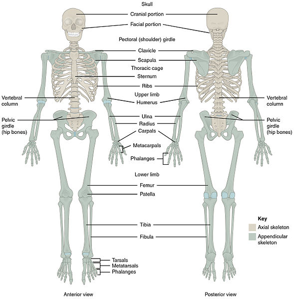
The skeletal system includes bones, joints, cartilages, and ligaments. It supports the body, facilitates movement with muscles, protects organs, produces blood cells, and stores and releases calcium to maintain homeostasis.[1] See Figure 10.1[2] for an illustration of the skeletal system. The skeletal system is subdivided into two major divisions called the axial skeleton and the appendicular skeleton. Each division is further discussed in the following sections, along with related medical terms.
The Axial Skeleton
The axial skeleton forms the central axis of the body and includes the bones of the head, neck, chest, and back. It serves to protect the brain, spinal cord, heart, and lungs. It also serves as the attachment site for muscles that move the head, neck, back, shoulders, and hip joints. The axial skeleton includes the cranium, hyoid, vertebral column, and thoracic cage.[3]
Cranium
The cranium is the bones that form the head, including the skull and the facial bones. It supports the face and protects the brain. See Figure 10.2[4] for an illustration of the anterior bones of the skull and the face.
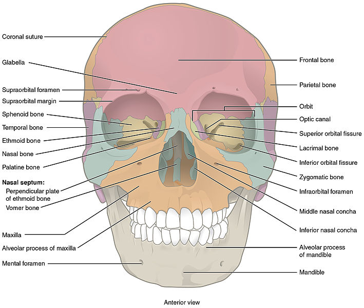
The major bones of the skull include the following:
- Frontal: Forehead
- Parietal: Upper lateral sides of the skull
- Temporal: Lower lateral sides of the skull
- Sphenoid: Posterior eye sockets and part of the base of the skull
- Ethmoid: Part of the nose and base of the skull
- Occipital: Posterior skull and base of the skull (See Figure 10.3[5] for an image of the occipital bone.)

The facial bones form the upper and lower jaws, the nose, nasal cavity and nasal septum, and the orbit. The facial bones include 14 bones, with six paired bones and two unpaired bones. The paired bones are the zygomatic, maxillary, palatine, nasal, lacrimal, and inferior conchae bones. The unpaired bones are the vomer and mandible bones.
- Zygomatic: Pair of cheekbones
- Maxillary: Upper jaw and hard palate
- Palatine: Pair of L-shaped bones between the maxilla and the sphenoid that form the hard palate, walls of the nasal cavity, and orbital floor of the eye
- Nasal: Pair of bones that form the bridge of the nose
- Lacrimal: Walls of the inner orbit (i.e., eye socket)
- Inferior conchae: Lower lateral walls of the nasal cavity
- Vomer: Bone that separates the left and right nasal cavity
- Mandible: Lower jawbone and only movable bone of the skull
The temporomandibular joint (TMJ) is a hinge joint between the temporal bone and the mandible that allows for the opening, closing, protrusion, retraction, and lateral movement of the lower jaw.
Hyoid
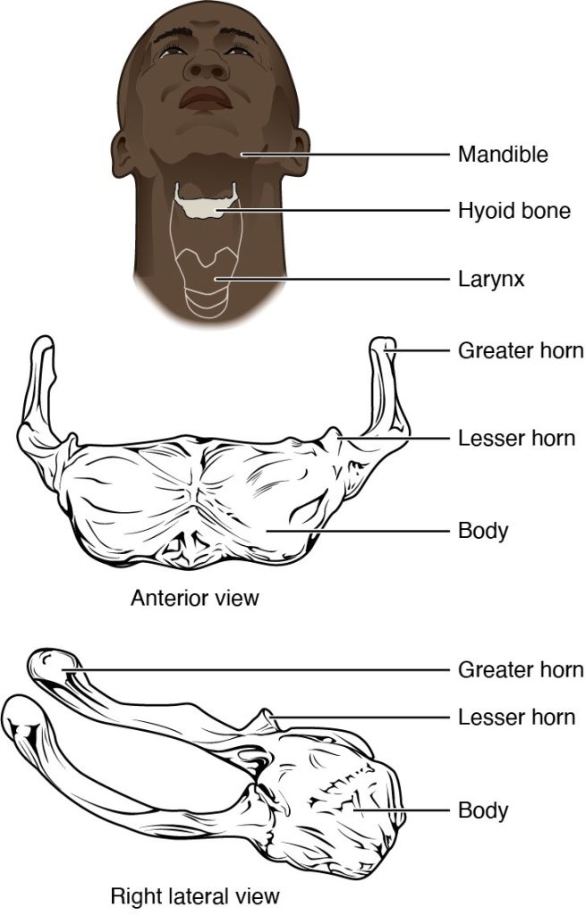
The hyoid bone is an independent bone that does not contact any other bone and, thus, is not part of the skull. It is a small U-shaped bone located in the upper neck near the level of the inferior mandible, with the tips of the “U” pointing posteriorly. The hyoid serves as the base for the tongue above and is attached to the larynx and the pharynx below. Movements of the hyoid are coordinated with movements of the tongue, larynx, and pharynx during swallowing and speaking. See Figure 10.4[6] for an illustration of the hyoid bone.
Vertebrae and Vertebral Column
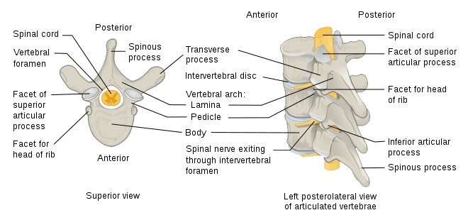
The vertebral column consists of vertebrae that are separated by intervertebral disks. Intervertebral disks are made up of cartilage that act as shock absorbers and allow for flexibility in the spine. The spinal cord runs through the center of the vertebrae. See Figure 10.5[7] for an illustration of the parts of a vertebra. A herniated disk refers to a condition in which a disk protrudes beyond the normal confines of the vertebrae.
Together, the vertebrae and intervertebral disks form the vertebral column, also known as the spinal column. It is a flexible column that supports the head, neck, and body and allows for their movements. It also protects the spinal cord, which passes down the back through openings in the vertebrae. View an illustration of the vertebral column in Figure 10.6.[8]
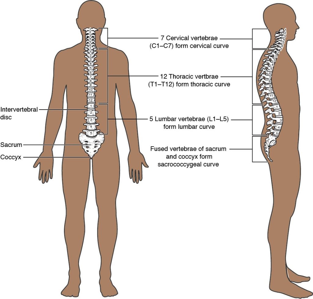
The vertebral column is normally curved, with two primary curvatures (thoracic and sacrococcygeal curves) and two secondary curvatures (cervical and lumbar curves).
Vertebrae Regions
The vertebrae are divided into five regions called the cervical, thoracic, lumbar, sacrum, and coccyx:
- Cervical: The first 7 vertebrae in the neck region, C1 to C7
- Thoracic: The next 12 vertebrae that form the outward curvature of the spine, T1 to T12
- Lumbar: The next 5 vertebrae that form the inner curvature of spine, L1 to L5
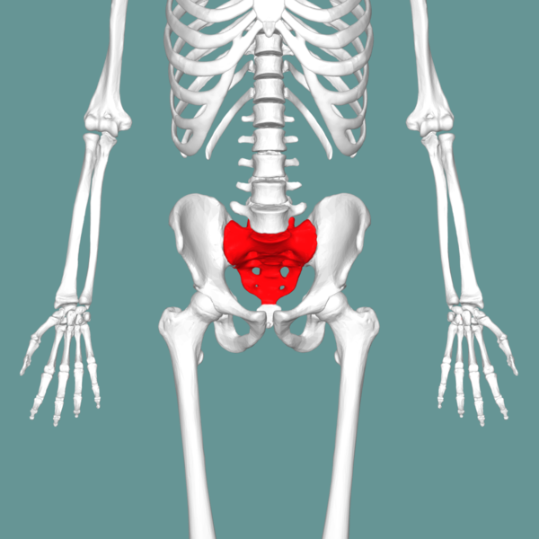
- Sacrum: The triangular-shaped bone at the base of the spine, formed by the fusion of five sacral vertebrae, a process that does not begin until after the age of 20 (See Figure 10.7[9] for an illustration of the sacrum.)
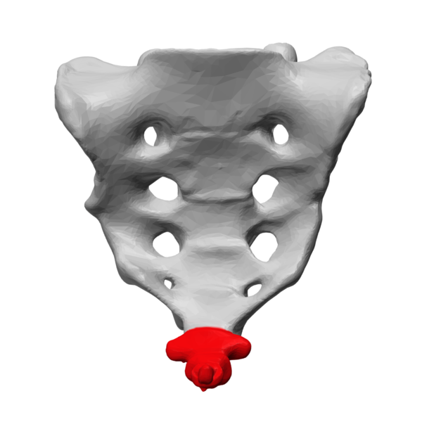
- Coccyx: The tailbone, formed by the fusion of four very small coccygeal vertebrae (See Figure 10.8[10] for an illustration of the coccyx.)
Thoracic Cage
The thoracic cage, commonly known as the rib cage, forms the chest (thorax). It consists of the sternum and 12 pairs of ribs, along with their costal cartilages. The ribs are anchored posteriorly to the 12 thoracic vertebrae (T1–T12). See Figure 10.9[11] for an illustration of the thoracic cage. The thoracic cage protects the heart and lungs.
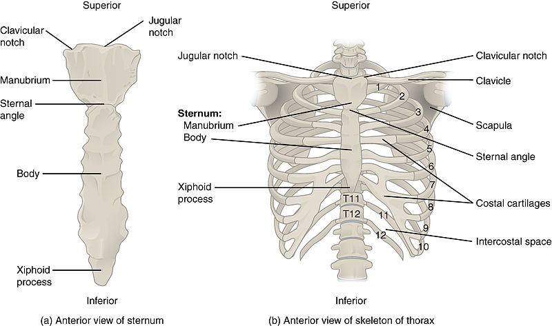
The sternum, also known as the breastbone, is divided into three parts:
- Manubrium: The upper portion of the sternum
- Body: The middle portion of the sternum
- Xiphoid process: The lower portion of the sternum made of cartilage
The 12 sets of ribs can be classified as true ribs, false ribs, and floating ribs:
- True ribs: Ribs 1-7 that are attached to the front of the sternum
- False ribs: Ribs 8, 9, and 10 that are attached to the cartilage that joins the sternum
- Floating ribs: Ribs 11 and 12 that are not attached to the front of the sternum
Intercostal means between the ribs. For example, people with labored breathing may experience intercostal retractions, where the muscles pull in between the ribs.
The Appendicular Skeleton
The appendicular skeleton includes the upper and lower limbs, plus the bones that attach each limb to the axial skeleton.[12]
Upper Limbs
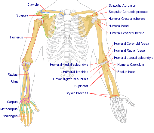
The bones of the upper limbs include the bones of the arms, wrists, and hands. The shoulder attaches the upper limbs to the axial skeleton. See Figure 10.10[13] for an illustration of the upper limbs, shoulder, clavicle, and scapula.
Arms
There are three bones in each arm:
View a supplementary YouTube video[14] on the radius and the ulna from UCDenver Anatomy Lab: Radius & ulna.
Wrists, Hands, and Fingers
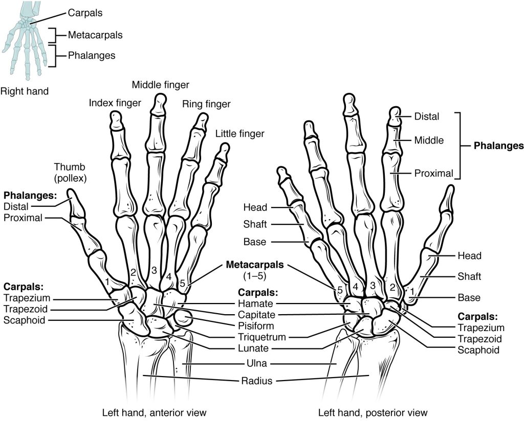
See Figure 10.11[15] for an illustration of the bones of the wrist, hand, and fingers:
- Carpals: Wrist bones
- Metacarpals: Wrist bones
- Phalanges: Fingers (and toes)
A single finger is called a phalanx and is composed of three bones called the distal phalanx, medial phalanx, and proximal phalanx. The exception is the thumb, which only has two bones, the distal and proximal.
The Shoulder
Refer back to Figure 10.10 to view the bones of the shoulder that connect the arms to the axial skeleton and include the clavicle, scapula, and acromion:
- Clavicle: Connects the sternum to the scapula, also known as the collarbone
- Scapula: Shoulder blade
- Acromion: An extension from the scapula that forms the bony tip of the shoulder
Together, the clavicle, acromion, and spine of the scapula form a V-shaped bony line that provides for the attachment of neck and back muscles that act on the shoulder, as well as muscles that pass across the shoulder joint to act on the arm.
Lower Limbs
The bones of the lower limbs include bones of the leg and the feet. The hip attaches the lower limbs to the axial skeleton.
Leg
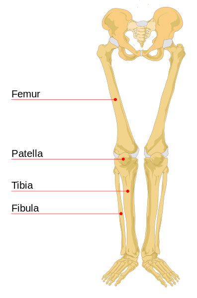
See Figure 10.12[16] for an illustration of the leg bones, including the femur, patella, tibia, and fibula:
- Femur: Thigh bone, the longest and strongest bone in the human body
- Patella: Kneecap
- Tibia: The medial bone and main weight-bearing bone of the lower leg, commonly called the shin. The distal end of the tibia forms the medial malleolus, the bony protrusion on the medial side of the ankle.
- Fibula: The smaller, lateral bone of the lower leg. The distal end of the fibula forms the lateral malleolus, the bony protrusion on the lateral side of the ankle.
Feet
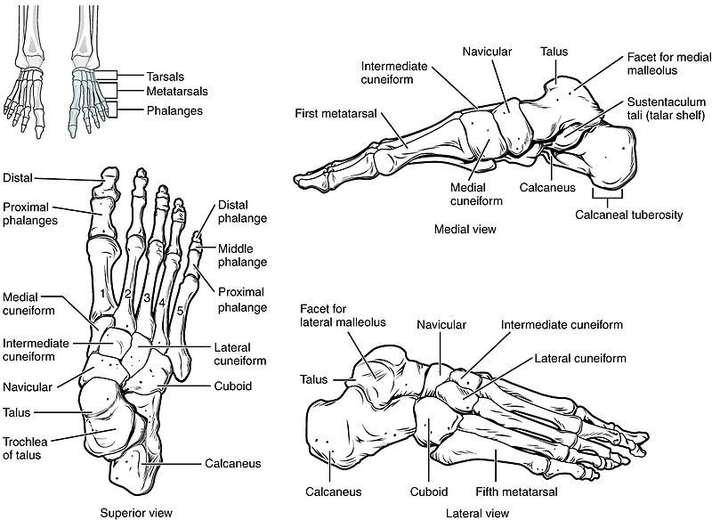
See Figure 10.13[17] for an illustration of the bones of the ankles and feet. There are several foot bones, but the major bones include the tarsals, metatarsals, phalanges, and calcaneus:
- Tarsals: Bones of the posterior half of the foot
- Metatarsals: Bones of the anterior half of the foot
- Phalanges: Toes (and fingers). The hallux is the great toe.
- Calcaneus: Heel bone
Like the fingers, the toes are composed of three bones called the distal phalanx, medial phalanx, and proximal phalanx, with the exception of the great toe, which only has two bones, the distal and proximal.
The Hip
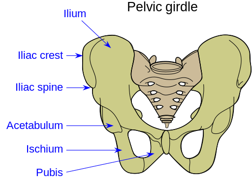
The hip serves as the attachment point for each lower limb at the acetabulum, the large socket that holds the head of the femur. Each adult hip bone is formed by three separate pelvic bones, called the ilium, ischium, and pubis, that fuse together during the late teenage years.
The ilium is the superior region that forms the largest part of the hip bone. It is attached to the sacrum at the sacroiliac joint. The ischium forms the posteroinferior region of each hip bone and supports the body when sitting. The pubis forms the anterior portion of the hip bone. The pubis curves medially, where it joins to the pubis of the opposite hip bone at a specialized joint called the pubic symphysis. The pelvis, also referred to as the pelvic girdle, refers to this entire structure formed by the two hip bones: the sacrum and the coccyx. It surrounds the pelvic cavity and connects the vertebral column to the lower limbs.
See Figure 10.14[18] for an illustration of the hip and pelvis.
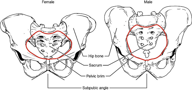
The shape of the pelvis is different for males and females. In general, the bones of the male pelvis are thicker and heavier because they have adapted to support a male’s typically heavier physical build. Because the female pelvis has adapted for childbirth, it is wider than the male pelvis. The shape and size of the pelvis can be used during forensic assessment to identify if skeletal remains are male or female. See Figure 10.15[19] for a comparison of the female and male pelvis.
Joints
Joints, also called articulations, are places where two bones or bone and cartilage come together and form a connection. Joints allow for movement and flexibility in the body. Dislocation refers to displacement of a bone from its normal position in a joint. Joints are categorized based on their structures and are referred to as fibrous joints, cartilaginous joints, or synovial joints.
Fibrous Joints
Fibrous joints are nonmoveable joints where two bones are attached by fibrous connective tissue. For example, a suture is the narrow fibrous joint found between the skull bones. During infancy, the space between skull bones is filled with flexible material that allows the skull to grow as the baby’s brain grows.
Cartilaginous Joints
Cartilaginous joints, also called amphiarthrosis or slightly movable joints, occur when two bones are connected by cartilage, a tough but flexible type of connective tissue. Examples of cartilaginous joints include the pubic symphysis and intervertebral disks.
Synovial Joints
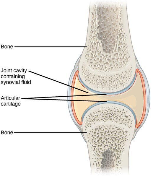
Synovial joints, also called diarthroses or fully movable joints, have a fluid-filled space where two bones come together, called a joint cavity. Because the bones in a synovial joint are not directly connected to each other with fibrous connective tissue or cartilage, they are able to move freely against each other, allowing for increased joint mobility. Synovial joints are the most common type of joint in the body. A synovial membrane is the lining or covering of synovial joints, and synovial fluid is the lubricating fluid found between synovial joints. See Figure 10.16[20] for an illustration of a synovial joint.
Types of Synovial Joints
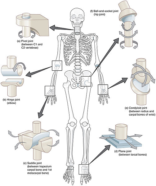
Synovial joints are categorized based on the shapes of the articulating surfaces of the bones that form each joint. The six types of synovial joints are pivot, hinge, condyloid, saddle, plane, and ball-and-socket joints. See Figure 10.17[21] for an illustration of the various types of synovial joints.
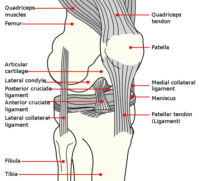
A few synovial joints have a fibrocartilage structure called an arterial disk or meniscus located between the articulating bones. Arterial disks are generally small and oval-shaped, whereas a meniscus is larger and C-shaped. For example, the knee connects the femur (upper leg bone) to the tibia (one of the lower leg bones) with a meniscus.
In addition to the meniscus, the knee also contains ligaments, tendons, and additional cartilage. Ligaments in the knee include the anterior cruciate ligament (ACL), lateral collateral ligament (LCL), medial collateral ligament (MCL), and posterior cruciate ligament (PCL). See Figure 10.18[22] for an illustration of these structures in the knee joint.
View a supplementary YouTube video[23] from Crash Course on joints: Joints: Crash Course Anatomy & Physiology #20.
Ligaments and Tendons
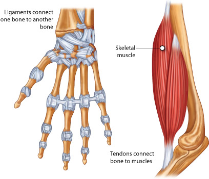
Other fibrous connective tissues in the musculoskeletal system include ligaments and tendons. Ligaments are narrow bands of fibrous connective tissue that connect a bone to a bone. A tendon is a narrow band of fibrous connective tissue that connects a muscle to a bone. Tendons are further discussed in the “Muscular System” subsection” below. See Figure 10.19[24] for an image that compares ligaments and tendons.
Bones
There are 206 bones in adults and 300 bones in children. As children grow, some bones fuse together to become 206 bones in adulthood.
There are three types of cells related to the growth and breakdown of bone. Osteoblasts are bone-forming cells, and osteocytes are mature bone cells. The dynamic nature of bone means that new bone tissue is constantly being formed; and old, injured, or unnecessary bone is dissolved for repair or for calcium release. The cells responsible for bone breakdown are osteoclasts. An equilibrium between osteoblasts and osteoclasts maintains healthy bone tissue. Two disorders caused by lack of equilibrium of these processes are osteopenia and osteoporosis. Osteopenia refers to abnormal reduction of bone mass, and during osteoporosis the bones become weak, brittle, and prone to fractures.
Bones contain more calcium (Ca+) than any other organ. When blood calcium levels decrease below normal levels, calcium is released from the bones by the osteoclasts, so there is an adequate supply for metabolic needs. In contrast, when blood calcium levels are increased, excess calcium is stored in bone by the osteoblasts. This dynamic process of releasing and storing calcium goes on continuously.
The bones of the skeletal system are composed of inner spongy tissue referred to as bone marrow. There are two types of bone marrow called red and yellow. Red bone marrow produces the red blood cells, white blood cells, and platelets. Yellow bone marrow contains adipose tissue, which can serve as a source of energy.
Review additional information about bone marrow and the production of red blood cells, white blood cells, and platelets in the “Hematological Alterations” chapter.
Muscular System
Types of Muscle
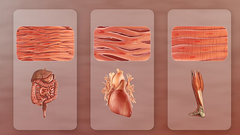
There are three major types of muscle tissue categorized as smooth, cardiac, and skeletal muscle. See Figure 10.20[25] for an illustration of these three types of muscle. Cardiac and skeletal muscles are striated, meaning they contain functional units called sarcomeres. Sarcomeres are composed of two protein filaments called actin and myosin that are responsible for muscular contraction.
Smooth Muscle
Smooth muscle is responsible for involuntary muscle movement. Smooth muscle is present in the following areas[26]:
- Walls of hollow organs, like the urinary bladder, uterus, stomach, and intestines, where muscle contractions cause the movement of fluids and other substances
- Walls of passageways, such as the arteries and veins of the circulatory system, where it causes vasodilation and vasoconstriction
- Tracts of the respiratory, urinary, and reproductive systems, where contraction and relaxation affect the movement of air, urine, and reproductive fluids
- Eyes, where it functions to change the size of the pupil
- Skin, where it causes hair to stand erect in response to cold temperature or fear, commonly called goose bumps
Cardiac Muscle
Cardiac muscle is only found in the heart. Highly coordinated contractions of cardiac muscle pump blood throughout the circulatory system. Cardiac muscle fiber cells are extensively branched and connected to one another at their ends to allow the heart to contract in a wavelike pattern and work as a pump.
Skeletal Muscle
Skeletal muscles are located throughout the body. They are under voluntary control and primarily produce movement of the arms, legs, back, and neck and maintain posture by resisting gravity. Small, constant adjustments of the skeletal muscles are needed for a person to hold their body upright or balanced in any position.
Skeletal muscles also have several additional functions. They are located throughout the body at the openings of internal tracts to control the movement of substances. These skeletal muscles allow voluntary control of functions such as swallowing, defecation, and urination in the digestive and urinary systems. Skeletal muscles also protect internal organs (particularly abdominal and pelvic organs) by acting as an external barrier against trauma and supporting the weight of the organs. They also contribute to maintaining homeostasis by generating heat. This heat generation is very noticeable during exercise, when sustained muscle movement causes a person’s body temperature to rise, or conversely during cold environmental temperatures when shivering produces random skeletal muscle contractions to generate heat. Skeletal muscles also play a role in blood flow. For example, the contraction of skeletal muscles during walking helps promote blood flow and reduces the risk of blood clot formation.
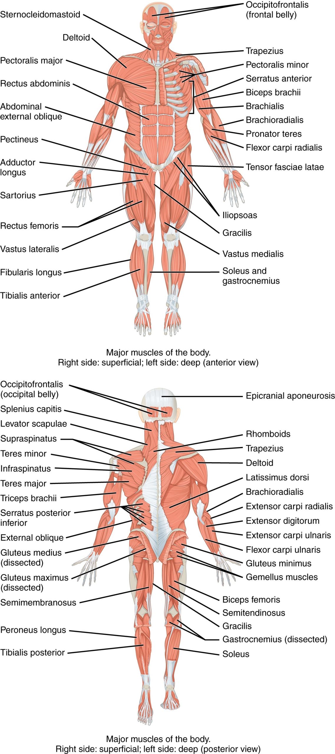
See Figure 10.21[27] for an illustration of the major skeletal muscles of the body. For the anterior and posterior views in this figure, superficial muscles are shown on the right side of the image, and deep muscles are shown on the left side of the image. For the legs, superficial muscles are shown in the anterior view while the posterior view shows both superficial and deep muscles.
Muscles are named based on various characteristics, such as the following:
- Body location: The area of the body, for example, biceps, triceps, and quadriceps
- Size: The size of the muscle, such as maximus (largest) and minimus (smallest)
- Shape: The shape of the muscle, such as deltoid (triangular) or trapezius (trapezoid)
- Action: The action of the muscle, such as flexor (i.e., to flex) or adductor (i.e., towards midline of body)
- Fiber direction: The direction of the muscle fibers, such as external oblique
Major Skeletal Muscles
Major skeletal muscles include the following:
- Biceps brachii: Muscle on the anterior upper arm
- Biceps brachialis: Muscle located in the arm that flexes the elbow joint and rotates the forearm
- Deltoid: A large triangular muscle covering the shoulder joint
- Gastrocnemius: The chief muscle of the calf of the leg
- Gluteus maximus: The largest and outermost of the three gluteal muscles in the buttocks
- Latissimus dorsi: A large muscle in the back
- Pectoralis major: A thick, fan-shaped muscle situated on the chest
- Quadriceps: A large muscle group on the front of the thigh
- Rectus abdominis: A paired muscle running vertically on each side of the anterior wall of the abdomen
- Triceps brachii: Muscle on the posterior of the upper arm
Tendons
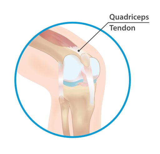
As previously mentioned in “The Appendicular Skeletal” subsection, tendons attach muscles to bones. For example, consider the Achilles tendon, hamstring, rotator cuff, and quadriceps tendons. The Achilles tendon attaches the calf muscles to the heel bone. The hamstring refers to five tendons at the back of a person’s knee that connect a group of three hamstring muscles to bones in the pelvis, knee, and lower leg. The rotator cuff is a group of muscles and tendons that stabilize the shoulder. The quadriceps tendon attaches the quadriceps muscle to the top of the kneecap. See Figure 10.22[28] for an illustration of the quadriceps tendon.
Function of Muscles
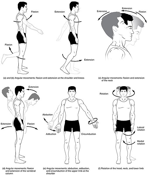
The main function of the muscular system is movement. Muscles work as antagonistic (opposing) pairs. As one muscle contracts, another muscle relaxes. This contraction pulls on the bones and assists with movement. Contraction is the shortening of muscle fibers whereas relaxation is the lengthening of fibers. This sequence of relaxation and contraction is stimulated by the nervous system.
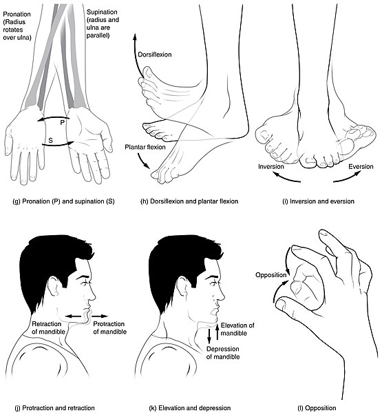
There are many types of actions that are caused by the contraction and relaxation of muscles, such as flexion, extension, abduction, adduction, rotation, dorsiflexion, plantar flexion, supination, and pronation. These muscle actions are summarized in Table 10.2. See Figures 10.23[29] and 10.24[30] for illustrations of these movements.
Table 10.2. Muscle Actions
| Action | Description |
|---|---|
| Flexion | Movement that decreases the angle between two bones, such as bending the arm at the elbow. |
| Extension | Movement that increases the angle between two bones, such as straightening the arm at the elbow. |
| Abduction | Movement of a limb away from the midline of the body. |
| Adduction | Movement of a limb toward the midline of the body. |
| Rotation | Circular movement around a central point. Internal rotation is toward the center of the body, and external rotation is away from the center of the body. |
| Dorsiflexion | Decreasing the angle of the foot and the leg (i.e., the foot moves upward toward the knee). This movement is the opposite of plantar flexion. |
| Plantar Flexion | Increasing the angle of the foot and leg (i.e., the moves foot downward toward the ground, such as when pressing down on a gas pedal in a car). |
| Supination | Movement of the hand or foot turning upward. When applied to the hand, it is the act of turning the palm upwards. When applied to the foot, it is the outward roll of the foot/ankle during normal movement. |
| Pronation | Movement of the hand or foot turning downward. When applied to the hand, it is the act of turning the palm downward. When applied to the foot, it is the inward roll of the foot/ankle during normal movement. |
| Eversion | Excessive movement when turning outward the sole of the foot away from the body’s midline, a common cause of an ankle sprain. |
| Inversion | Excessive movement when turning inward the sole of the foot towards the median plane, a common cause of an ankle sprain. |
- National Cancer Institute. (n.d.). Introduction to the skeletal system. National Institutes of Health. https://training.seer.cancer.gov/anatomy/skeletal ↵
- “701 Axial Skeleton-01.jpg” by OpenStax is licensed under CC BY 3.0 ↵
- Betts, J. G., Young, K. A., Wise, J. A., Johnson, E., Poe, B., Kruse, D. H., Korol, O., Johnson, J. E., Womble, M., & DeSaix, P. (2022). Anatomy and Physiology 2e. OpenStax. https://openstax.org/books/anatomy-and-physiology-2e/pages/1-introduction ↵
- “704_Skull-01.jpg” by OpenStax College is licensed under CC BY 3.0 ↵
- This work is derivative of “Occipital_bone_lateral4.png” by Anatomography is licensed under CC BY-SA 2.1 ↵
- “Hyoid Bone” by OpenStax is licensed under CC BY 4.0. Access for free at https://openstax.org/books/anatomy-and-physiology-2e/pages/7-2-the-skull ↵
- “718_Vertebra-en.svg” by Jmarchn is licensed under CC BY-SA 3.0 ↵
- “Vertebral Column” by OpenStax is licensed under CC BY 4.0. Access for free at https://openstax.org/books/anatomy-and-physiology-2e/pages/7-3-the-vertebral-column ↵
- “Sacrum_-_anterior_view02.png” by BodyParts3D, made by DBCLS is licensed under CC BY 2.1 Japan ↵
- “Coccyx_-_anterior_view01.png” by BodyParts3D, made by DBCLS is licensed under CC BY 2.1 Japan ↵
- “721_Rib_Cage.jpg” by OpenStax College is licensed under CC BY 3.0 ↵
- Betts, J. G., Young, K. A., Wise, J. A., Johnson, E., Poe, B., Kruse, D. H., Korol, O., Johnson, J. E., Womble, M., & DeSaix, P. (2022). Anatomy and Physiology 2e. OpenStax. https://openstax.org/books/anatomy-and-physiology-2e/pages/1-introduction ↵
- “Human_arm_bones_diagram.svg” by LadyofHats Mariana Ruiz Villarreal is licensed in the Public Domain ↵
- UCDenver Anatomy Lab 3244. (2014, August 31). Radius & ulna [Video]. YouTube. All rights reserved. https://www.youtube.com/watch?v=ThmvWFvHOmI ↵
- “541c7cf2586e9daaea40767bbc68011fdb8573b1.jpg" OpenStax is licensed under CC BY 4.0. Access for free at https://openstax.org/books/anatomy-and-physiology-2e/pages/8-2-bones-of-the-upper-limb ↵
- “Human_leg_bones_labeled.svg” by LadyofHats Mariana Ruiz Villarreal is licensed in the Public Domain ↵
- “812_Bones_of_the_Foot.jpg” by OpenStax College is licensed under CC BY 3.0 ↵
- “Pelvic_girdle_illustration.svg” by Fred the Oyster is licensed under CC BY 4.0 ↵
- “809_Male_Female_Pelvic_Girdle.jpg” by OpenStax College is licensed under CC BY 3.0 ↵
- “Figure_38_03_03.jpg” by CNX OpenStax is licensed under CC BY 4.0 ↵
- “909_Types_of_Synovial_Joints.jpg” by OpenStax College is licensed under CC BY 3.0 ↵
- “Knee_diagram.svg” by Mysid is licensed in the Public Domain ↵
- CrashCourse. (2015, May 26). Joints: Crash Course Anatomy & Physiology #20 [Video]. All rights reserved. https://www.youtube.com/watch?v=DLxYDoN634c ↵
- “tendon-lig.jpg” by unknown author is licensed under CC BY-SA 4.0. Access for free at https://pressbooks.ccconline.org/bio106 ↵
- “Types_Of_Muscle.jpg” by www.scientificanimations.com is licensed under CC BY-SA 4.0 ↵
- Betts, J. G., Young, K. A., Wise, J. A., Johnson, E., Poe, B., Kruse, D. H., Korol, O., Johnson, J. E., Womble, M., & DeSaix, P. (2022). Anatomy and Physiology 2e. OpenStax. https://openstax.org/books/anatomy-and-physiology-2e/pages/1-introduction ↵
- “1105_Anterior_and_Posterior_Views_of_Muscles.jpg” by OpenStax is licensed under CC BY 4.0 ↵
- “Quadriceps_tendon.svg” by InjuryMap is licensed under CC BY-SA 4.0 ↵
- “Body_Movements_I.jpg” by Tonye Ogele CNX is licensed under CC BY-SA 3.0 ↵
- “Body_Movements_II.jpg” by Tonye Ogele CNX is licensed under CC BY-SA 3.0 ↵
- RegisteredNurseRN. (2021, March 3). Flexion and extension anatomy: Shoulder, hip, forearm, neck, leg, thumb, wrist, spine, finger [Video]. YouTube. Used with permission. All rights reserved. https://www.youtube.com/watch?v=p4xbehGmkmk ↵
- RegisteredNurseRN. (2021, March 29). Abduction and adduction of wrist, thigh, fingers, thumb, arm | Anatomy body movement terms [Video]. YouTube. Used with permission. All rights reserved. https://www.youtube.com/watch?v=qcxE9uql8g4 ↵
- RegisteredNurseRN. (2021, January 11). Inversion and eversion of the foot, ankle | Anatomy body movement terms [Video]. YouTube. Used with permission. All rights reserved. https://youtu.be/4HuxLWQykxk?feature=shared. ↵
- RegisteredNurseRN. (2020, December 29). Dorsiflexion and plantar flexion of foot | Anatomy body movement terms [Video]. YouTube. Used with permission. All rights reserved. https://www.youtube.com/watch?v=e6WugOzgFIM ↵
- Dr. Matt & Dr. Mike. (2021, February 7). Joint movements [Video]. YouTube. All rights reserved. https://www.youtube.com/watch?v=tAJjXvumL7E ↵
The bones that form the head including the skull and the facial bones.
Bone covering the forehead area of the skull.
Upper lateral sides of the skull.
Lower lateral sides of the skull.
Posterior eye sockets and part of the base of the skull.
Part of the nose and base of the skull.
Posterior skull and base of the skull.
Pair of cheekbones.
Upper jaw and hard palate.
Pair of L-shaped bones between the maxilla and the sphenoid that form the hard palate, walls of the nasal cavity, and orbital floor of the eye.
Pair of bones that form the bridge of the nose.
Walls of the inner orbit (i.e., eye socket).
Lower lateral walls of the nasal cavity.
Bone that separates the left and right nasal cavity.
Lower jawbone and only movable bone of the skull.
A hinge joint between the temporal bone and the mandible that allows for the opening, closing, protrusion, retraction, and lateral movement of the lower jaw.
An independent bone that does not contact any other bone and, thus, is not part of the skull.
Vertebrae that are separated by intervertebral disks.
back bones
Cartilage that act as shock absorbers and allow for flexibility in the spine.
A condition in which a disk protrudes beyond the normal confines of the vertebrae.
The first 7 vertebrae in the neck region, C1 to C7.
The next 12 vertebrae that form the outward curvature of the spine, T1 to T12.
The 5 vertebrae that form the inner curvature of spine, L1 to L5.
The triangular-shaped bone at the base of the spine, formed by the fusion of five sacral vertebrae, a process that does not begin until after the age of 20.
The tailbone, formed by the fusion of four very small coccygeal vertebrae.
The breastbone.
The upper portion of the sternum.
Reference to the middle portion of the sternum.
The lower portion of the sternum made of cartilage.
Ribs 1-7 that are attached to the front of the sternum.
Ribs 8, 9, and 10 that are attached to the cartilage that joins the sternum.
Ribs 11 and 12 that are not attached to the front of the sternum.
Area between the ribs.
Upper arm.
The thumb side of the forearm.
The fifth finger side of the forearm.
Wrist bones.
Wrist bones.
Fingers (and toes).
Single finger.
Connects the sternum to the scapula, also known as the collarbone.
Shoulder blade.
An extension from the scapula that forms the bony tip of the shoulder.
Thigh bone, the longest and strongest bone in the human body
Kneecap
The medial bone and main weight-bearing bone of the lower leg
The bony protrusion on the medial side of the ankle.
The smaller, lateral bone of the lower leg
The bony protrusion on the lateral side of the ankle.
Bones of the posterior half of the foot
Bones of the anterior half of the foot
Toes (and fingers)
The great toe.
Heel bone
The large socket that holds the head of the femur.
The superior region that forms the largest part of the hip bone.
Forms the posteroinferior region of each hip bone and supports the body when sitting.
Forms the anterior portion of the hip bone.
Structure formed by the two hip bones, the sacrum, the coccyx.
Places where two bones or bone and cartilage come together and form a connection.
Displacement of a bone from its normal position in a joint.
Non-moveable joints where two bones are attached by fibrous connective tissue.
The narrow fibrous joint found between the skull bones.
When two bones are connected by cartilage, a tough but flexible type of connective tissue.
Fully movable joints, have a fluid-filled space where two bones come together.
The lining or covering of synovial joints.
The lubricating fluid found between synovial joints.
Narrow bands of fibrous connective tissue that connect a bone to a bone.
Narrow bands of fibrous connective tissue that connect a muscle to a bone.
Bone-forming cells.
Mature bone cells.
Cells responsible for bone breakdown.
Abnormal reduction of bone mass.
A progressive bone disease characterized by a decrease in bone density and deterioration of bone tissue, leading to bone fragility and increased risk of fractures in the hips, spine, and wrist.
Muscle cells that contain functional units called sarcomeres.
Responsible for involuntary muscle movement.
Specialized muscle located in the heart.
Voluntary muscle which produce movement of the arms, legs, back, neck, and maintain posture by resisting gravity.
Muscle on the anterior upper arm.
Muscle located in the arm that flexes the elbow joint and rotates the forearm.
A large triangular muscle covering the shoulder joint.
The chief muscle of the calf of the leg.
The largest and outermost of the three gluteal muscles in the buttocks.
A large muscle in the back.
A thick, fan-shaped muscle situated on the chest.
A large muscle group on the front of the thigh.
A paired muscle running vertically on each side of the anterior wall of the abdomen.
Muscle on the posterior of the upper arm.
Attach muscles to bones.
Attaches the calf muscles to the heel bone.
Five tendons at the back of a person’s knee that connect a group of three hamstring muscles to bones in the pelvis, knee, and lower leg.
A group of muscles and tendons that stabilize the shoulder.

