21.2 Basic Concepts
Before discussing specific procedures related to facilitating bowel and bladder function, let’s review basic concepts related to urinary and bowel elimination. When facilitating alternative methods of elimination, it is important to understand the anatomy and physiology of the gastrointestinal and urinary systems, as well as the adverse effects of various conditions and medications on elimination. Use the information below to review information about these topics.
For more information about the anatomy and physiology of the gastrointestinal system and medications used to treat diarrhea and constipation, visit the “Gastrointestinal” chapter of the Open RN Nursing Pharmacology textbook.
For more information about the anatomy and physiology of the kidneys and diuretic medications used to treat fluid overload, visit the “Cardiovascular and Renal System” chapter in Open RN Nursing Pharmacology textbook.
For more information about applying the nursing process to facilitate elimination, visit the “Elimination” chapter in Open RN Nursing Fundamentals.
Urinary Elimination Devices
This section will focus on the devices used to facilitate urinary elimination. Urinary catheterization is the insertion of a catheter tube into the urethral opening and placing it in the neck of the urinary bladder to drain urine. There are several types of urinary elimination devices, such as indwelling catheters, intermittent catheters, suprapubic catheters, and external devices. Each of these types of devices is described in the following subsections.
Indwelling Catheter
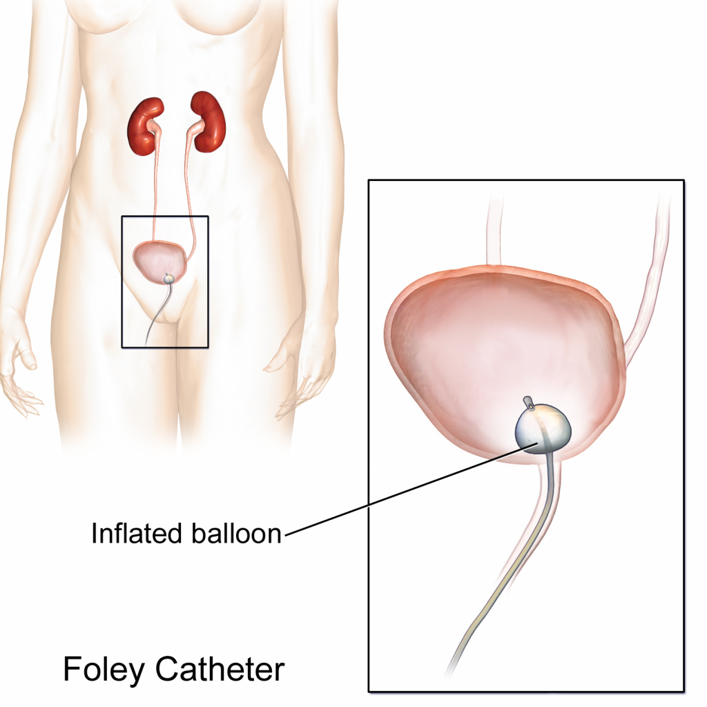
An indwelling catheter, often referred to as a “Foley catheter,” refers to a urinary catheter that remains in place after insertion into the bladder for the continual collection of urine. It has a balloon on the insertion tip to maintain placement in the neck of the bladder. The other end of the catheter is attached to a drainage bag for the collection of urine. See Figure 21.1[1] for an illustration of the anatomical placement of an indwelling catheter in the bladder neck.
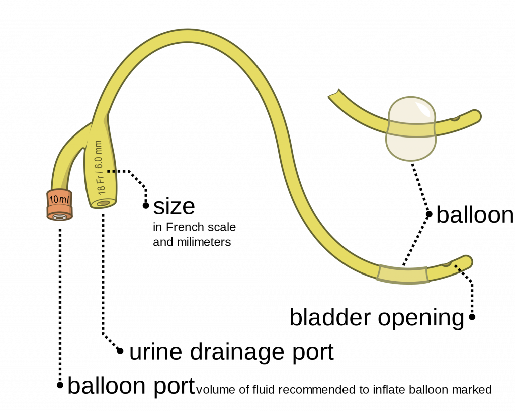
The distal end of an indwelling catheter has a urine drainage port that is connected to a drainage bag. The size of the catheter is marked at this end using the French catheter scale. A balloon port is also located at this end, where a syringe is inserted to inflate the balloon after it is inserted into the bladder. The balloon port is marked with the amount of fluid required to fill the balloon. See Figure 21.2[2] for an image of the parts of an indwelling catheter.
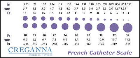
Catheters have different sizes, with the larger the number indicating a larger diameter of the catheter. See Figure 21.3[3] for an image of the French catheter scale.
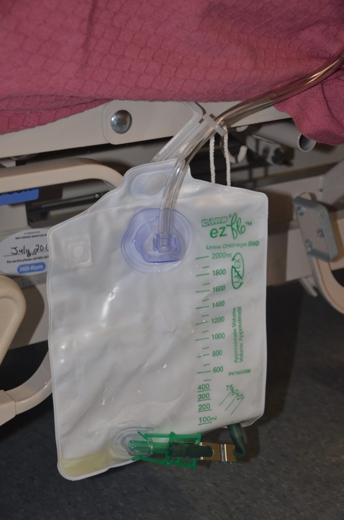
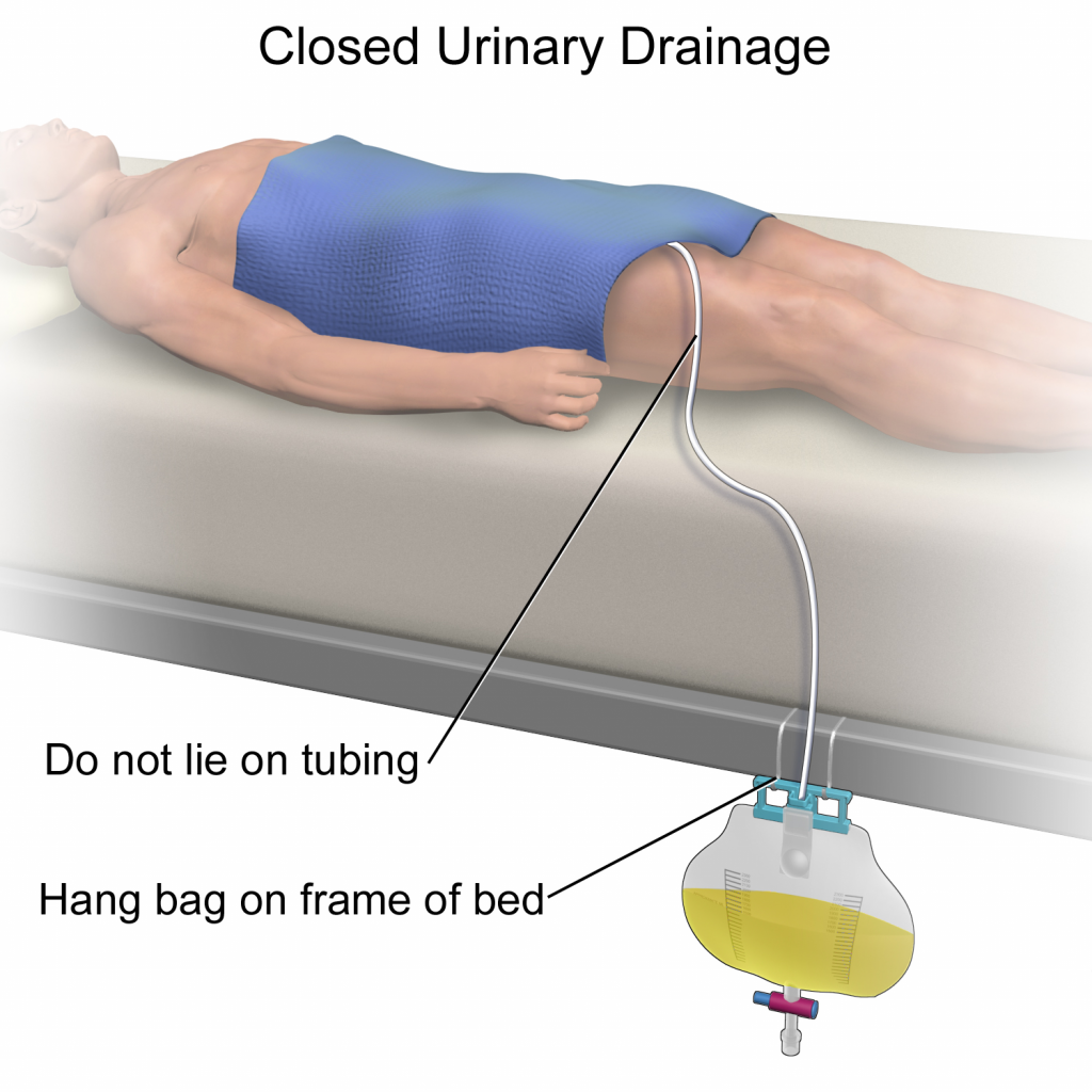
There are two common types of bags that may be attached to an indwelling catheter. During inpatient or long-term care, larger collection bags that can hold up to two liters of fluid are used. See Figure 21.4[4] for an image of a typical collection bag attached to an indwelling catheter. These bags should be emptied when they are half to two-thirds full to prevent traction on the urethra from the bag. Additionally, the collection bag should always be placed below the level of the patient’s bladder so that urine flows out of the bladder and urine does not inadvertently flow back into the bladder. Ensure the tubing is not kinked or compressed so that urine can flow unobstructed into the bag. Slack should be maintained in the tubing to prevent injury to the patient’s urethra. To prevent the development of a urinary tract infection, the bag should not be permitted to touch the floor.
See Figure 21.5[5] for an illustration of the placement of the urine collection bag when the patient is lying in bed.
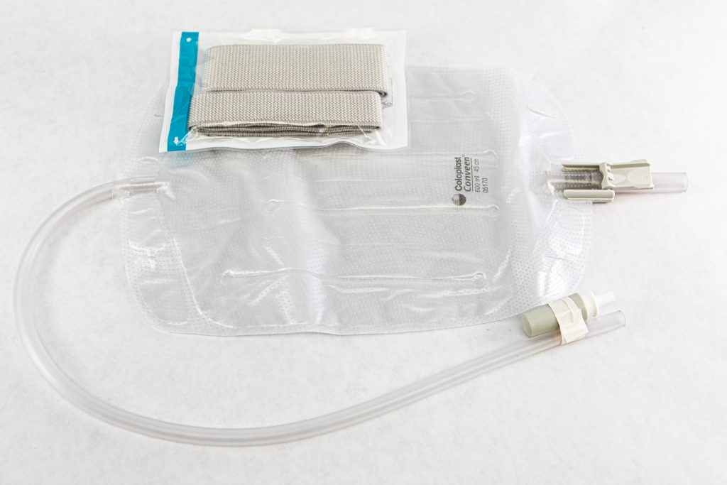
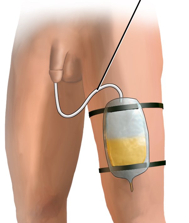
A second type of urine collection bag is a leg bag. Leg bags provide discretion when the patient is in public because they can be worn under clothing. However, leg bags are small and must be emptied more frequently than those used during inpatient care. Figure 21.6[6] for an image of leg bag and Figure 21.7[7] for an illustration of an indwelling catheter attached to a leg bag.
Straight Catheter
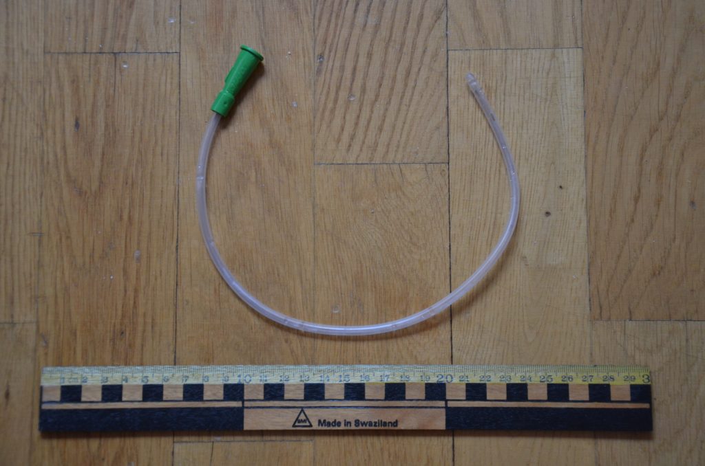
A straight catheter is used for intermittent urinary catheterization. The catheter is inserted to allow for the flow of urine and then immediately removed, so a balloon is not required at the insertion tip. See Figure 21.8[8] for an image of a straight catheter. Intermittent catheterization is used for the relief of urinary retention. It may be performed once, such as after surgery when a patient is experiencing urinary retention due to the effects of anesthesia, or performed several times a day to manage chronic urinary retention. Some patients may also independently perform self-catheterization at home to manage chronic urinary retention caused by various medical conditions. In some situations, a straight catheter is also used to obtain a sterile urine specimen for culture when a patient is unable to void into a sterile specimen cup. According to the Centers for Disease Control and Prevention (CDC), intermittent catheterization is preferred to indwelling urethral catheters whenever feasible because of decreased risk of developing a urinary tract infection.[9]
Other Types of Urinary Catheters
Coude Catheter Tip
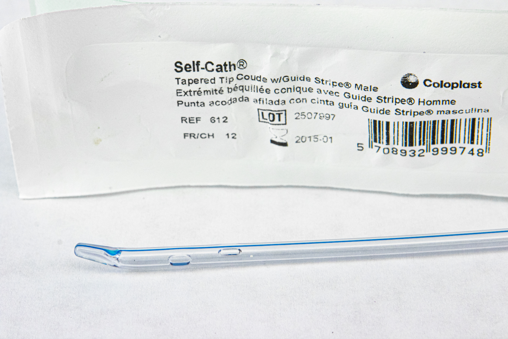
Coude catheter tips are curved to follow the natural curve of the urethra during catheterization. They are often used when catheterizing male patients with enlarged prostate glands. See Figure 21.9[10] for an example of a urinary catheter with a coude tip. During insertion, the tip of the coude catheter must be pointed anteriorly or it can cause damage to the urethra. A thin line embedded in the catheter provides information regarding orientation during the procedure; maintain the line upwards to keep it pointed anteriorly.
Irrigation Catheter
Irrigation catheters are typically used after prostate surgery to flush the surgical area. These catheters are larger in size to allow for irrigation of the bladder to help prevent the formation of blood clots and to flush them out. See Figure 21.10[11] for an image comparing a larger 20 French catheter (typically used for irrigation) to a 14 French catheter (typically used for indwelling catheters).

Suprapubic Catheters
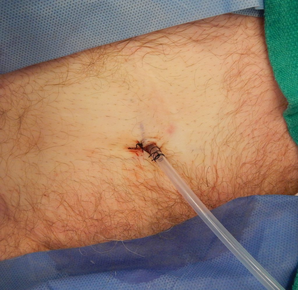
Suprapubic catheters are surgically inserted through the abdominal wall into the bladder. This type of catheter is typically inserted when there is a blockage within the urethra that does not allow the use of a straight or indwelling catheter. Suprapubic catheters may be used for a short period of time for acute medical conditions or may be used permanently for chronic conditions. See Figure 21.11[12] for an image of a suprapubic catheter. The insertion site of a suprapubic catheter must be cleaned regularly according to agency policy with appropriate steps to prevent skin breakdown.
Male Condom Catheter
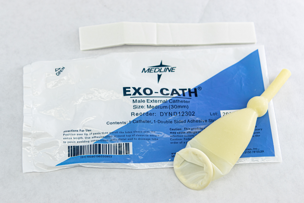
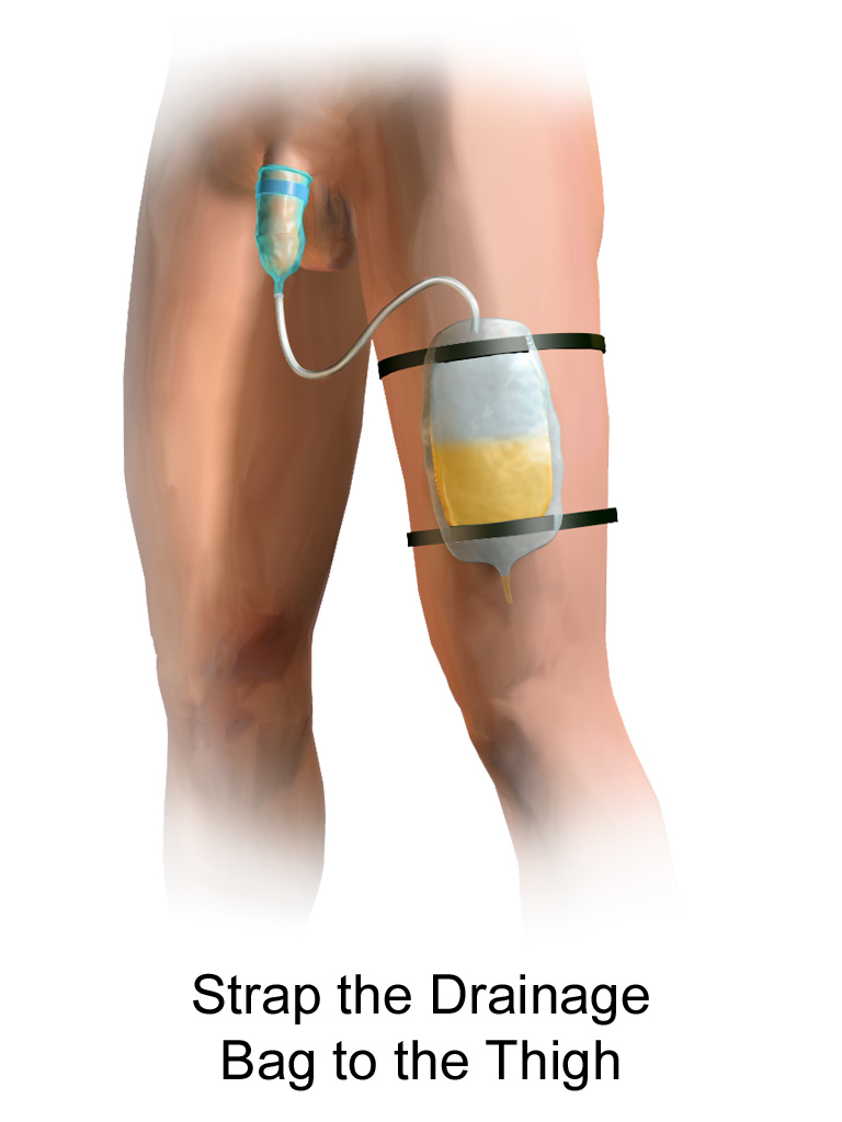
A condom catheter is a noninvasive device used for males with incontinence. It is placed over the penis and connected to a drainage bag. This device protects and promotes healing of the skin around the perineal area and inner legs and is used as an alternative to an indwelling urinary catheter. See Figure 21.12[13] for an image of a condom catheter and Figure 21.13[14] for an illustration of a condom catheter attached to a leg bag.
Female External Urinary Catheter
Female external urinary catheters (FEUC) have been recently introduced into practice to reduce the incidence of catheter-associated urinary tract infection (CAUTI) in women.[15] The external female catheter device is made of a purewick material that is placed externally over the female’s urinary meatus. The wicking material is attached to a tube that is hooked to a low-suction device. When the wick becomes saturated with urine, it is suctioned into a drainage canister. Preliminary studies have found that utilizing the FEUC device reduced the risk for CAUTI.[16],[17]
View these supplementary YouTube videos on female external urinary catheters:
Students demonstrate use of PureWick female external catheter[18]
How to use the use the PureWick – a female external catheter[19]
- “Foley Catheter.png” by BruceBlaus is licensed under CC BY-SA 4.0 ↵
- “Foley catheter inflated and deflated EN.svg” by Olek Remesz (wiki-pl: Orem, commons: Orem) is licensed under CC BY-SA 3.0 ↵
- “French catheter scale.gif” by Glitzy queen00 is licensed under CC BY-SA 3.0 ↵
- “DSC_2104.jpg” by British Columbia Institute of Technology is licensed under CC BY 4.0. Access for free at https://ecampusontario.pressbooks.pub/clinicalskills/chapter/2-2-head-to-toe-assessment-checklist/ ↵
- “Closed Urinary Drainage.png” by BruceBlaus is licensed under CC BY-SA 4.0 ↵
- “Leg Bag_3I3A0667.jpg” by Deanna Hoyord, Chippewa Valley Technical College is licensed under CC BY 4.0 ↵
- “Foley Catheter Drainage (cropped).png” by BruceBlaus is licensed under CC BY-SA 4.0 ↵
- “Urinary catheter.JPG” by Bengt Oberger is licensed under CC BY-SA 3.0 ↵
- Centers for Disease Control and Prevention. (2015, November 15). Catheter-associated urinary tract infections (CAUTI). https://www.cdc.gov/infectioncontrol/guidelines/cauti/ ↵
- “Self Cath_3I3A0676.jpg” by Deanna Hoyord, Chippewa Valley Technical College is licensed under CC BY 4.0 ↵
- “Irrigation Catheter and a 14 Fr. Catheter - 3I3A0753.jpg” by Deanna Hoyord, Chippewa Valley Technical College is licensed under CC BY 4.0 ↵
- “NewlyPlaceSubprapubic.jpg” by James Heilman, MD is licensed under CC BY-SA 4.0 ↵
- “Male External Cath 1 .jpg” by Deanna Hoyord, Chippewa Valley Technical College is licensed under CC BY 4.0 ↵
- “Condom Cather Drainage.png” by BruceBlaus is licensed under CC BY-SA 4.0 ↵
- Eckert, L., Mattia, L., Patel, S., Okumura, R., Reynolds, P., & Stuiver, I. Reducing the risk of indwelling catheter-associated urinary tract infection in female patients by implementing an alternative female external urinary collection device: A quality improvement project. Journal of Wound Ostomy Continence Nursing, 47(1), 50-53. https://doi.org/10.1097/won.0000000000000601 ↵
- Eckert, L., Mattia, L., Patel, S., Okumura, R., Reynolds, P., & Stuiver, I. Reducing the risk of indwelling catheter-associated urinary tract infection in female patients by implementing an alternative female external urinary collection device: A quality improvement project. Journal of Wound Ostomy Continence Nursing, 47(1), 50-53. https://doi.org/10.1097/won.0000000000000601 ↵
- Glover, E., Bleeker, E., Bauermeister, A., Koehlmoos, A., & Van Whye, M. (2018). External catheters and reducing adverse effects in the female inpatient. Northwestern College Department of Nursing. https://nwcommons.nwciowa.edu/cgi/viewcontent.cgi?article=1026&context=celebrationofresearch ↵
- Madrid, S. (2019, June 19). Purewick [Video]. YouTube. All rights reserved. https://youtu.be/1rnQaHvIMBc ↵
- Newton, C. (2016, August 4). PureWick user instructions [Video]. YouTube. All rights reserved.https://youtu.be/xSOuvcShikw ↵
The insertion of a catheter tube into the urethral opening and placing it in the neck of the urinary bladder to drain urine.
A device often referred to as a “Foley catheter” that is inserted into the neck of the bladder and remains in place for continual collection of urine into a collection bag.
A catheter used for intermittent urinary catheterization; it does not have a balloon at the insertion end.
The insertion and removal of a straight catheter for relief of urinary retention.
A catheter is specifically designed to maneuver around obstructions or blockages in the urethra such as with enlarged prostate glands in males.

