20.10 Checklist for Intermittent Suture Removal
Sutures are tiny threads, wire, or other material used to sew body tissue and skin together. They may be placed deep in the tissue and/or superficially to close a wound. The most commonly seen suture is the intermittent suture.
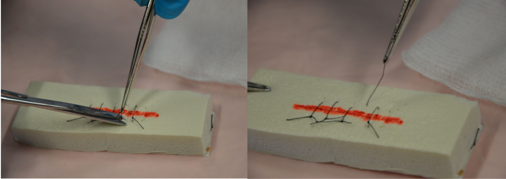
Sutures may be absorbent (dissolvable) or nonabsorbent (must be removed). Nonabsorbent sutures are usually removed within 7 to 14 days. Suture removal is determined by how well the wound has healed and the extent of the surgery. See Figure 20.32[1] for an example of suture removal. Sutures must be left in place long enough to establish wound closure with enough strength to support internal tissues and organs. If sutures are removed too early in the wound healing process, dehiscence can occur. The wound line must be observed for separations during the process of suture removal and the procedure stopped if there are any concerns.
The health care provider must assess the wound to determine whether or not to remove the sutures and provide an order for the removal of sutures. Alternate sutures (every second suture) may be removed first, and then the remaining sutures removed after adequate approximation of the skin tissue is determined. If the wound is well-healed, all the sutures may be removed at the same time, but if there are concerns about approximation, the removal of the remaining sutures may be delayed for several days to avoid dehiscence. Steri-Strips may be applied prior to suture removal to lessen the chance of wound dehiscence. See Figure 20.33.[2]


Checklist for Intermittent Suture Removal
Use the checklist below to review the steps for completion of “Intermittent Suture Removal.”
Steps
Disclaimer: Always review and follow agency policy regarding this specific skill.
- Gather supplies: sterile suture scissors, sterile dressing tray (to clean incision site prior to suture removal), nonsterile gloves, normal saline, Steri-Strips, and sterile outer dressing.
- Perform safety steps:
- Perform hand hygiene.
- Check the room for transmission-based precautions.
- Introduce yourself, your role, the purpose of your visit, and an estimate of the time it will take.
- Confirm patient ID using two patient identifiers (e.g., name and date of birth).
- Explain the process to the patient and ask if they have any questions.
- Be organized and systematic.
- Use appropriate listening and questioning skills.
- Listen and attend to patient cues.
- Ensure the patient’s privacy and dignity.
- Assess ABCs.
- Confirm provider order and explain procedure to patient. Inform the patient that the procedure is not painful, but they may feel some pulling of the skin during suture removal.
- Prepare the environment, position the patient, adjust the height of the bed, and turn on the lights. Ensuring proper lighting allows for good visibility to assess the wound. Ensure proper body mechanics for yourself and create a comfortable position for the patient.
- Perform hand hygiene and put on nonsterile gloves.
- Place a clean, dry barrier on the bedside table. Add necessary supplies.
- Remove dressing and inspect the wound. Visually assess the wound for uniform closure of the wound edges, absence of drainage, redness, and swelling. After assessing the wound, decide if the wound is sufficiently healed to have the sutures removed. If there are concerns, discuss the order with the appropriate health care provider.
- Remove gloves and perform hand hygiene.
- Put on a new pair of nonsterile or sterile gloves, depending on the patient’s condition and the type, location, and depth of the wound.
- Irrigate the wound with sterile normal saline solution to remove surface debris or exudate (and to help prevent specimen contamination if collecting a specimen). Alternatively, commercial wound cleanser may be used. This step reduces risk of infection from microorganisms on the wound site or surrounding skin. Cleaning also loosens and removes any dried blood or crusted exudate from the sutures and wound bed.
- To remove intermittent sutures, hold the scissors in your dominant hand and the forceps in your nondominant hand for dexterity with suture removal.
- Place a sterile 2″ x 2″ gauze close to the incision site to collect the removed suture pieces.
- Grasp the knot of the suture with the forceps and gently pull up the knot while slipping the tip of the scissors under the suture near the skin. Examine the knot.
- Cut under the knot as close as possible to the skin at the distal end of the knot:
- Never snip both ends of the knot as there will be no way to remove the suture from below the surface.
- Do not pull the contaminated suture (suture on top of the skin) through tissue.
- Grasp the knotted end of the suture with forceps, and in one continuous action pull the suture out of the tissue and place it on the sterile 2″ x 2″ gauze.
- Remove every second suture until the end of the incision line. Assess wound healing after removal of each suture to determine if each remaining suture will be removed. If the wound edges are open, stop removing sutures, apply Steri-Strips (using tension to pull wound edges together), and notify the appropriate health care provider. Remove remaining sutures on the incision line if indicated.
- Using the principles of no-touch technique, cut and place Steri-Strips along the incision line:
- Cut Steri-Strips so that they extend 1.5 to 2 inches on each side of the incision.
- Remove gloves and perform hand hygiene.
- Assist the patient to a comfortable position, ask if they have any questions, and thank them for their time.
- Ensure safety measures when leaving the room:
- CALL LIGHT: Within reach
- BED: Low and locked (in lowest position and brakes on)
- SIDE RAILS: Secured
- TABLE: Within reach
- ROOM: Risk-free for falls (scan room and clear any obstacles)
- Document the procedure and related assessment findings of the incision. Report any concerns according to agency policy.
- “DSC_0262.jpg” and “DSC_0263.jpg” by British Columbia Institute of Technology are licensed under CC BY 4.0. Access for free at https://opentextbc.ca/clinicalskills/chapter/4-3-suture-care-and-removal/ ↵
- “DSC_1658.jpg” and “DSC_09811.jpg” by British Columbia Institute of Technology are licensed under CC BY 4.0. Access for free at https://opentextbc.ca/clinicalskills/chapter/4-3-suture-care-and-removal/ ↵

