9.6 Diseases and Disorders of the Cardiovascular System
Aneurysm
An aneurysm (AN-yŭ-rizm) is a dilation or bulging of a blood vessel caused by weakened vessel walls. Aneurysms can occur in vessels throughout the body but are more commonly found in arteries due to their increased pressure. Aneurysms can be caused by a variety of factors, including smoking, atherosclerosis (fatty plaque buildup in a vessel), and hypertension.[1] See Figure 9.9[2] for an illustration of thoracic and abdominal aneurysms.
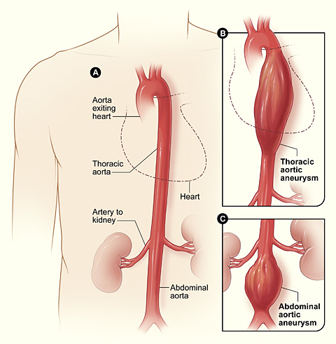
Aneurysms are often asymptomatic and detected incidentally during diagnostic tests that are being done for other reasons. The force of blood pumping through the vessel with an aneurysm can split the layers of the artery wall, allowing blood to leak in between them, called a dissection. An aneurysm can burst completely, called a rupture, and cause severe, life-threatening bleeding. Aneurysms are managed with antihypertensive medications or may require surgical repair.[3]
Arrhythmia
A heart rhythm that follows the regular conduction system of the heart is called normal sinus rhythm (SĪ-nŭs RITH-ŭm). For example, bradycardia and tachycardia that occurs during normal sinus rhythm have abnormally slow or fast rates but have a regular rhythm.
- Bradycardia (brad-ē-KAR-dē-ă) refers to a slow resting heart rate of less than 60 beats per minute. A normal resting heart rate for an adult is 60 to 100 beats per minute (bpm). However, heart rates can vary widely among individuals based on several factors like age, fitness level, overall health, and medication side effects. Physically fit athletes may have a heart rate of 50 to 60 bpm at rest with no symptoms. However, individuals with bradycardia whose heart is not pumping enough blood to sufficiently supply the brain and body tissues with oxygen may have symptoms, such as feeling tired, dizzy, short of breath, or confused. Individuals with symptomatic bradycardia may experience syncope (SING-kŏ-pē), a medical term for a brief lapse in consciousness, commonly referred to as fainting. Bradycardia is a common side effect of many cardiac medications and can also be caused by several cardiac diseases and disorders. Treatment of symptomatic bradycardia includes medications to increase heart rate or surgical implantation of a pacemaker in the heart to maintain a normal heart rate.
- Tachycardia (tak-ē-KAR-dē-ă) is a rapid heart rate greater than 100 bpm. Tachycardia can be caused by stress, side effects of medications, and several medical conditions. When the heart beats too rapidly, its chambers do not have time to sufficiently fill with blood, resulting in a decreased amount of oxygenated blood being pumped to body tissues and organs. For this reason, symptoms of tachycardia are similar to symptoms of bradycardia. The individual may also have palpitations (pal-pĭ-TĀ-shŏnz), a sensation that the heart is racing, pounding, fluttering, or skipping a beat. Treatment is based on the underlying cause and may include medications or surgery.
Arrhythmia (ā-RITH-mē-ă) does not mean an absence of a heartbeat, but rather means the absence of a normal heart rhythm due to disrupted electrical activity of the conduction system. Examples of common arrhythmias are atrial fibrillation, heart block, ventricular tachycardia, and ventricular fibrillation.
- Atrial fibrillation (Ā-trē-ăl fĭb-rĭ-LĀ-shŏn) (A Fib) is characterized by atrial quivering instead of contracting. A Fib can cause a decrease in the volume of blood propelled with each heartbeat because the ventricles do not completely fill with blood before contracting. A Fib also increases the risk of a cerebrovascular accident (also known as a stroke).
- Heart block (härt blŏk) refers to a disruption in the normal conduction pathway of electrical signals through the atria and ventricles. Heart block can vary in severity from first-degree heart block, which is the least severe and often has no symptoms, to third-degree heart block that can be life-threatening. Symptoms of heart block may include bradycardia, missed heart beats, fatigue, dizziness, chest pain, and shortness of breath.
- Ventricular tachycardia (ven-TRĬK-yū-lăr tak-ĭ-KAR-dē-ă) (V Tach) refers to a very rapid heartbeat that originates in the ventricles. Ventricular fibrillation (ven-TRĬK-yū-lăr fĭb-rĭ-LĀ-shŏn) (V Fib) is a disorganized heart rhythm that causes the ventricles to quiver instead of contract. Ventricular tachycardia and ventricular fibrillation are life-threatening arrhythmias that require rapid emergency response and the use of a defibrillator to resolve the abnormal rhythm.
A defibrillator (dē-FIB-rĭ-lā-tŏr) is a device used by trained medical personnel who apply an electrical charge to the heart in an attempt to restart the SA node and establish a normal sinus rhythm. Automated external defibrillators (aw-tŏ-māt′-ĕd ĕks-tĕr′năl dē-fĭb′-rĭ-lā-tŏrs) (AEDs) are life-saving devices available in public areas to help individuals who are experiencing a cardiac arrest (KAR-dē-ăk ăr-REST) survive. Cardiac arrest is caused by arrhythmias that cause the heart to suddenly stop pumping blood. The average response time after calling 911 is 8 to 12 minutes, and for each minute defibrillation is delayed, the odds of survival are reduced by approximately 10%, so having access to an AED and knowing how to perform cardiopulmonary resuscitation (CPR) are critical. CPR refers to repeated compressions of the chest in an attempt to restore blood circulation following a cardiac arrest. While CPR is being performed, special pads are applied to the individual’s chest. The AED automatically analyzes the heart’s rhythm and, if indicated, delivers an electrical shock to help the heart reestablish an effective rhythm until paramedics arrive. See Figure 9.10[4] for an image of an AED.
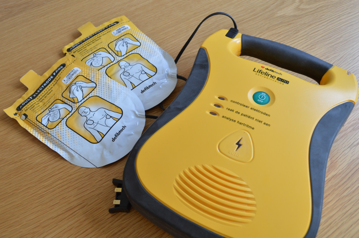
Individuals who have experienced a cardiac arrest due to life-threatening arrhythmias may have an implantable cardioverter defibrillator (ĭm-plănt′-ă-bŭl kär′dē-ō-vûr′-tŏr dē-fĭb′-rĭ-lā-tŏr) (ICD) surgically placed in their chest. An ICD continuously analyzes the heart rhythm and provides an automatic electric shock to convert a dangerous heart rhythm to a normal heart rhythm.
Atherosclerosis
Atherosclerosis (ă-thĕr-ō-sklĕ-RO-sĭs) refers to hardening and narrowing of arteries due to the buildup of cholesterol and other fats called plaque within the lining of the arteries. Plaque creates inflammation and decreases the amount of blood flow through an artery, also referred to as decreased perfusion. Plaque can also grow so large that is causes a blockage of blood flow in the artery. When this accumulation of plaque occurs in the coronary arteries of the heart, it is called coronary artery disease (KOR-ŏ-nā-rē AR-tĕr-ē dĭ-ZĒZ) (CAD) or atherosclerotic heart disease. Read more information about coronary artery disease in a later subsection of this section. See Figure 9.11[5] for an illustration of the narrowing of a coronary artery by an atherosclerotic plaque.
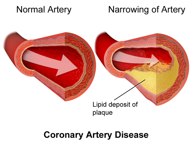
Cerebrovascular disease (sĕr′-brō-VAS-kyū-lăr dĭ-ZĒZ) (CVD) occurs when arteries in the brain or neck become thick, hard, or full of plaque and blood flow is restricted, which can cause a cerebrovascular accident (commonly known as a stroke). Peripheral artery disease (pĕr-IF-ĕr-ăl AR-tĕr-ē dĭ-ZĒZ) (PAD) occurs when blood vessels are obstructed in peripheral regions of the body, commonly causing pain during activity and possibly causing ulcers in the legs and feet.
A clinician may hear a bruit (brwē) when auscultating over an artery if atherosclerosis is present. A bruit sounds like an abnormal blowing, swishing sound. For example, a carotid bruit indicates the individual is at risk for a stroke.
Cardiomyopathy
Cardiomyopathy (kar-dē-ō-my-OP-ă-thē) refers to disease of the heart muscle. When cardiomyopathy occurs, the normal muscle in the heart can thicken, enlarge, stiffen, or thin out. As a result, the heart muscle’s ability to pump blood is reduced, which can lead to irregular heartbeats or the backup of blood into the lungs or rest of the body. Cardiomyopathy can be acquired (i.e., developed because of another disorder), inherited, or occur due to an unknown cause. Categories of cardiomyopathy include dilated, hypertrophic, and restrictive.[6] See Figure 9.12[7] for an illustration of common types of cardiomyopathy.
![]"Major_categories_of_cardiomyopathy.png" by Npatchett is licensed under CC BY-SA 4.0 Illustration showing Categories of Cardiomyopathy](https://wtcs.pressbooks.pub/app/uploads/sites/46/2023/05/Major_categories_of_cardiomyopathy.png)
Congenital Heart Conditions
A congenital (kŏn-JĔN-ĭ-tăl) condition means it is present at birth. There are several types of congenital heart disorders. Some are caused by openings present in the heart that do not close as they should after birth. See Figure 9.13[8] for an illustration of common congenital cardiac disorders.
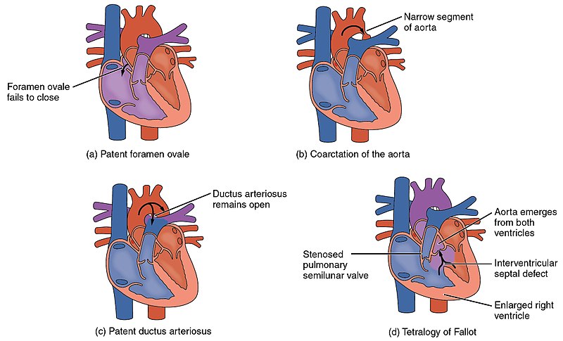
- Patent ductus arteriosus (PĀ-tĕnt DŬK-tŭs är-tēr′-ē-ō-sŭs) occurs when the ductus arteriosus fails to close after birth. The ductus arteriosus is a vessel in the fetal heart that connects the pulmonary trunk and aorta, bypassing the pulmonary circulation.
- Patent foramen ovale (PĀ-tĕnt fŏ-rā′-mĕn ō-vā′-lē) is a hole in the septum between the atria of the heart that does not close at birth.
- Coarctation (kō-ark-TĀ-shŏn) of the aorta is a narrowing of the aorta that forces the heart to pump harder to move blood through the aorta.
- Atrial septal defect (Ā-trē-ăl SĔP-tăl dĭ-FĔKT) (ASD) refers to an opening that exists in the septum (wall) between the left and right atria.
- Ventricular septal defect (ven-TRĬK-yū-lăr SĔP-tăl dĭ-FĔKT) (VSD) is an opening that exists in the septum (wall) between the left and right ventricles.
Tetralogy of Fallot (tĕt′ră-lō-jē ŏf fă-LŌ) is a serious congenital disorder with high mortality. It includes several abnormal conditions in the heart that allow unoxygenated blood from the right ventricle to flow into the left ventricle and mix with the blood that is relatively high in oxygen. Symptoms of Tetralogy of Fallot include a heart murmur, dyspnea, low blood oxygen saturation, clubbing of the fingers and toes, difficulty in eating, and failure to grow and develop. A murmur (MUR-mŭr) is an abnormal heart sound heard on auscultation by a stethoscope that may sound like a “whooshing” sound. Tetralogy of Fallot is diagnosed by echocardiography. Treatment involves extensive surgical repair.[9]
Coronary Artery Disease
Coronary artery disease (KOR-ŏ-nā-rē AR-tĕr-ē dĭ-ZĒZ) (CAD) occurs when atherosclerosis (i.e., buildup of plaque) in the coronary arteries obstructs the flow of oxygenated blood to the heart muscle. As oxygenated blood flow to the cardiac muscle cells is reduced, referred to as ischemia (ĭs-KEE-mē-ă), individuals often experience chest pain called angina (an-JĪ-nă). See Figure 9.14[10] for an image of a blockage of coronary arteries on an angiogram.
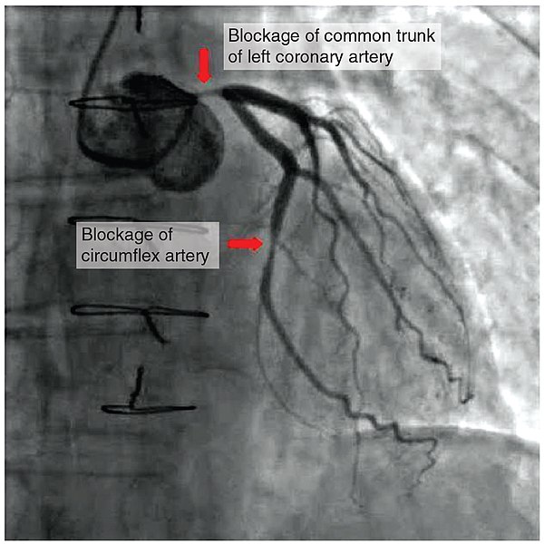
CAD is progressive and, if left untreated, can cause a myocardial infarction (MI). Risk factors include smoking, family history, hypertension, obesity, diabetes, lack of physical activity, stress, hyperlipidemia, and excessive alcohol use. Treatments may include medication, changes to diet and exercise, angioplasty with insertion of a stent, or coronary artery bypass graft (CABG).[11] Read more about these procedures in the “Medical Specialists, Diagnostic Testing, and Procedures Related to the Cardiovascular System” section.
Deep Vein Thrombosis
A deep vein thrombosis (dēp vān THROM-bō-sĭs) (DVT) is a blood clot in a deep vein that most often occurs in the lower extremities. DVTs can break free and become life-threatening emboli that travel to other areas of the body and cause blockages. For example, a pulmonary embolus (PE) is a medical emergency caused by a DVT that became an embolus and lodged in the lung.
Symptoms of a DVT often include calf pain in one leg with swelling, redness, and warmth of that leg. A DVT is diagnosed with a Doppler ultrasound that identifies altered blood flow in a vessel due to a clot.[12]
Edema
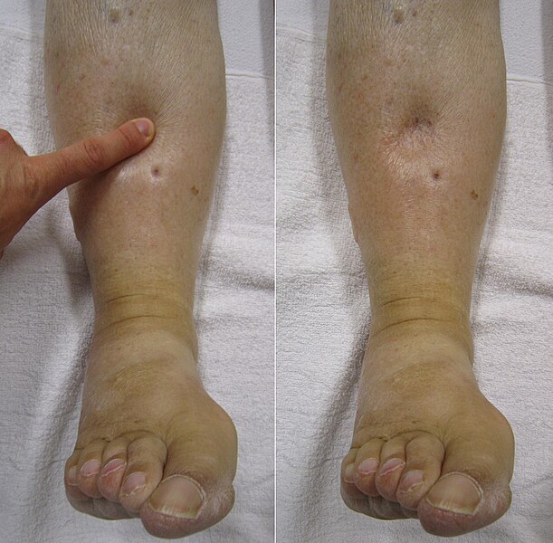
Despite the presence of valves within veins that prevent the backflow of blood, over the course of a day, some blood will inevitably pool in the lower limbs, due to the pull of gravity. Any blood that accumulates in a vein will increase the pressure within it. Increased pressure will promote the flow of fluids out of the capillaries and into the interstitial fluid. The presence of excess tissue fluid around the cells leads to a condition called edema (e-DĒ-ma). Medications, such as diuretics, and compression stockings are used to help minimize the edema. See Figure 9.15[13] for an image of a patient with edema.
Heart Failure
Heart failure (HART FĀL-yŭr) (HF) occurs when the heart loses its effectiveness in pumping blood. When the heart is not pumping effectively, symptoms of fatigue, shortness of breath, edema, or lung congestion can occur. Chronic HF is a progressive disorder that can be represented on a continuum. The continuum ranges from individuals who are at risk for HF but are asymptomatic, to individuals who have end-stage heart failure and require end-of-life care. Treatments may include medication, a low sodium diet, and fluid restrictions.[14] See Figure 9.16[15] for an illustration of common symptoms of heart failure.
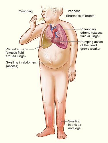
There are many potential causes of HF, including coronary artery disease (CAD), myocardial infarction (MI), hypertension, valve disorders, and cardiomyopathy. Treatment for heart failure may include medications to eliminate excess fluid, a low salt diet, fluid restrictions, implanted devices that assist the heart to pump blood, and heart transplant surgery.[16]
Hypertension
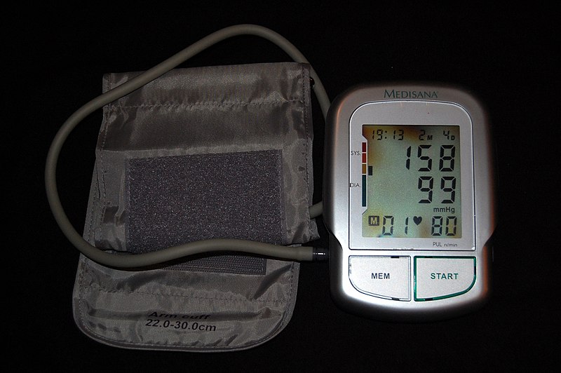
Hypertension (hī-pĕr-TEN-shŏn) refers to elevated blood pressure greater than 120/80 mm Hg in an adult. Hypertension is very common, affecting millions of individuals worldwide. See Figure 9.17[17] for an image of a sphygmomanometer (sfig-mō-mă-NOM-ĕt-ĕr) used to obtain blood pressure readings.
Risk factors for developing hypertension include age, ethnicity, family history, and lifestyle factors. Individuals with obesity, diets high in sodium, excessive alcohol intake, chronic stress, and limited physical activity are at higher risk for developing hypertension. Many people with hypertension do not have symptoms, so they are unaware they have the disorder until a complication like a myocardial infarction, stroke, or heart failure occurs. Treatment includes medications and healthy lifestyle changes like adopting a heart-healthy diet, exercising regularly, stopping smoking, getting good sleep, losing weight, and managing stress.[18]
Myocardial Infarction
Myocardial infarction (mī-ŏ-kar′dē-ăl in-FARK-shŏn) (MI), commonly called a heart attack, is caused by blockage of blood flow to the heart tissue, resulting in death of the cardiac muscle cells. Acute coronary syndrome (ə-KYŪT KOR-ŏ-nā-rē SĬN-drōm) (ACS) is a general term used for medical situations in which blood supplied to the heart muscle is suddenly insufficient. An MI is commonly caused by atherosclerosis causing a blockage in a coronary artery. It can also occur when a piece of an atherosclerotic plaque breaks off and travels through the coronary arterial system until it lodges in one of the smaller vessels. See Figure 9.18[19] for a depiction of an individual experiencing an MI and the damage occurring to the cardiac muscle as a result of the coronary artery blockage.
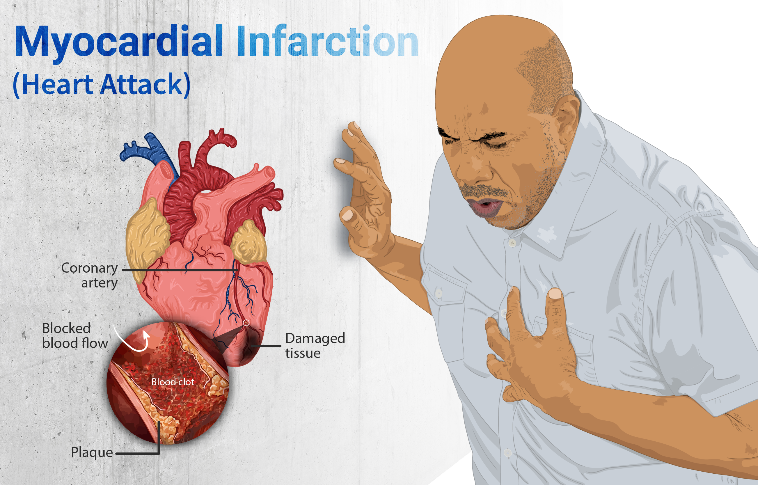
A classic symptom of an MI includes sudden crushing pain beneath the sternum called angina (an-JĪ-nă) that may radiate down the left arm. Individuals often describe angina as “it feels as if something is sitting on my chest.” Other common symptoms include dyspnea, diaphoresis, nausea, and light-headedness. Diaphoresis (dī-ă-fŏ-RĒ-sĭs) means profuse (excessive) sweating. However, females may not experience these symptoms and only have a feeling similar to indigestion.[20]
An MI is a medical emergency. The faster the response, the more cardiac muscle cells that can be saved, so anyone who is suspected of having an MI should call 911. An MI is diagnosed with an electrocardiogram (ē-lĕk-trō-KĂR-dē-ō-grăm) (ECG) and blood tests called cardiac enzymes. Cardiac enzymes include creatine kinase and cardiac troponin, both of which are released by damaged cardiac muscle cells.[21]
Immediate treatment typically includes administration of oxygen, aspirin (to stop the plaque from growing), and a medication called nitroglycerin (to help dilate the blood vessels to get more oxygenated blood to the cardiac muscle). Based on the results of the ECG and cardiac enzyme tests, the individual may undergo thrombolysis (thrŏm-BOL-ĭ-sĭs), a procedure that involves administering a clot-dissolving medication to restore blood flow in a coronary artery. An alternative to thrombolysis is angioplasty (AN-jee-ō-plas-tē) or coronary artery bypass graft surgery (KOR-ŏ-nā-rē AR-tĕr-ē bī-păs graft Sŭr-jĕr-ē) (CABG). Read more about these procedures in the “Medical Specialists, Diagnostic Testing, and Procedures Related to Cardiovascular System” section of this chapter.[22]
Valvular Heart Disease
The heart has four valves that open and close at specific times during the cardiac cycle to control or regulate the blood flowing into and out of the heart. See Figure 9.19[23] for an illustration of the heart valves including the tricuspid, pulmonary, mitral, and aortic. Three of the heart valves (tricuspid, pulmonary, and aortic valves) are composed of three leaflets or flaps that work together to open and close to allow blood to flow forward and not backwards through the opening. The mitral valve (also known as the bicuspid valve) only has two flaps that open and close.
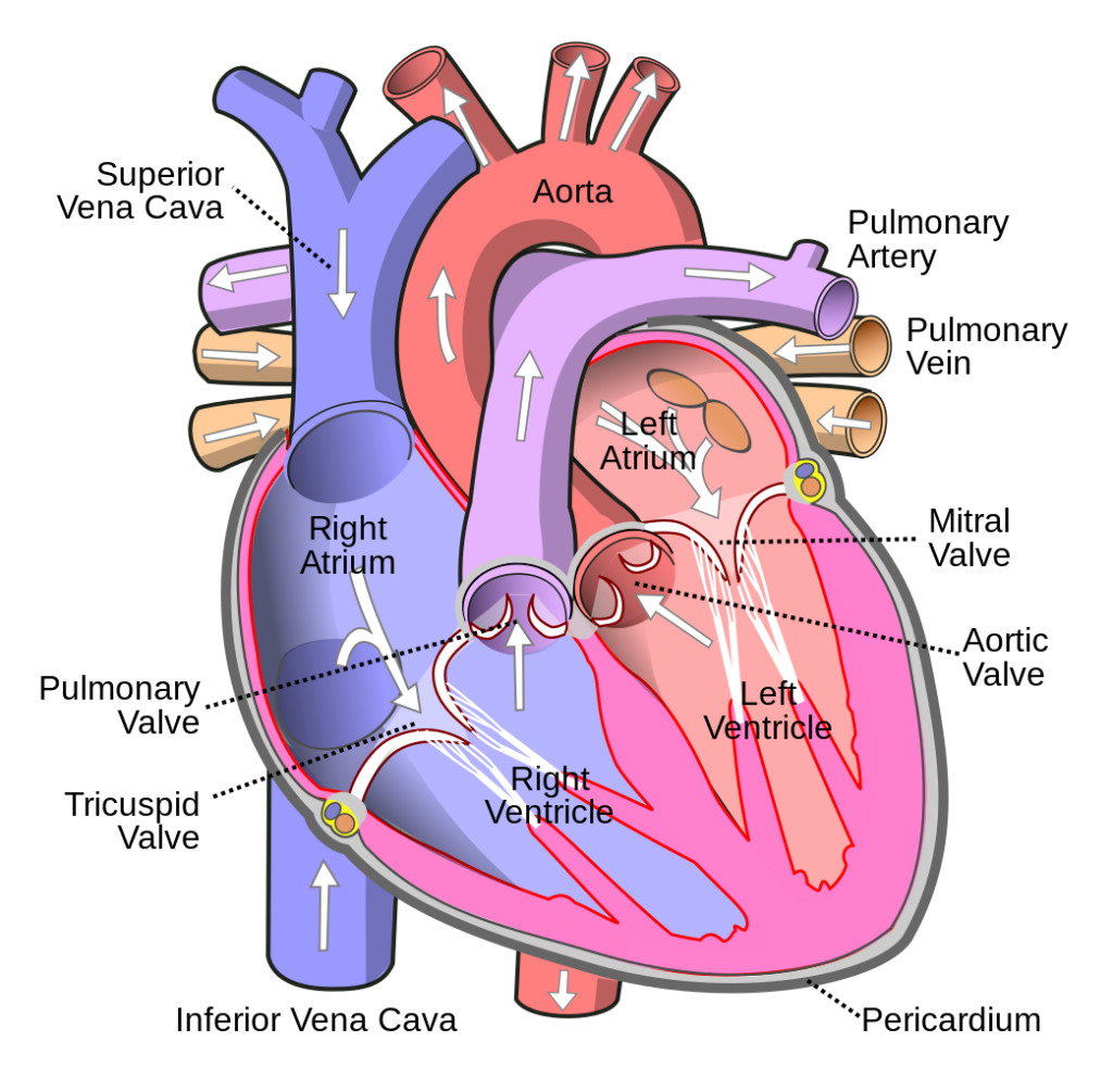
Healthy heart valves fully open and close during the heartbeat, but patients with valvular heart disease (văl′-vyū-lăr härt dĭ-zēz′) have diseased valves that do not fully open and close. Diseased valves that become “leaky” and don’t completely close cause a condition called regurgitation (rē-gŭr′-jĭ-tā′-shŭn). When regurgitation occurs, blood flows backwards and not enough blood is pushed forward through the heart. Aortic insufficiency (ā-OR-tĭk in-sŭ-FISH-ĕn-sē) (AI) is a valvular heart disease that occurs when the aortic valve does not close properly and allows regurgitation of blood back into the left ventricle.
Another type of valve disorder occurs when the opening of the valve is narrowed and stiff, causing it to not open fully when blood is trying to pass through. This disorder is called stenosis (stĕ-NŌ-sĭs).[24]
There are several causes of valvular heart disease, including congenital heart conditions, infections, degenerative conditions (i.e., they wear out with age), and conditions linked to other types of heart disease. A murmur (MUR-mŭr), an abnormal heart sound heard on auscultation by a stethoscope, is a classic sign of a valvular heart disorder. Other symptoms include shortness of breath, chest pain, fatigue, dizziness, and palpitations.[25]
Valvular heart disease is diagnosed by echocardiography. Treatment may include medications to treat the symptoms or surgery to repair or replace the valves.[26] Read more about these procedures in the “Medical Specialists, Diagnostic Testing, and Procedures Related to the Cardiovascular System.”
Varicose Veins
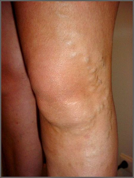
Varicose veins (VAR-i-kōs VĀNZ) are a common condition caused by weak or damaged vein walls and valves. Veins have one-way valves that open and close to keep blood flowing toward the heart. Weak or damaged valves or walls in the veins can cause blood to pool and even flow backward.[27] See Figure 9.20[28] for an image of varicose veins.
Symptoms include bulging, bluish veins, and a feeling of heaviness or discomfort in the legs and feet. Without treatment, they tend to grow worse over time. The use of compression stockings (commonly known as TED hose), as well as elevating the feet and legs whenever possible, may be helpful in alleviating the symptoms of varicose veins. However, severe cases may require resolution through medical procedures.[29]
Procedures used to treat varicose veins include the following[30]:
- Endovenous laser ablation (ĕn′-dō-vē′-nŭs lā′-zŏr ăb-lā′-shŭn) to close off a varicose vein. A health care provider uses laser or radiofrequency energy to heat the inside of the vein and close it off.
- Sclerotherapy (sklĕr-ō-THER-ă-pē) to seal off a varicose vein. The health care provider injects liquid or foam chemicals into the vein to create a plug that seals it shut.
- Phlebectomy (flĕ-BĔK-tŏ-mē) to remove small varicose veins. The health care provider makes small cuts to remove smaller veins near the skin.
- Surgery to tie off and remove large varicose veins in a procedure called vein ligation or stripping.
- Centers for Disease Control and Prevention. (2021, September 27). Aortic aneurysm. Department of Health & Human Services. https://www.cdc.gov/heartdisease/aortic_aneurysm.htm ↵
- “Aortic_aneurysm.jpg” by en:National Institutes of Health is licensed in the Public Domain. ↵
- Centers for Disease Control and Prevention. (2021, September 27). Aortic aneurysm. Department of Health & Human Services. https://www.cdc.gov/heartdisease/aortic_aneurysm.htm ↵
- “aed_resuscitation_cardiac_arrest_first_aid_seh_acute_death_rescue-888373.jpg” by unknown author via Pxhere is licensed under CC0, Public Domain. ↵
- “Blausen_0259_CoronaryArteryDisease_02.png” by Blausen.com staff (2014). “Medical gallery of Blausen Medical 2014.” WikiJournal of Medicine is licensed under CC BY 3.0 ↵
- Centers for Disease Control and Prevention. (2023, February 21). Cardiomyopathy. Department of Health & Human Services. https://www.cdc.gov/heartdisease/cardiomyopathy.htm ↵
- “Major_categories_of_cardiomyopathy.png” by Npatchett is licensed under CC BY-SA 4.0 ↵
- “2009_Congenital_Heart_Defects.jpg” by OpenStax College is licensed under CC BY 3.0 ↵
- This work is a derivative of Anatomy & Physiology by OpenStax and is licensed under CC BY 4.0. Access for free at https://openstax.org/details/books/anatomy-and-physiology-2e ↵
- “2016_Occluded_Coronay_Arteries.jpg” by OpenStax College is licensed under CC BY 3.0 ↵
- Centers for Disease Control and Prevention. (2023, May 15). About heart disease. Department of Health & Human Services. https://www.cdc.gov/heartdisease/about.htm ↵
- This work is a derivative of StatPearls by Baker, Anjum, & dela Cruz and is licensed under CC BY 4.0 ↵
- “Combinpedal.jpg” by James Heilman, MD is licensed under CC BY-SA 3.0 ↵
- Centers for Disease Control and Prevention. (2023, April 14). Heart disease. Department of Health & Human Services. https://www.cdc.gov/heartdisease/about.htm ↵
- “Heartfailure.jpg” by National Heart, Lung, and Blood Institute, National Institutes of Health is licensed in the Public Domain. ↵
- Centers for Disease Control and Prevention. (2023, April 14). Heart disease. Department of Health & Human Services. https://www.cdc.gov/heartdisease/about.htm ↵
- “Grade_1_hypertension.jpg” by Steven Fruitsmaak is licensed under CC BY 3.0 ↵
- National Heart, Lung, and Blood Institute. (2022, March 24). What is high blood pressure. National Institutes of Health. https://www.nhlbi.nih.gov/health/high-blood-pressure ↵
- “Depiction_of_a_person_suffering_from_a_heart_attack_(Myocardial_Infarction).png” by https://www.myupchar.com/en is licensed under CC BY-SA 4.0 ↵
- American Heart Association. (n.d.). Heart attack. https://www.heart.org/en/health-topics/heart-attack ↵
- American Heart Association. (n.d.). Heart attack. https://www.heart.org/en/health-topics/heart-attack ↵
- American Heart Association. (n.d.). Heart attack. https://www.heart.org/en/health-topics/heart-attack ↵
- “Diagram_of_the_human_heart_(cropped).svg” by Wapcaplet is licensed under CC BY 3.0 ↵
- Centers for Disease Control and Prevention. (2019, December 9). Valvular heart disease. Department of Health & Human Services. https://www.cdc.gov/heartdisease/valvular_disease.htm ↵
- Centers for Disease Control and Prevention. (2019, December 9). Valvular heart disease. Department of Health & Human Services. https://www.cdc.gov/heartdisease/valvular_disease.htm ↵
- Centers for Disease Control and Prevention. (2019, December 9). Valvular heart disease. Department of Health & Human Services. https://www.cdc.gov/heartdisease/valvular_disease.htm ↵
- National Heart, Lung, and Blood Institute. (2023). Varicose veins. https://www.nhlbi.nih.gov/health/varicose-veins ↵
- “Varicose_ASV1.jpg” by Nini00 is licensed under CC BY-SA 3.0 ↵
- National Heart, Lung, and Blood Institute. (2023). Varicose veins. https://www.nhlbi.nih.gov/health/varicose-veins ↵
- National Heart, Lung, and Blood Institute. (2023). Varicose veins. https://www.nhlbi.nih.gov/health/varicose-veins ↵
