12.7 Medical Specialists, Diagnostic Testing, and Procedures Related to the Digestive System
Medical Specialists
Gastroenterology
Gastroenterology (găs-trō-ĕn-tĕr-ŎL-ă-jē) is the study of the normal function and diseases of the esophagus, stomach, small intestine, colon and rectum, pancreas, gallbladder, bile ducts, and liver. A physician who specializes in the diagnosis and treatment of gastrointestinal disorders is known as a gastroenterologist (găs-trō-ĕn-tĕr-ŎL-ŏ-jĭst).
A colorectal surgeon (kō-lō-REK-tăl SUR-jĕn) is a surgeon who specializes in conditions affecting the large intestine (i.e., colon, rectum, and anus.) A proctologist (prok-TOL-ŏ-jĭst) is a physician who specializes in the diagnosis and treatment of diseases of the rectum.
To learn more about gastroenterology, visit the American College of Gastroenterology web page.
Diagnostic Tests
Barium Enema
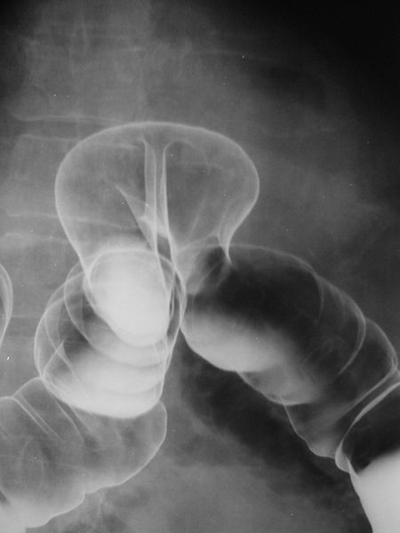
A barium enema (BĀR-ē-ŭm EN-ĕ-mă) is a diagnostic procedure involving the use of a contrast material known as barium. Barium is a chalky material that appears white on X-ray and coats the inner wall of the large intestine for visualization. During the procedure, barium is administered into the rectum, along with air (that appears black on X-ray). Fluoroscopy is often used during a barium enema. During fluoroscopy, a continuous X-ray is passed through the large intestine and is transmitted to a monitor so the movement of the barium through the large intestine can be visualized as it is instilled through the rectum. After the procedure, if the barium isn’t completely eliminated from the body, constipation or fecal impaction may occur.[1] See Figure 12.24[2] for an image of a barium enema showing herniation of the colon. The barium causes the colon to appear white on the left side of the image, and air in the colon causes it to appear black on the right side of the image.
Colonoscopy
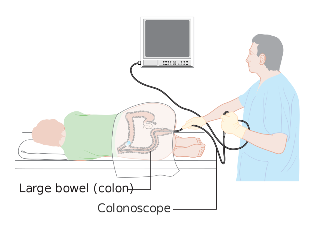
A colonoscopy (kŏl-ŏn-OS-kŏ-pē) is a procedure that examines the inside of the large intestine, including the colon, rectum, and anus. A sigmoidoscopy (sig-moy-DOS-kŏ-pē) is a similar procedure but only examines the sigmoid colon. During both procedures, the physician uses an endoscope (EN-dŏ-skōp), a flexible tube with a lighted camera on the end that is inserted into the anus and colon. Along the way, it sends pictures of the inside of the large intestine to a screen. The physician can take biopsies during these procedures and also remove polyps.[3] See Figure 12.25[4] for an illustration of a colonoscopy.
The American Cancer Society recommends routine colonoscopy screening for colon cancer for individuals of average risk starting at age 45.[5] Colonoscopies are also used to diagnose medical conditions like inflammatory bowel disease or colon cancer.
Colonoscopy prep is very important to the success of the procedure. The health care provider provides detailed instructions to the patient to follow in the days leading up to the procedure. The purpose of these preparations is to make sure the large intestine is as clear as possible for visualization of the tissue. Typically, a low-fiber diet is followed for two or three days, followed by a clear liquid diet on the day before the procedure. The afternoon before the colonoscopy, a strong laxative is taken to purge the bowels.[6]
Upper GI Endoscopy
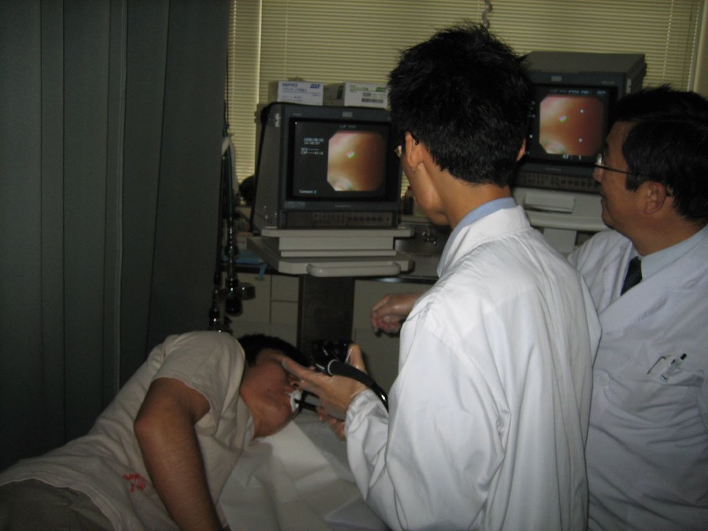
During an upper GI endoscopy (ŬP-ĕr Jī ĕn-DŎS-kŏ-pē en-DOS-kŏ-pē), also called an esophagogastroduodenoscopy (ē-sof-ă-gō-gas-trō-doo-ŏ-dē-NOS-kŏ-pē) (EGD), a doctor uses a flexible tube with a camera to see the lining of the upper gastrointestinal tract, including the esophagus, stomach, and duodenum. During upper GI endoscopy, biopsies may be taken by passing an instrument through the endoscope to take small pieces of tissue from the lining of the esophagus. A pathologist examines the tissue under a microscope.[7] See Figure 12.26[8] for an image of an upper GI endoscopy.
Upper GI Series
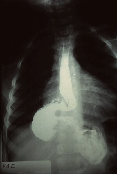
An upper gastrointestinal series (ŬP-ĕr GAS-trō-in-TĔS-tĭ-năl SĪR-ēz) is an X-ray examination of the upper gastrointestinal (GI) tract. The esophagus, stomach, and duodenum are made visible on X-ray film by a liquid suspension that may be barium or a water-soluble contrast. If only the pharynx and esophagus are examined with barium, the procedure is called a barium swallow (BĀR-ē-ŭm SWŎL-ō). Fluoroscopy is often used during an upper GI series to allow the radiologist to visualize the movement of the barium through the esophagus, stomach, and duodenum as a person drinks. After the procedure, if barium is not completely eliminated from the body, constipation and fecal impaction can occur.[9] See Figure 12.27[10] for an image of an upper GI series that shows a hernia and an obstruction. The barium causes the stomach and esophagus to appear white.
Stool-Based Tests
The U.S. Preventive Services Task Force recommends that adults aged 45 to 75 be screened for colorectal cancer. Several stool-based tests can be used to screen for polyps or colorectal cancer[11]:
- Fecal occult blood test (FĒ-kăl ŏ-KULT blŭd tĕst) (FOBT) or (gFOBT): Uses a chemical called guaiac to detect blood in the stool. For this test, the patient receives a test kit from their health care provider. At home, they use a small stick or brush to obtain a small amount of stool and place it on a card. The test kit is returned to a lab, where the stool sample is checked for the presence of blood.
- Fecal immunochemical test (FĒ-kăl im-yŭn-Ō-kĕm-ĭ-kăl tĕst): Uses antibodies to detect blood in the stool.
- Stool DNA test (stōōl dē-ĕn-ā tĕst): Combines the fecal immunochemical test with a test that detects altered DNA in the stool. For this test, an entire bowel movement is collected and sent to a lab, where it is checked for altered DNA and the presence of blood.
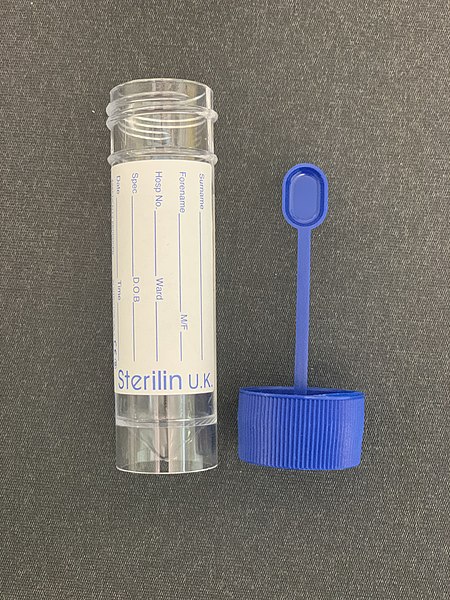
If a stool test is positive, then a colonoscopy is scheduled for follow-up testing. See Figure 12.28[12] for an image of a stool specimen container.
Stool Culture
A stool culture (stōōl KUL-chŭr) is a test that detects and identifies bacteria that cause infections of the lower digestive tract. The test distinguishes between the types of bacteria that cause disease (pathogenic) and the types that are normally found in the digestive tract (normal flora). The test helps to determine if pathogenic bacteria are the cause of a person’s gastrointestinal symptoms.[13]
- Johns Hopkins Medicine. (n.d.). Barium enema. https://www.hopkinsmedicine.org/health/treatment-tests-and-therapies/barium-enema ↵
- “Colonic_Herniation_08787.jpg” by Nevit Dilmen (talk) is licensed under CC BY-SA 3.0 ↵
- Cleveland Clinic. (2022, November 30). Colonoscopy. https://my.clevelandclinic.org/health/diagnostics/4949-colonoscopy ↵
- “Diagram_showing_a_colonoscopy_CRUK_060.svg” by Cancer Research UK. Wikimedia Commons is licensed under CC BY-SA 4.0 ↵
- American Cancer Society. (2020, November 17). American Cancer Society guideline for colorectal cancer screening. https://www.cancer.org/cancer/types/colon-rectal-cancer/detection-diagnosis-staging/acs-recommendations.html ↵
- Cleveland Clinic. (2022, November 30). Colonoscopy. https://my.clevelandclinic.org/health/diagnostics/4949-colonoscopy ↵
- National Institute of Diabetes and Digestive and Kidney Diseases. (2020, July). Diagnosis of GER & GERD. National Institutes of Health. https://www.niddk.nih.gov/health-information/digestive-diseases/acid-reflux-ger-gerd-adults/diagnosis ↵
- “180619235_391dec588d_b.jpg” by Yuya Tamai is licensed under CC BY 2.0 ↵
- Johns Hopkins Medicine. (n.d.). Upper gastrointestinal series. https://www.hopkinsmedicine.org/health/treatment-tests-and-therapies/upper-gastrointestinal-series ↵
- “Radiology_0014_Nevit.jpg” by Nevit Dilmen (talk) is licensed under CC BY-SA 3.0 ↵
- Centers for Disease Control and Prevention. (2023, February 23). Colorectal cancer screening tests. U.S. Department of Health & Human Services. https://www.cdc.gov/cancer/colorectal/basic_info/screening/tests.htm ↵
- “Stool_specimen_container.jpg” by Whispyhistory is licensed under CC BY-SA 4.0 ↵
- Testing.com. (2021, November 9). Stool culture. https://www.testing.com/tests/stool-culture ↵

