10.4 Anatomy of the Hematology System
The hematology system consists of the blood and the bone marrow that create the cellular elements of the blood, as well as accessory organs, including the spleen and the liver.
Blood
Blood is made of formed elements (cells) and plasma. The formed elements include erythrocytes (ĕr-Ĭ-thrō-sītz), leukocytes (LOO-kō-sītz), and thrombocytes (THRŌM-bō-sītz), all suspended in a liquid called plasma. In diagnostic reports, erythrocytes are referred to as red blood cells (RBCs), leukocytes are referred to as white blood cells (WBCs), and thrombocytes are referred to as platelets.
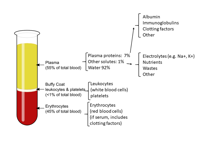
When a blood sample is taken from a patient and sent to the laboratory for analysis, the sample is centrifuged to separate the components of blood from one another so they can be measured. As the heaviest elements in blood, red blood cells (RBCs) settle at the very bottom of the tube. RBCs make up about 45% of the volume of the blood, measured in a blood test called hematocrit. Hematocrit (hē-MAT-ō-krit) (HCT) is the ratio of the volume of red blood cells to the total volume of blood. The normal hematocrit for men is 40 to 54%, and for women it is 36 to 48%.[1]
Above the RBC layer in a centrifuged sample is a layer of white blood cells (WBCs) and platelets, which together make up less than 1% of the sample of whole blood. Above this buffy layer is plasma (PLAZ-mă), the liquid part of blood. Plasma is a pale, straw-colored fluid that constitutes 55% of a blood sample. Plasma is mostly water with dissolved proteins, including albumin, immunoglobulins, and clotting factors. It also contains nutrients, electrolytes, and cellular wastes. Plasma suspends RBCs, WBCs, and platelets, enabling them to circulate throughout the body within the cardiovascular system.[2] See Figure 10.1[3] for an illustration of the composition of blood in a blood sample.
Phlebotomy (flĕ-BOT-ŏ-mē) refers to the opening or puncture of a vein in order to withdraw a blood sample for analysis. Hemolysis (hē-MOL-ĭ-sĭs) refers to the breaking down of red blood cells due to the mishandling of blood samples during routine blood collection and transport.
Bone Marrow
Formation of the formed elements of the blood takes place primarily in the red bone marrow. Bone marrow (bōn MĀR-ō) is the soft tissue inside bones that produces red blood cells (RBC), white blood cells (WBC), and platelets. Red bone marrow is primarily located in flat bones such as the sternum, skull, and pelvis.[4]
In the red bone marrow, stem cells are undifferentiated and have the remarkable capacity to become any type of cell. As these stem cells differentiate, they require specific growth factors for specialization. For example, erythropoietin (ĕr-ĭ-thrō-poi-Ē-tin) (EPO) is a hormone that stimulates the production of erythrocytes (red blood cells) that is produced by the kidneys.[5] During a stem cell transplant, donated healthy stem cells are provided to a patient and, if successful, take over production of their blood cells in their bone marrow. Read more about stem cell transplants in the “Medical Specialists, Diagnostic Testing, and Procedures Related to Blood.”
Red Blood Cells
Red blood cells (rĕd blŭd sĕls) (RBCs) transport oxygen and carbon dioxide between tissues and the lungs. RBCs are packed with hemoglobin (HĒM-ō-glō-bin) (Hgb), a protein molecule that carries oxygen. For this reason, deficiency in RBCs results in symptoms related to decreased oxygenation of the body’s tissues and organs.[6] See Figure 10.2[7] for an illustration of RBCs containing hemoglobin that carries oxygen molecules.
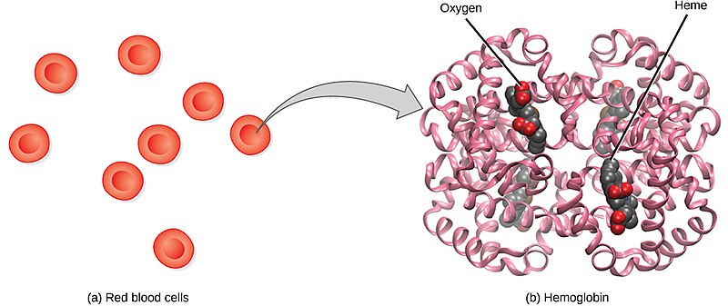
Production of RBCs in the red bone marrow occurs at the staggering rate of more than 2 million cells per second. For this production to occur, raw materials, including iron, folic acid, and B12, must be present in adequate amounts. RBCs live only 120 days on average and must be continually replaced.[8]
Old RBCs and hemoglobin are broken down by white blood cells in a process called phagocytosis. When hemoglobin breaks down, a yellow substance called bilirubin (bil-ĭ-ROO-bin) is released. If the body cannot adequately metabolize bilirubin, a symptomatic yellow discoloration called jaundice occurs in the skin, mucus membranes, and eyes.[9]
White Blood Cells
White blood cells (wīt blŭd sĕls) (WBCs), also called leukocytes, provide immune functions and defenses against disease. WBCs protect the body against invading microorganisms, as well as its own cells that have mutated DNA, and they also clean up cellular debris. There are five main types of WBCs called neutrophils (NŪ-trō-filz), lymphocytes (LĬM-fō-sītz), monocytes (MON-ō-sītz), eosinophils (ē-ō-SIN-ō-filz), and basophils (BAS-ō-filz).[10] See Figure 10.3[11] for an illustration of these types of WBCs.
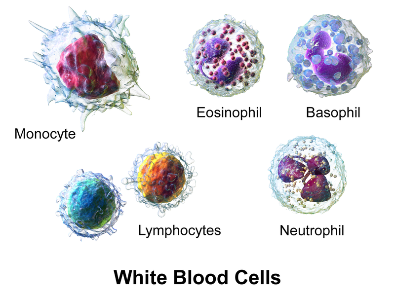
Table 10.4 summarizes the main functions of the different types of WBCs.
Table 10.4. Types of WBCs and Functions[12]
| Type of WBC | Percentages in Total WBC Count | Function |
|---|---|---|
| Neutrophils | 40 -70% | Engulf and destroy bacteria during acute bacterial infections and inflammation. |
| Lymphocytes | 20-45% | Important role in adaptive immunity with the production of antibodies. |
| Monocytes | 2-10% | Transformed into macrophages in acute infections to clear debris and pathogens. |
| Eosinophils | 1-6% | Defend against parasites and cause allergic reactions by releasing histamine and other chemicals. |
| Basophils | 0.5-1% | Release histamine and other inflammatory mediators during an allergic reaction. |
Platelets
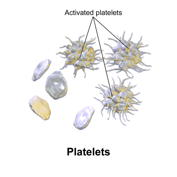
Platelets (PLĀT-lĭtz), also called thrombocytes, help form a clot when there is bleeding. Platelets serve a critical function in hemostasis (hē-mō-STĀ-sĭs), the process by which the body seals a broken blood vessel and prevents further blood loss. During hemostasis, platelets become activated and create a platelet plug.[13] See Figure 10.4[14] for an illustration of platelets and activated platelets. Hemostasis and blood clot formation are further discussed in the “Physiology of the Hematology System.”
Accessory Organs
The spleen and liver are accessory organs for blood production and clotting regulation.
Spleen
The spleen (splēn) produces and stores WBCs, filters and stores RBCs and platelets, and plays a role in the filtration and destruction of old RBCs. There are two types of tissue in the spleen: white pulp and red pulp. White pulp, the lymphatic tissue of the spleen, produces some WBCs, although most WBCs are produced in the red bone marrow. Red pulp serves as a storage site for RBCs and platelets. The spleen also filters antigens and plays a role in the destruction of old RBCs and the breakdown of hemoglobin.[15] See Figure 10.5[16] for an illustration of the location of the spleen.
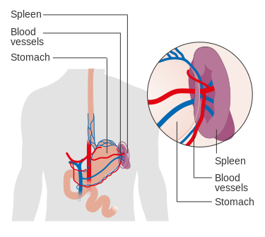
Because the spleen serves as a reservoir for blood, injury to the spleen can result in excessive, life-threatening bleeding called hemorrhage (HEM-ŏ-rāj). For this reason, patients who have experienced blunt abdominal trauma undergo rapid diagnostic testing to check for and rapidly treat splenic damage and prevent hemorrhage.
Enlargement of the spleen, called splenomegaly (sple-nō-MĒG-ă-lē), can occur due to a variety of reasons, such as systemic infection, sickle cell disease, or cancer. Patients who undergo splenectomy (splē-NEK-tŏ-mē), the removal of the spleen, experience reduced immune function and have an increased risk of infection.[17]
Liver
The liver (LIV-ĕr) serves as a storage site for blood and blood cells. It also produces prothrombin and other clotting factors. See Figure 10.6[18] for an illustration of the location of the liver.
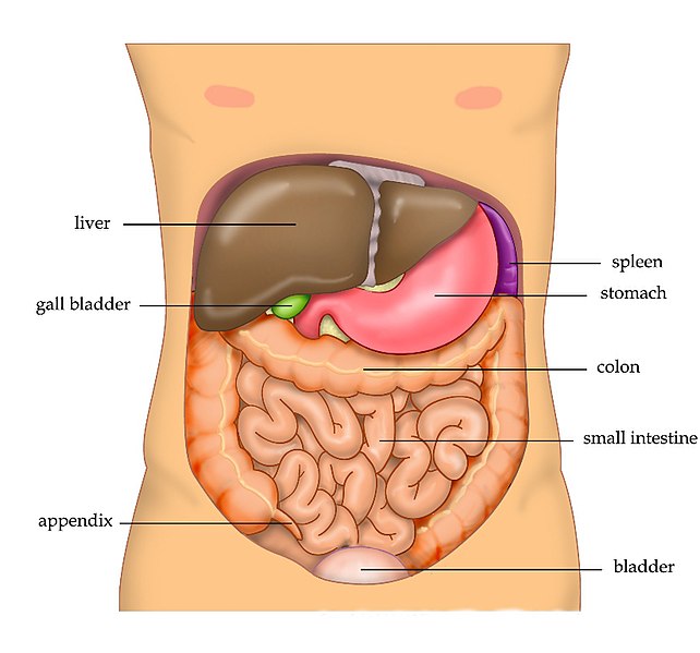
The liver also plays a significant role in the breakdown of hemoglobin. The liver receives bilirubin from the breakdown of RBCs, where it undergoes a chemical change called conjugation so it can be filtered and excreted by the kidneys. If the liver becomes impaired and cannot adequately metabolize bilirubin, jaundice occurs.[19] Jaundice (JAWN-dis) refers to yellowish discoloration of the skin and eyes.
Enlargement of the liver is called hepatomegaly (hep-a-tō-MĒG-ă-lē). Hepatomegaly can be caused by liver disease, cancer, and excess alcohol intake.
Blood Types
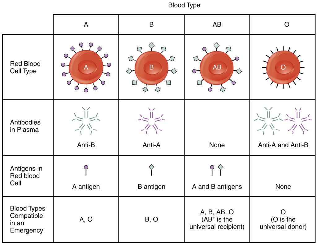
There are four blood types called A, B, AB, and O. These blood types are genetically determined for every individual. Each type is determined by the presence or absence of specific antigens called “A” or “B” on the individual’s red blood cells.[20]
Based on the presence or lack of A or B antigens on an individual’s red blood cells, antibodies are present in their blood that recognize these antigens as foreign and stimulate their immune response. For example, an individual with Type A blood has an A antigen on their RBC but has antibodies to the B antigen in their blood. For this reason, if an individual requires a blood transfusion, it is vital for the donated blood to be compatible with their blood type or a life-threatening reaction will occur.[21] A blood transfusion (blŭd tran-SFŪ-zhŏn) is a procedure that enables the transfer of blood products from one person to another. Figure 10.7[22] shows the different blood types and associated antigens and antibodies, as well as their compatible blood types.
Rh Blood Group
In addition to the A, B, AB, or O blood type, individuals may have another antigen on their red blood cells called Rh factor (Ār āch FAK-tŏr). Individuals who have the antigen on their RBCs are described as Rh positive (Ār-āch PŌZ-ĭ-tĭv) (Rh+), and those who lack the antigen are described as Rh negative (Ār-āch NĔG-ă-tĭv) (Rh−). When identifying a patient’s blood type, the Rh factor is designated by adding the word positive or negative to the ABO type. For example, A positive (A+) means A blood type with the Rh antigen present, and AB negative (AB−) means AB blood type without the Rh antigen. Individuals receiving blood transfusions must receive donated blood that matches their Rh type, or a serious, life-threatening reaction will occur.[23]
During pregnancy, the fetus can be significantly affected if the mother has a different Rh factor. If an Rh- mother conceives a Rh+ baby, her Rh antibodies can cross the placenta and destroy the fetal RBCs. This condition is known as hemolytic disease of the newborn (hē-mō-LĬT-ik dĭ-zēz əv thə NŪ-bôrn) and can be so severe that without treatment the fetus may die in the womb or shortly after birth. A medication known as RhoGAM (RŌ-găm) can temporarily prevent the development of Rh antibodies in an Rh- mother, thereby averting this potentially serious disease for the fetus if it is Rh+. RhoGAM is administered to Rh- mothers during weeks 26−28 of pregnancy and within 72 hours following birth.[24]
- Billett, H. H. (1990). Hemoglobin and hematocrit. In Walker, H. K., Hall, W. D., & Hurst, J. W. (Eds), Clinical Methods: The history, physical, and laboratory examinations (3rd ed., Chapter 151). Butterworths. https://www.ncbi.nlm.nih.gov/books/NBK259/ ↵
- This work is a derivative of Anatomy & Physiology by OpenStax and is licensed under CC BY 4.0. Access for free at https://openstax.org/details/books/anatomy-and-physiology-2e ↵
- “1913_ABO_Blood_Groups.jpg” by Alan Sved is licensed under CC BY-SA 4.0. ↵
- This work is a derivative of Anatomy and Physiology by OpenStax licensed under CC BY 4.0. Access for free at https://openstax.org/books/anatomy-and-physiology/pages/1-introduction ↵
- This work is a derivative of Anatomy & Physiology by OpenStax and is licensed under CC BY 4.0. Access for free at https://openstax.org/details/books/anatomy-and-physiology-2e ↵
- This work is a derivative of Anatomy & Physiology by OpenStax and is licensed under CC BY 4.0. Access for free at https://openstax.org/details/books/anatomy-and-physiology-2e ↵
- “Figure_39_04_01.jpg” by CNX OpenStax is licensed under CC BY 4.0 ↵
- This work is a derivative of Anatomy and Physiology by OpenStax licensed under CC BY 4.0. Access for free at https://openstax.org/books/anatomy-and-physiology/pages/1-introduction ↵
- This work is a derivative of StatPearls by Kalakonda, Jenkins, & John and is licensed under CC BY 4.0 ↵
- This work is a derivative of Anatomy & Physiology by OpenStax and is licensed under CC BY 4.0. Access for free at https://openstax.org/details/books/anatomy-and-physiology-2e ↵
- “Blausen_0909_WhiteBloodCells.png” by Blausen.com staff (2014). “Medical gallery of Blausen Medical 2014.” WikiJournal of Medicine is licensed under CC BY 3.0 ↵
- American Society of Hematology. (n.d.). Blood basics. https://www.hematology.org/education/patients/blood-basics ↵
- This work is a derivative of Anatomy & Physiology by OpenStax and is licensed under CC BY 4.0. Access for free at https://openstax.org/details/books/anatomy-and-physiology-2e ↵
- “Blausen_0740_Platelets.png” by Blausen.com staff (2014). “Medical gallery of Blausen Medical 2014.” WikiJournal of Medicine is licensed under CC BY 3.0 ↵
- This work is a derivative of StatPearls by Kapila, Wehrle, & Tuma and is licensed under CC BY 4.0 ↵
- “Diagram_showing_the_position_of_the_spleen_CRUK_417.svg” by Cancer Research UK uploader is licensed under CC BY-SA 4.0 ↵
- This work is a derivative of StatPearls by Kapila, Wehrle, & Tuma and is licensed under CC BY 4.0 ↵
- “Anatomy_Abdomen_Tiesworks.jpg” by Tvanbr is licensed in the Public Domain. ↵
- This work is a derivative of StatPearls by Kalra, Yetiskul, Wehrle, & Tuma and is licensed under CC BY 4.0 ↵
- This work is a derivative of Anatomy and Physiology by OpenStax licensed under CC BY 4.0. Access for free at https://openstax.org/books/anatomy-and-physiology/pages/1-introduction ↵
- This work is a derivative of Anatomy and Physiology by OpenStax licensed under CC BY 4.0. Access for free at https://openstax.org/books/anatomy-and-physiology/pages/1-introduction ↵
- “1913_ABO_Blood_Groups.jpg” by OpenStax College is licensed under CC BY 3.0 ↵
- This work is a derivative of Anatomy & Physiology by OpenStax and is licensed under CC BY 4.0. Access for free at https://openstax.org/details/books/anatomy-and-physiology-2e ↵
- This work is a derivative of Anatomy & Physiology by OpenStax and is licensed under CC BY 4.0. Access for free at https://openstax.org/details/books/anatomy-and-physiology-2e ↵

