4.6 Diseases and Disorders of the Respiratory System
This section provides an overview of common respiratory disorders and diseases.
Allergies
Allergies (ĂL-ĕr-jēz) occur when a person’s immune system reacts to a substance and makes antibodies that identify that substance as harmful. Substances identified as allergens can cause inflammation of the skin, sinuses, nasal passages, airways, or digestive system. The severity of allergies varies from person to person and can range from minor irritation to a potentially life-threatening emergency called anaphylaxis (ăn-ă-fĭ-LĂK-sĭs). While most allergies can’t be cured, allergy medications can help relieve symptoms.[1]
There are many types of allergies. Allergic rhinitis, commonly referred to as “hay fever,” can cause sneezing; pruritus (PRŪ-rī-tŭs), itching of the skin, as well as itching of the nose, eyes, or roof of the mouth; rhinorrhea; and watery, red, or swollen eyes. A food allergy can cause tingling in the mouth; swelling of the lips, tongue, face, or throat; hives; and anaphylaxis. Read more information about anaphylaxis in the following subsection. An insect sting allergy can cause swelling at the sting site, itching or hives all over the body, cough, chest tightness, wheezing, shortness of breath, and anaphylaxis. A drug allergy can cause hives, pruritus, rash, swelling in the respiratory tract, and anaphylaxis.
Anaphylaxis
Severe allergies, including allergies to foods, insect stings, medications, and blood transfusions, can trigger a severe reaction known as anaphylaxis. As a life-threatening medical emergency, anaphylaxis can cause a patient to go into anaphylactic shock, a potentially fatal condition. Signs and symptoms of anaphylaxis are as follows[2]:
- Loss of consciousness
- Drop in blood pressure
- Severe shortness of breath
- Skin rash
- Light-headedness
- Rapid, weak pulse
- Nausea and vomiting
Treatment of anaphylaxis often requires injection of a medication called epinephrine to rapidly reduce the body’s allergic response and restore adequate respiratory status.
Asthma
Asthma (AZ-muh) is a common chronic respiratory condition that can affect all age groups. Asthma is characterized by episodes of inflammation and edema of the airways and bronchospasms that prevent air from entering the lungs. Excessive mucus secretion can also occur, which further contributes to blockage of the airway and shortness of breath. Bronchospasms can lead to asthma attacks (ĂZ-mă ă-TĂKS) that can range from mild to severe and can be life-threatening. An asthma attack may be triggered by environmental factors such as dust, pollen, pet hair, or dander; changes in the weather; mold; tobacco smoke; respiratory infections; or by exercise and stress.[3] Between episodes of asthma attacks, many individuals with asthma are asymptomatic.
See Figure 4.11[4] for an illustration of how asthma affects the airways.
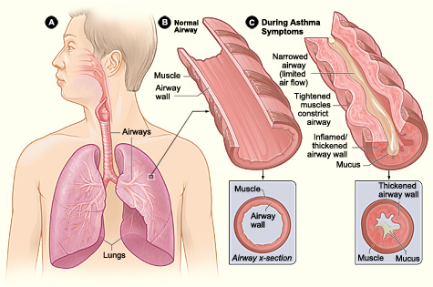
Symptoms of an asthma attack involve coughing, dyspnea (shortness of breath), wheezing, and tightness of the chest. Severe asthma attacks can be fatal and require immediate medical attention. Symptoms of a severe asthma attack include worsening dyspnea that can cause cyanotic (sī-ă-NŎT-ĭk) or blue lips or fingertips, worsening wheezing, confusion, drowsiness, a rapid pulse, sweating, and severe anxiety.[5]
Chronic asthma is typically diagnosed by health care providers by using pulmonary function tests. Read more about pulmonary function tests in the “Medical Specialists, Diagnostic Testing, and Procedures Related to the Respiratory System” section of this chapter. Individuals with asthma are often prescribed a peak flow meter (pēk flō mē-tĕr), a portable instrument used to measure air flow during forced exhalation, to help manage their symptoms.
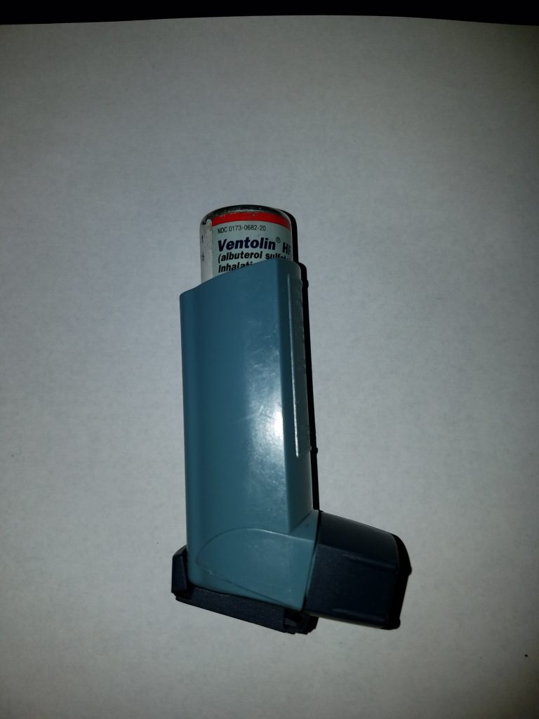
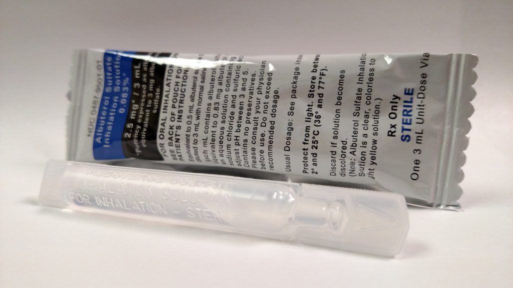
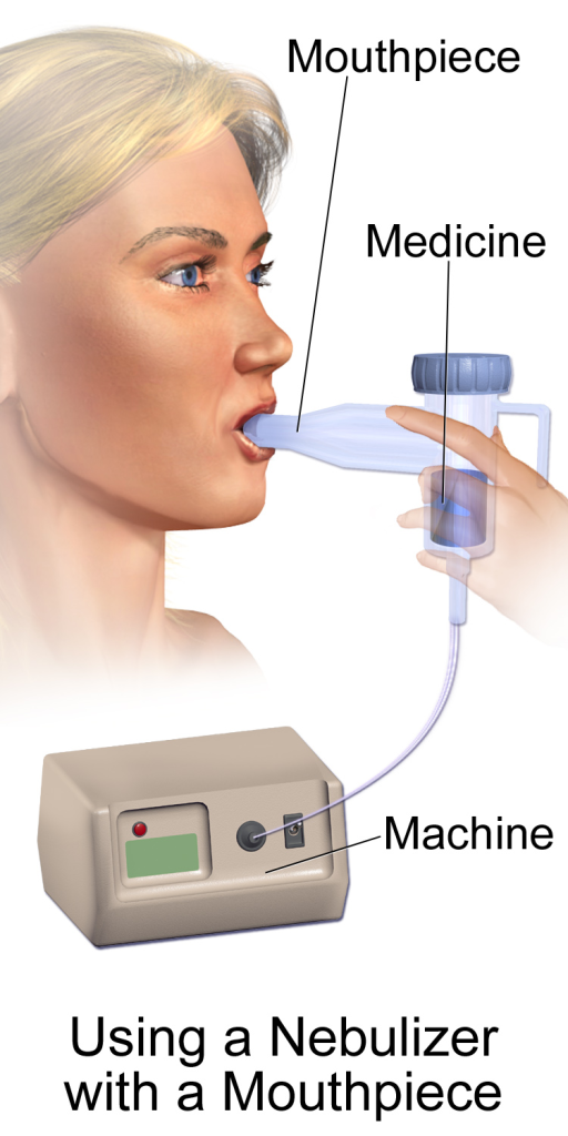
Asthma is treated by respiratory medications given via an inhaler and/or nebulizer (NEB-yŭ-lī-zĕr). A nebulizer is a medical device that creates a mist for delivering respiratory medication. Inhalers may be referred to as dry powder inhalers (DPI) or metered dose inhalers (MDI). The severity of the condition, frequency of attacks, and identified triggers influence the type of medication that an individual may require. Long-term treatments for patients with severe asthma include oral and/or injectable medications.[6] See Figures 4.12,[7] 4.13,[8] and 4.14[9] for images of an albuterol inhaler, respiratory medication used in a nebulizer, and a nebulizer.
To learn more about asthma, visit the Centers for Disease Control and Prevention (CDC) web page on asthma.
View the following YouTube video[10] for additional information about asthma: How Does Asthma Work? – Christopher E. Gaw
Bronchitis
Bronchitis is an inflammation of the lining of the bronchial tubes, which carry air to and from the lungs. People who have bronchitis often cough up thickened mucus, which can be discolored. Bronchitis may be either acute or chronic.
Often developing from a cold or other respiratory infection, acute bronchitis is very common. Acute bronchitis, also called a chest cold, usually improves within a week to ten days without lasting effects, although the cough may linger for weeks.
Chronic bronchitis (KRŎN-ĭk brŏng-KĪ-tĭs), a more serious condition, is a constant irritation or inflammation of the lining of the bronchial tubes, often due to smoking. Chronic bronchitis is one of the conditions included in chronic obstructive pulmonary disease (COPD).[11] Read more about COPD in the following subsection.
Symptoms for either acute bronchitis or chronic bronchitis may include the following:
- Cough
- Production of mucus (sputum), which can be clear, white, yellowish-gray, or green in color — rarely, it may be streaked with blood
- Fatigue
- Shortness of breath
- Slight fever and chills
- Chest discomfort
Chronic Obstructive Pulmonary Disease
Chronic obstructive pulmonary disease (KRŎN-ĭk ŏb-STRŬK-tĭv PŬL-mō-nĕ-rē dĭ-ZĒZ) (COPD) is an inflammatory lung disease that causes obstructed airflow out of the lungs. COPD is a chronic condition with most symptoms appearing in people in their middle 50s. COPD is most often caused by smoking but can also be caused by long-term exposure to irritating gases or dust. People with COPD are at increased risk of developing heart disease, lung cancer, and a variety of other conditions.
Emphysema and chronic bronchitis are the two types of COPD that often occur together. Emphysema (ĕm-fŭ-SĒ-mă) is a disorder affecting the alveoli where they become abnormally inflated, damaging their walls and making it harder to breathe. Chronic bronchitis is inflammation of the lining of the bronchial tubes and characterized by chronic cough and mucus (sputum) production. See Figure 4.15 for an illustration of normal lungs compared to lungs with COPD.[12]
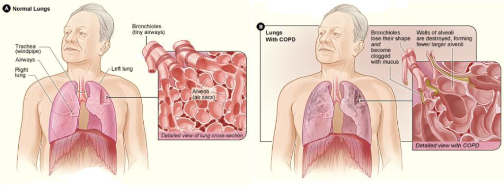
COPD symptoms often don’t appear until significant lung damage has occurred, and they usually worsen over time, particularly if smoke exposure continues. Common signs and symptoms of COPD include the following:
- Dyspnea (shortness of breath), especially during physical activities
- Wheezing (HWĒ-zĭng) (breathing with a whistling or rattling sound in the chest)
- Chronic cough
- Excessive sputum (mucus from the respiratory tract) that may be clear, white, yellow, or greenish
- Frequent respiratory infections
- Lack of energy
- Barrel chest (enlarged, rounded chest)
- Unintended weight loss (in later stages)
Signs and symptoms of COPD worsen during exacerbations (ĕg-ză-sĕr-BĀ-shŭns) or flare-ups and can result in hospitalization. Symptoms of COPD exacerbation may include worsening dyspnea, green or brown mucus, fever, cyanosis (sī-ă-NŌ-sĭs) or blue lips or fingers caused by lack of oxygenation of tissues, and increased fatigue.
COPD is diagnosed by health care providers using spirometry. Spirometry is further described in pulmonary function tests in the “Medical Specialists, Diagnostic Testing, and Procedures Related to the Respiratory System” section of this chapter.
COPD is progressive disease that is treatable but not curable. Shortness of breath may be controlled with a medication called a bronchodilator (brŏng-kō-dī-LĀ-tŭr), a drug that relaxes and expands the bronchi. The best way to avoid exacerbations is to avoid triggers, avoid people who are sick, get the annual flu shot, and reduce exposure to cigarette smoke and pollution.[13] The majority of cases of COPD are caused by cigarette smoking, and the best way to prevent COPD is to never smoke or stop smoking.[14]
To learn more about COPD, visit the Centers for Disease Control and Prevention’s web page on COPD.
The Effects of Second-Hand Tobacco Smoke
The burning of a tobacco cigarette creates multiple chemical compounds that are released through mainstream smoke, which is inhaled by the smoker, and through sidestream smoke, which is the smoke that is given off by the burning cigarette. Second-hand smoke, which is a combination of sidestream smoke and the mainstream smoke that is exhaled by the smoker, has been demonstrated by numerous scientific studies to cause disease. At least 40 chemicals in sidestream smoke have been identified that negatively impact human health, leading to the development of COPD, cancer, or other conditions, such as immune system dysfunction, liver toxicity, cardiac arrhythmias, pulmonary edema, and neurological dysfunction. Furthermore, second-hand smoke has been found to harbor at least 250 compounds that are known to be toxic, carcinogenic, or both. Some major classes of carcinogens in second-hand smoke are polyaromatic hydrocarbons (PAHs), N-nitrosamines, aromatic amines, formaldehyde, and acetaldehyde.
Tobacco and second-hand smoke are considered to be carcinogenic. Exposure to second-hand smoke can cause lung cancer in individuals who are not tobacco users themselves. It is estimated that the risk of developing lung cancer is increased by up to 30 percent in nonsmokers who live with an individual who smokes in the house, as compared to nonsmokers who are not regularly exposed to second-hand smoke. Children are especially affected by second-hand smoke. Children who live with an individual who smokes inside the home have a larger number of lower respiratory infections, which are associated with hospitalizations, and higher risk of sudden infant death syndrome (SIDS). Second-hand smoke in the home has also been linked to a greater number of ear infections in children, as well as worsening symptoms of asthma.[15]
Common Cold
The common cold, also known as an upper respiratory infection (URI) is a viral infection of the upper respiratory tract. Many types of viruses can cause a common cold. Children younger than six are at greatest risk of colds, but healthy adults can also expect to have two or three colds annually. Most people recover from a common cold within a week to ten days. Symptoms might last longer in people who smoke.
Symptoms of a common cold usually appear one to three days after exposure to a cold-causing virus. Signs and symptoms, which can vary from person to person, are as follows[16]:
- Runny or stuffy nose
- Sore throat
- Cough
- Congestion
- Slight body aches or a mild headache
- Sneezing
- Low-grade fever
- Generally feeling unwell (malaise)
Coronavirus (COVID-19)
Influenza and COVID-19 are both highly contagious respiratory illnesses, but they are caused by different viruses. COVID-19 is caused by infection with a coronavirus named SARS-CoV-2. You cannot tell the difference between the common cold, influenza, and COVID-19 by symptoms alone because all have similar symptoms. Individuals with symptoms should get tested for both influenza and COVID-19 and take appropriate precautions to prevent the spread of infection.[17]
To read current information about COVID-19 symptoms and treatment, go to the Centers for Disease Control and Prevention (CDC) website on COVID-19.
Cystic Fibrosis
Cystic fibrosis (SĬS-tĭk fī-BRŌ-sĭs) is a genetic disease that causes problems with breathing and digestion. People with cystic fibrosis have thick, sticky mucus that builds up in the lungs and digestive tract and other areas of the body. This excessive mucus blocks airways, traps germs, makes respiratory infections more likely, and leads to lung damage. It also affects the pancreas, with the increased secretions preventing digestive enzymes from reaching the intestines, which decreases the body’s ability to absorb nutrients from food.[18]
Influenza
Influenza (in-floo-EN-ză), commonly referred to as the “flu,” is a highly contagious viral infection affecting the respiratory tract. Symptoms of influenza include fever, cough, sore throat, rhinorrhea (runny nose), muscle aches, headache, and fatigue (severe tiredness). Most people who get influenza will recover in less than two weeks, but some people develop complications (such as pneumonia) that can be life-threatening. Anyone can get influenza, even healthy people, and serious problems related to the flu can happen to anyone at any age, but some people are at higher risk of developing serious flu-related complications if they get sick. This includes people 65 years and older, people of any age with certain chronic medical conditions (such as asthma, diabetes, or heart disease), pregnant women, and children younger than five years old. Everyone aged six months and older should get an annual flu vaccine, ideally by the end of October, to prevent influenza and its potential complications.[19]
Lung Cancer
Cancer (KĂN-sŏr) is a disease in which cells in the body grow out of control. When cancer begins in the lungs, it is called lung cancer (lŭng KĂN-sŏr), although it may spread to lymph nodes or other organs in the body, such as the brain. Cancer from other organs also may spread to the lungs. When cancer cells spread from one organ to another, they are called metastases (mĕ-TĂS-tă-sēz).[20]
Cigarette smoking is the number one risk factor for lung cancer. In the United States, cigarette smoking is linked to about 80% to 90% of lung cancer deaths. People who quit smoking at any age have a lower risk of lung cancer than if they had continued to smoke, but their risk is higher than the risk for people who never smoked.[21]
After smoking, radon is the second leading cause of lung cancer in the United States. Radon is a naturally occurring gas that forms in rocks, soil, and water. It cannot be seen, tasted, or smelled. When radon gets into homes or buildings through cracks or holes, it can get trapped and build up in the air inside. People who live or work in these homes and buildings breathe in high radon levels. Over long periods of time, radon can cause lung cancer.[22]
Most people with lung cancer don’t have symptoms until the cancer is advanced and has metastasized. Lung cancer symptoms may include the following[23]:
- Coughing that gets worse or doesn’t go away
- Chest pain
- Shortness of breath
- Wheezing
- Hemoptysis (hē-MŎP-tĭ-sĭs) (coughing up blood)
- Fatigue (feeling very tired all the time)
- Weight loss with no known cause
- Repeated episodes of pneumonia
- Swollen or enlarged lymph nodes inside the chest in the area between the lungs
Lung cancer may initially be suspected after routine tests like chest X-rays (chĕst ĕks-rās), also referred to as radiographs (RĀ-dē-ŏ-grăfs). Additional diagnostic testing is performed if lung cancer is suspected. Read more about diagnostic tests in the “Medical Specialists, Diagnostic Testing, and Procedures Related to the Respiratory System” section of this chapter.
Treatments for lung cancer may include surgery, chemotherapy, targeted therapy, immunotherapy, and radiation therapy.
Obstructive Sleep Apnea
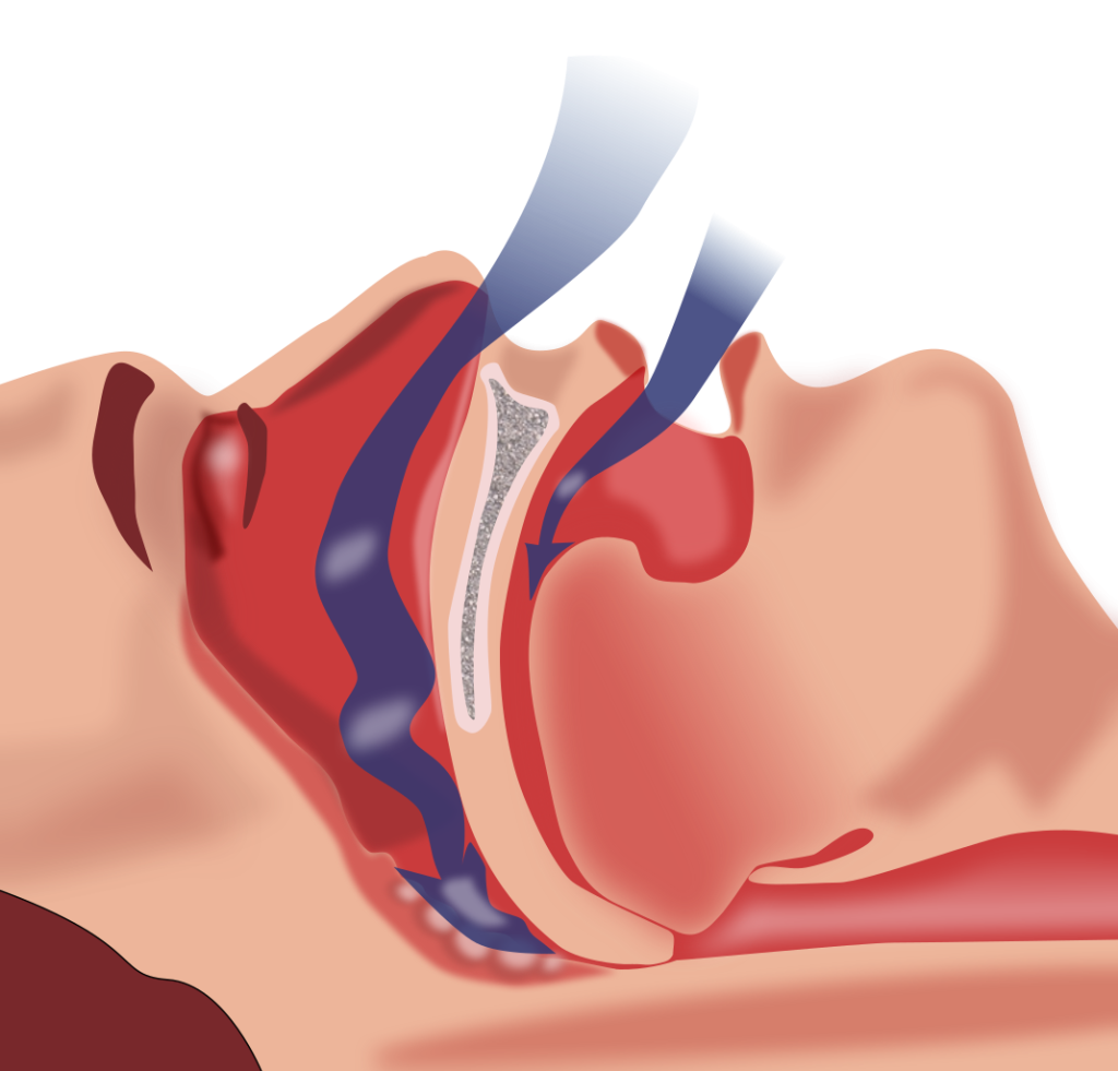
Obstructive sleep apnea (ŏb-STRŬK-tĭv slēp ăp-NĒ-ă) is a chronic disorder that can occur in children and adults. It is characterized by periods of no breathing during sleep. These episodes may last for several seconds or several minutes and may differ in the frequency with which they are experienced. Sleep apnea leads to poor sleep. Symptoms of sleep apnea include fatigue, evening napping, irritability, memory problems, morning headaches, and excessive snoring. A diagnosis of sleep apnea is usually done during a sleep study, where the patient is monitored in a sleep laboratory or with home testing. Treatment of sleep apnea commonly includes the use of a device called a continuous positive airway pressure device (kŏn-TĬN-yū-ŭs POZ-ĭ-tĭv AIR-wā PRESS-ŭr dĭ-VĪS) (CPAP) during sleep. The CPAP machine has a mask that covers the nose, or the nose and mouth, and forces air into the airway at regular intervals. This pressurized air can help to gently force the airway to remain open, allowing for more normal ventilation to occur.[24] Read more about a CPAP device in the “Medical Specialists, Diagnostic Testing, and Procedures Related to the Respiratory System” section. See Figure 4.16[25] for an illustration of obstructive sleep apnea. As soft tissue falls to the back of the throat, it impedes the passage of air (blue arrows) through the trachea and is characterized by repeated episodes of complete or partial obstructions of the upper airway during sleep.
Figure 4.16 Obstructive Sleep Apnea
Pneumonia
Pneumonia is an infection of the alveoli of the lungs caused by microorganisms like bacteria, viruses, or fungi that can cause mild to severe illness in people of all ages.
Symptoms of pneumonia are as follows[26]:
- Cough, which may produce greenish or yellow mucus (often called purulent sputum)
- Fever and shaking chills
- Dyspnea (shortness of breath)
- Rapid, shallow breathing
- Sharp or stabbing chest pain that gets worse when breathing deeply or coughing
- Loss of appetite, low energy, and fatigue
See Figure 4.17[27] for an image of purulent sputum.

Common diagnostic tests for pneumonia include sputum cultures and chest X-rays. Read more about these diagnostic tests in the “Medical Specialists, Diagnostic Testing, and Procedures Related to the Respiratory System” section of this chapter.
There are several categories of pneumonia that are treated with different types of antibiotics[28]:
- Aspiration pneumonia: Pneumonia that occurs when food or liquid is breathed into the airways or lungs, instead of being swallowed.
- Community-acquired pneumonia: Pneumonia that is diagnosed in someone in the community (not in a hospital).
- Healthcare-associated pneumonia: Pneumonia that is diagnosed in someone during or following a stay in a health care setting.
- Ventilator-associated pneumonia: Pneumonia that is diagnosed in someone who has been on a ventilator.
Incentive Spirometry
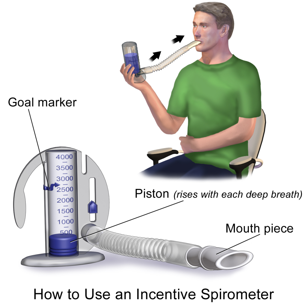
Patients who have undergone surgery are at risk for developing atelectasis and pneumonia. Atelectasis means the alveoli have become deflated, which can cause a partial or complete collapsed lung and/or lead to pneumonia. An incentive spirometer is a medical device often prescribed as a preventative measure after surgery or for patients with atelectasis. While sitting upright, the patient should breathe in slowly and deeply through the tubing with the goal of raising the piston to a specified level. See Figure 4.18[29] for an image of a patient using an incentive spirometer. The patient should attempt to hold their breath for five seconds, or as long as tolerated, and then rest for a few seconds. This technique should be repeated by the patient ten times every hour while awake.[16]
Pulmonary Edema
Pulmonary edema is the accumulation of fluid in the alveoli causing decreased gas exchange, resulting in increased levels of carbon dioxide and decreased levels of oxygen in the blood. Pulmonary edema can be caused by several medical conditions, such as pneumonia, heart failure, kidney failure, and liver failure. It is treated with medications that help eliminate excess fluid from the body, called diuretics.
Pulmonary Embolism
A pulmonary embolism (PULL-muh-nair-ee EM-boh-liz-uhm) (PE) is a blood clot or other substance, such as fat or an air bubble, that has traveled through the bloodstream and lodged in a smaller vessel within the pulmonary circulation in the lungs and obstructed blood flow. This blockage causes lack of oxygen delivery to the tissues supplied by that blood vessel. A PE is medical emergency that requires rapid treatment to prevent severe damage to the lungs or heart and death. Symptoms of a PE may include sudden, severe dyspnea; sharp pain in the chest or arm; and pale, clammy skin. Treatment includes medications to dissolve the clot or procedures to remove the clot in order to quickly restore adequate pulmonary circulation.[30]
Respiratory Syncytial Virus
Respiratory syncytial virus (rĕs-pĭ-RĂ-tōr-ē SĬN-sĭ-shē-ăl VĪ-rŭs) (RSV) is a very common respiratory virus that usually causes mild, cold-like symptoms. Most people recover in a week or two, but RSV can be very serious in infants and older adults. RSV is a common cause of pneumonia and bronchiolitis. Bronchiolitis (brŏng-kē-ŎL-ĭ-tĭs) refers to inflammation of the small airways in the lung. Older adults and infants younger than six months of age with RSV may require hospitalization if they have problems breathing or become dehydrated. Severe cases may require endotracheal intubation and mechanical ventilation.[31]
Tuberculosis
Tuberculosis (tū-bĕr-kyŭ-LŌ-sĭs) (TB) is a serious infectious disease that affects the lungs. It is caused by bacteria that can be spread through the air from person to person. Symptoms include a chronic cough, hemoptysis (hē-MŎP-tĭ-sĭs) or coughing up blood, weight loss, night sweats, and fever. Patients with TB require a long course of treatment involving multiple antibiotics.[32]
View a supplementary YouTube video[33] on tuberculosis: CDC Tuberculosis (TB) Tansmission and Pathogenesis Video
- Mayo Clinic Staff. (2022, August 5). Allergies. https://www.mayoclinic.org/diseases-conditions/allergies/symptoms-causes/syc-20351497 ↵
- Mayo Clinic. (2021). Anaphylaxis. https://www.mayoclinic.org/diseases-conditions/anaphylaxis/symptoms-causes/syc-20351468 ↵
- This work is a derivative of Anatomy and Physiology by OpenStax licensed under CC BY 4.0. Access for free at https://openstax.org/books/anatomy-and-physiology/pages/1-introduction ↵
- “Asthma_attack-illustration_NIH.jpg” by United States-National Institute of Health: National Heart, Lung, Blood Institute is in the Public Domain ↵
- This work is a derivative of Anatomy and Physiology by OpenStax licensed under CC BY 4.0. Access for free at https://openstax.org/books/anatomy-and-physiology/pages/1-introduction ↵
- This work is a derivative of Anatomy and Physiology by OpenStax licensed under CC BY 4.0. Access for free at https://openstax.org/books/anatomy-and-physiology/pages/1-introduction ↵
- “Ventolin® HFA (Albuterol Sulfate) Inhaler.jpg” by MisterNarwhal is licensed under CC BY SA 4.0 ↵
- “Albuterol 2.jpg” by Mark Oniffrey is licensed under CC BY SA 4.0 ↵
- “Nebulizer_Mouthpiece.png” by BruceBlaus is licensed under CC BY-SA 4.0 ↵
- TED-Ed. (2017, May 11). How does asthma work? - Christopher E. Gaw [Video]. YouTube. https://youtu.be/PzfLDi-sL3w ↵
- Mayo Clinic Staff. (2017, April 11). Bronchitis. https://www.mayoclinic.org/diseases-conditions/bronchitis/symptoms-causes/syc-20355566 ↵
- “Copd 2010Side.JPG” by National Heart, Lung, and Blood Institute is licensed in the Public Domain. ↵
- Centers for Disease Control and Prevention. (2023, June 30). What is COPD? https://www.cdc.gov/copd/index.html ↵
- Mayo Clinic Staff. (2020, April 15). COPD. https://www.mayoclinic.org/diseases-conditions/copd/symptoms-causes/syc-20353679 ↵
- This work is a derivative of Anatomy and Physiology by OpenStax licensed under CC BY 4.0. Access for free at https://openstax.org/books/anatomy-and-physiology/pages/1-introduction ↵
- Mayo Clinic Staff. (2023, May 24). Common cold. https://www.mayoclinic.org/diseases-conditions/common-cold/symptoms-causes/syc-20351605 ↵
- Centers for Disease Control and Prevention. (n.d.). COVID-19. https://www.cdc.gov/coronavirus/2019-ncov/ ↵
- Centers for Disease Control and Prevention. (2022, May 9). Cystic fibrosis. https://www.cdc.gov/genomics/disease/cystic_fibrosis.htm ↵
- Centers for Disease Control and Prevention. (2022, October 3). Flu symptoms & complications. https://www.cdc.gov/flu/symptoms/symptoms.htm ↵
- Centers for Disease Control and Prevention. (2023, July 31). What is lung cancer? https://www.cdc.gov/cancer/lung/basic_info/what-is-lung-cancer.htm ↵
- Centers for Disease Control and Prevention. (2023, July 31). What is lung cancer? https://www.cdc.gov/cancer/lung/basic_info/what-is-lung-cancer.htm ↵
- Centers for Disease Control and Prevention. (2023, July 31). What are the risk factors for lung cancer? https://www.cdc.gov/cancer/lung/basic_info/risk_factors.htm ↵
- Centers for Disease Control and Prevention. (2023, July 31). What are the symptoms of lung cancer? https://www.cdc.gov/cancer/lung/basic_info/symptoms.htm ↵
- Slowik, J. M., Sankari, A., & Collen, J. F. (2022). Obstructive sleep apnea. [Updated 2022 Dec 11]. In: StatPearls [Internet]. https://www.ncbi.nlm.nih.gov/books/NBK459252/ ↵
- “Obstruction ventilation apnée sommeil.svg” by Habib M’henni is in the Public Domain. ↵
- American Lung Association. (2023, August 3). Pneumonia symptoms and diagnosis. https://www.lung.org/lung-health-diseases/lung-disease-lookup/pneumonia/symptoms-and-diagnosis ↵
- “Sputum.JPG” by Zhangmoon618 is licensed under CC0 ↵
- Centers for Disease Control and Prevention. (2022, September 30). Causes of pneumonia. https://www.cdc.gov/pneumonia/causes.html ↵
- “Incentive Spirometer.pngsommeil.svg” by BruceBlaus is licensed under CC BY-SA 4.0 ↵
- Cleveland Clinic. (2022). Pulmonary embolism. https://my.clevelandclinic.org/health/diseases/17400-pulmonary-embolism ↵
- Centers for Disease Control and Prevention. (2023). Respiratory syncytial virus. https://www.cdc.gov/rsv/about/symptoms.html ↵
- Centers for Disease Control and Prevention. (2023). Tuberculosis. https://www.cdc.gov/tb/default.htm ↵
- Centers for Disease Control and Prevention. (2020, May 6). CDC tuberculosis (TB) transmission and pathogenesis video [Video]. YouTube. All rights reserved. https://youtube.com/watch?v=UKV8Zn7x0wM ↵

