4.5 Physiology of the Respiratory System
The main function of the respiratory system is gas exchange (găs ĭk-SCHĀNJ), meaning providing a constant supply of oxygen to the body and removing carbon dioxide. To achieve gas exchange, the structures of the respiratory system create the mechanical movement of air into and out of the lungs called ventilation (vĕn-tĭ-LĀ-shŭn) (i.e., breathing).[1]
Ventilation and the Mechanics of Breathing
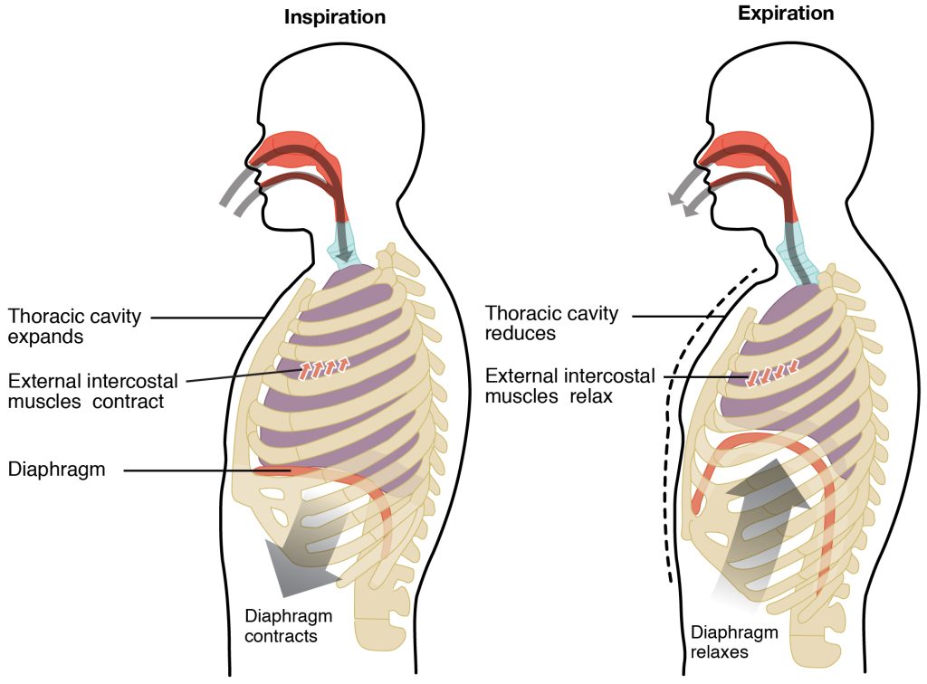
The lungs bring oxygen to the cells of our body through inhalation. Inhalation (i-hā-LĀ-shŭn), also called inspiration, is the act of breathing in. During inhalation, the diaphragm contracts and flattens, creating a larger lung cavity, which decreases the pressure in the lungs. At the same, the intercostal muscles (the muscles between the ribs) pull downward, also causing the thoracic cavity to expand. The thoracic cavity (thuh-RAS-ik KA-vah-tee) is the space inside the chest that contains the heart, lungs, and other organs. As the thoracic cavity expands, a negative pressure (i.e., vacuum) is created inside the chest cavity, causing air to rush into the lungs (because air always moves from high pressure to low pressure).
During exhalation (ĕks-hā-LĀ-shŭn), also called expiration, the act of breathing out, the diaphragm relaxes and the thoracic cavity springs back to its original position. This causes the volume of the thoracic cavity to decrease and pressure to increase, causing air to leave the lungs.[2] See Figure 4.10[3] for an illustration of inhalation and exhalation.

Health care professionals use an instrument called a stethoscope (STETH-ŏ-skōp) to listen to internal body sounds like lung sounds. Lung sounds are caused by the movement of air from the trachea to the bronchioles to the alveoli and can be impacted by the presence of sputum, bronchoconstriction, or fluid in the alveoli. These sounds are referred to as rhonchi (coarse crackles), rales (fine crackles), wheezes, stridor, and pleural rub[4]:
- Rhonchi (rŏng-kahy), also referred to as coarse crackles, are low-pitched, continuous sounds heard on expiration that are a sign of turbulent airflow through mucus in the large airways.
- Rales (rāylz), also called fine crackles, are popping or crackling sounds heard on inspiration. They are associated with medical conditions, such as heart failure or pneumonia, that cause fluid accumulation within the alveolar and interstitial spaces. The sound is similar to that produced by rubbing strands of hair together close to your ear.
- Wheezes (wēz-ĕz) are whistling noises produced when air is forced through airways narrowed by bronchoconstriction or mucosal edema. For example, patients with asthma commonly have wheezing.
- Stridor (strī-door) is heard only on inspiration. It is associated with obstruction of the trachea/upper airway.
- Pleural rub (plur-uhl ruhb) sounds like the rubbing together of leather and can be heard on inspiration and expiration. It is caused by inflammation of the pleura membranes that results in friction as the surfaces rub against each other.
View the following YouTube video[5] to review the mechanics of breathing: Mechanics of Breathing AIDA Freediving
Forced breathing, also known as hyperpnea (hī-pĕrp-NĒ-ă), is a type of breathing that can occur during exercise or actions that require the active manipulation of breathing, such as singing. During forced breathing, muscle contractions of accessory muscles, in addition to the diaphragm, are used for inspiration and expiration. These additional muscle contractions during inspiration also occur during labored breathing (LĀ-bŏrd BRĒ-thĭng), a symptom of many respiratory disorders.[6]
The word root pnea refers to breathe. Therefore, tachypnea (tak-ip-NĒ-ă) refers to rapid breathing, bradypnea (brăd-ĬP-nē-ă) refers to slow breathing, hypopnea (hī-POP-ne-ă) refers to deficient breathing, and apnea (AP-nē-ă) refers to the absence of breathing. Dyspnea (disp-NĒ-ă) is a common symptom of respiratory disorders and refers to difficulty breathing.
Control of Breathing
Respiratory rate (RES-pĭr-ă-tō-rē rāt) is the number of breaths taken per minute. The normal respiratory rate for adults is 12-20 breaths per minute. A child under 1 year of age has a normal respiratory rate between 30 and 60 breaths per minute. By the time a child is about 10 years old, the normal rate is closer to 18 to 30. Respiratory rate can be an important indicator of a respiratory disorder because the rate may increase or decrease during illness or disease.[7]
The respiratory rate is controlled by the respiratory center located within the medulla oblongata and the pons in the brain stem, which responds primarily to changes in carbon dioxide, oxygen, and pH levels in the blood. These changes are sensed by central chemoreceptors, which are located in the brain, and peripheral chemoreceptors, which are located in the aortic arch and carotid arteries. The major factor that drives breathing is surprisingly not hypoxemia (hī-pŏk-SĒ-mē-ă), low levels of oxygen in the blood, rather the concentration of carbon dioxide. Carbon dioxide is a waste product of cellular respiration and is toxic at high levels in the blood, referred to as hypercapnia (hī-pĕr-KAP-nē-ă). As carbon dioxide levels in the blood increase, the central chemoreceptors stimulate the contraction of the diaphragm and intercostal muscles (i.e., the muscles between the ribs), and the rate and depth of respiration increase to help rid the body of carbon dioxide. Hyperventilation (hī-pĕr-vĕn-tĭ-LĀ-shŭn) refers to rapid and deep breathing. In contrast, low levels of carbon dioxide in the blood stimulate shallow, slow breathing to help the body retain carbon dioxide. Hypoventilation (hī-pō-vĕn-tĭ-LĀ-shŭn) refers to slow and shallow breathing.[8]
Gas Exchange
Ventilation (i.e., the mechanics of breathing) provides air to the alveoli in the lungs for gas exchange. Respiration (rĕs-pĭ-RĀ-shŏn) refers to the exchange of gases in the lungs between the alveoli and the pulmonary capillaries or in the tissues between the systemic capillaries and cells/tissues.[9]
Gas exchange refers to the exchange of oxygen and carbon dioxide through capillary walls of the alveoli and the pulmonary capillaries, called external respiration. During external respiration, oxygen from the air we breathe diffuses into the blood. Carbon dioxide (waste) diffuses out of the blood and into the alveoli where it can be exhaled. Throughout the rest of the body, gas exchange also occurs between the systemic capillaries and body cells/tissues, called internal respiration. During internal respiration, oxygen diffuses out of the systemic capillaries and into the surrounding cells and tissues, and carbon dioxide diffuses from the cells/tissues into the systemic capillaries where it is carried to the lungs. It is through this process that cells in the body are oxygenated and carbon dioxide, the waste product of cellular respiration, is removed from the body.[10]
Asphyxia (ăs-FIK-sē-ă) refers to deprivation of oxygen to the tissues, commonly referred to as suffocation.
Perfusion
In addition to adequate ventilation, the second important aspect of gas exchange is perfusion. Perfusion (pĕr-FYŪ-zhŭn) refers to the flow of blood. In the lungs, perfusion occurs in the pulmonary circulation (PŬL-mŭ-năr-ē sĕr-kyŏŏ-LĀ-shŭn), as it moves from the heart to the lungs and then back to the heart for distribution to the body. The pulmonary arteries (PŬL-mō-nĕ-rē ăr-tĕ-rēs) carry deoxygenated blood from the heart into the lungs, where they branch and eventually become the capillary network composed of pulmonary capillaries. These pulmonary capillaries create the respiratory membrane with the alveoli. As the blood is pumped through this capillary network, gas exchange occurs.[11]
Although a small amount of the oxygen is able to dissolve directly into the blood from the alveoli, most of the oxygen binds to hemoglobin (HĒ-mō-glō-bĭn) within red blood cells (erythrocytes). The more oxygen the hemoglobin in red blood cells carry, the brighter red the color of the blood. Oxygenated blood returns to the heart through the pulmonary veins to the left atrium and ventricle, where it is pumped out to the body via the aorta. The hemoglobin on the red blood cells transports the oxygen to the tissues throughout the body.[12]
Hypoxia
Diseases and disorders affecting the respiratory system can cause hypoxia (hī-PŎK-sē-ă), low levels of oxygen in the tissues. A patient’s oxygenation status is routinely assessed by health care professionals using pulse oximetry. Pulse oximetry (pŭls ŏk-SĬM-ĭ-trē) is an estimated oxygenation level based on the saturation of hemoglobin measured by a pulse oximeter. Because the majority of oxygen carried in the blood is attached to hemoglobin within the red blood cells, pulse oximetry estimates how much hemoglobin is “saturated” with oxygen. The normal range for pulse oximetry is 94-100%.[13]
Hypoxia can occur due to inadequate ventilation or impaired perfusion. For example, a medical condition called pulmonary edema (PŬL-mō-nĕ-rē ĕ-DĒ-mă) refers to fluid accumulation in alveoli, often caused by heart failure or kidney failure. As a result of the fluid, oxygen cannot move across the alveolar membrane into the blood, and carbon dioxide cannot be removed from the blood. As a result, hypoxia and hypercapnia (high levels of carbon dioxide) may occur, requiring urgent medical interventions to sustain life by decreasing carbon dioxide levels and increasing oxygen levels.[14]
View supplementary YouTube videos for additional review of the respiratory system:
Respiratory System[15]
Overview of the Respiratory System, Animation[16]
Respiratory System, Part 1: Crash Course A&P #31[17]
- This work is a derivative of Anatomy and Physiology by OpenStax licensed under CC BY 4.0. Access for free at https://openstax.org/books/anatomy-and-physiology/pages/1-introduction ↵
- This work is a derivative of Anatomy and Physiology by OpenStax licensed under CC BY 4.0. Access for free at https://openstax.org/books/anatomy-and-physiology/pages/1-introduction ↵
- “2316_Inspiration_and_Expiration.jpg” by OpenStax College is licensed under CC BY 3.0 ↵
- This work is a derivative of Open RN Nursing Skills 2e by Chippewa Valley Technical College with CC BY 4.0 licensing. ↵
- Chandra, S. (2017, November 1). Mechanics of breathing AIDA freediving [Video]. YouTube. All rights reserved. https://youtu.be/baYZ_dgGIWw?si=_Vwrlr9J8vNSZuwA ↵
- This work is a derivative of Anatomy and Physiology by OpenStax licensed under CC BY 4.0. Access for free at https://openstax.org/books/anatomy-and-physiology/pages/1-introduction ↵
- This work is a derivative of Anatomy and Physiology by OpenStax licensed under CC BY 4.0. Access for free at https://openstax.org/books/anatomy-and-physiology/pages/1-introduction ↵
- This work is a derivative of Anatomy and Physiology by OpenStax licensed under CC BY 4.0. Access for free at https://openstax.org/books/anatomy-and-physiology/pages/1-introduction ↵
- This work is a derivative of Anatomy and Physiology by OpenStax licensed under CC BY 4.0. Access for free at https://openstax.org/books/anatomy-and-physiology/pages/1-introduction ↵
- This work is a derivative of Anatomy and Physiology by OpenStax licensed under CC BY 4.0. Access for free at https://openstax.org/books/anatomy-and-physiology/pages/1-introduction ↵
- This work is a derivative of Anatomy and Physiology by OpenStax licensed under CC BY 4.0. Access for free at https://openstax.org/books/anatomy-and-physiology/pages/1-introduction ↵
- This work is a derivative of Anatomy and Physiology by OpenStax licensed under CC BY 4.0. Access for free at https://openstax.org/books/anatomy-and-physiology/pages/1-introduction ↵
- This work is a derivative of Anatomy and Physiology by OpenStax licensed under CC BY 4.0. Access for free at https://openstax.org/books/anatomy-and-physiology/pages/1-introduction ↵
- This work is a derivative of Anatomy and Physiology by OpenStax licensed under CC BY 4.0. Access for free at https://openstax.org/books/anatomy-and-physiology/pages/1-introduction ↵
- Amoeba Sisters. (2022, February 28). Respiratory system [Video]. YouTube. All rights reserved. https://www.youtube.com/watch?v=v_j-LD2YEqg ↵
- Alila Medical Media. (2019, April 15). Overview of the respiratory system, animation [Video]. YouTube. All rights reserved. https://youtu.be/03qvN5pjCTU?si=LJRgMq6RwLiUhcXF ↵
- CrashCourse. (2015, August 24). Respiratory system, Part 1: Crash Course Anatomy & Physiology #31 [Video]. YouTube. All rights reserved. https://youtu.be/bHZsvBdUC2I ↵

