11.4 Anatomy of the Lymphatic and Immune Systems
Anatomy of the Lymphatic and Immune Systems
The lymphatic system (lĭmf-ă-TĬK sĭs-tŭm) is the system of vessels, cells, and organs that transports fluid called lymph to the bloodstream and also filters pathogens from the blood. The immune system (ĭ-MŪN SĬS-tĕm) is the complex collection of cells and organs that destroys or neutralizes pathogens that would otherwise cause infection, disease, or death. The lymphatic system, for most people, is associated with the immune system to such a degree that the two systems are virtually indistinguishable.[1]
Lymph Vessels
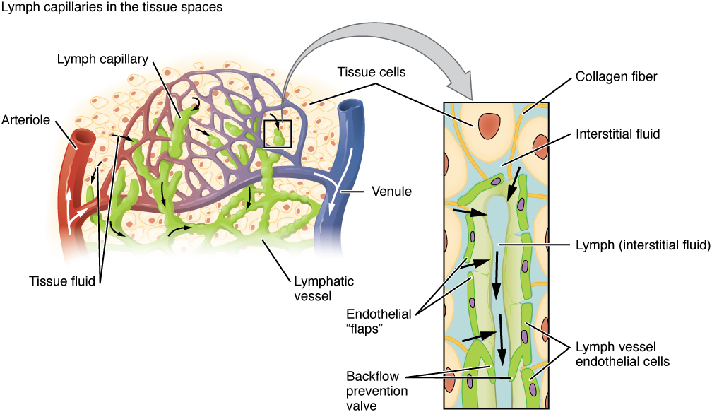
Lymph capillaries are interlaced in interstitial space (in-tĕr-STISH-ăl spās), the space between individual cells in the tissues. Interstitial fluid enters the lymphatic capillaries and becomes lymph. Lymph (limf) is a clear-to-white fluid that transports immune system cells, as well as dietary lipids and fat-soluble vitamins absorbed in the small intestine called chyle (kīl).[2] See Figure 11.1[3] for an image of lymph capillaries.

Lymph capillaries empty into larger lymphatic vessels that become larger and larger until they ultimately empty into the bloodstream at the left and right subclavian veins.
Lymph Nodes
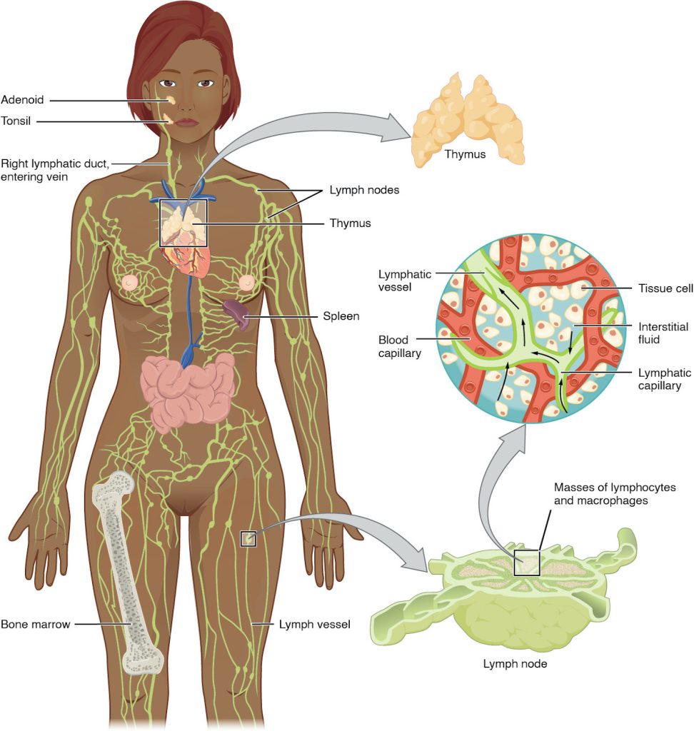
Immune system cells not only use lymphatic vessels to make their way from interstitial space into the circulation, but also use lymph nodes as major staging areas for the development of critical immune responses. Afferent lymphatic vessels (AF-ĕ-rĕnt lim-FAT-ik VES-ĕls) are vessels that lead into a lymph node, and efferent lymphatic vessels (EF-ĕ-rĕnt lim-FAT-ik VES-ĕls) are vessels that lead out of a lymph node.[4]
Lymph nodes (limf nōdz) are small, bean-shaped organs located throughout the lymphatic system. Lymph nodes store immune system cells (B cells and T cells) that help the body fight infection and also filter the lymph fluid to remove foreign material such as bacteria and cancer cells. Lymph nodes are also the sites of adaptive immune responses that are discussed in the “Physiology of the Lymphatic and Immune Systems.”
Lymph travels through the lymph nodes near the groin, armpits, neck, chest, and abdomen. Humans have about 500–600 lymph nodes throughout the body.[5] See Figure 11.2[6] for an illustration of lymph vessels and nodes.

A major distinction between the lymphatic and cardiovascular systems is that lymph is not actively pumped by the heart but is forced through the vessels by the contraction of skeletal muscles during body movements and breathing. One-way valves in lymphatic vessels keep the lymph moving toward the heart.[7]
When the lymphatic system is damaged in some way, such as by being blocked by cancer cells or destroyed by injury, lymph “backs up” from the lymph vessels into interstitial spaces. This inappropriate accumulation of fluid, referred to as lymphedema (limf-e-DĒ-ma), can cause serious medical consequences.
Primary Lymphoid Organs: Bone Marrow and Thymus
The primary lymphoid organs are the bone marrow and thymus gland. The lymphoid organs are where lymphocytes mature, proliferate, and are selected to attack pathogens.
A lymphocyte (lim-fa- SĪT) is a type of white blood cell that fights infection. There are two main types of lymphocytes: B cells and T cells. B cells (B sĕls) produce antibodies that are used to attack invading bacteria, viruses, and toxins. T cells (T sĕls) destroy the body’s own cells that have been taken over by viruses or become cancerous.[8]
Recall that all blood cells, including lymphocytes, are formed in the red bone marrow. The B cell undergoes nearly all of its development in the bone marrow, whereas the immature T cell, called a thymocyte, leaves the bone marrow and matures in the thymus gland. The thymus gland (THĪ-mŭs gland) is an organ found in the space between the sternum and the aorta of the heart (refer to Figure 11.2).[9]
Secondary Lymphoid Organs
Lymphocytes develop and mature in the primary lymphoid organs, but they mount immune responses from the secondary lymphoid organs. Secondary lymphoid organs include the spleen, tonsils, and lymph nodes.[10]
Spleen
The spleen (splēn) is sometimes called the “filter of the blood.” As discussed in the “Anatomy of the Hematology System” in the “Blood Terminology” chapter, the spleen stores and filters red blood cells and also produces white blood cells and antibodies to fight infection.
Tonsils
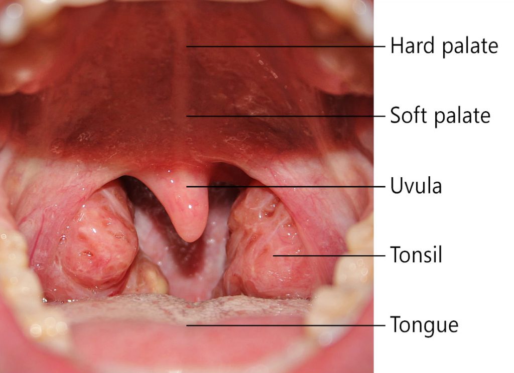
The tonsils (TON-sĭls) are lymphoid nodules located along the inner surface of the pharynx (throat) and are important in developing immunity to oral pathogens. There are five tonsils, including the pharyngeal tonsil (also called the adenoid) located behind the nasal cavity in the nasopharynx, two palatine tonsils located at the back of the mouth in the oropharynx, and two lingual tonsils attached to the tongue. See Figure 11.3[11] for an illustration of the palatine tonsils. Swelling or enlargement of the tonsils is an indication of an active immune response to infection. A major function of tonsils is to help children’s bodies recognize, destroy, and develop immunity to common environmental pathogens so that they will be protected in their later lives. However, tonsils are often removed for individuals who have recurring throat infections because swollen tonsils can interfere with breathing and/or swallowing.[12]

- This work is a derivative of Open Stax Anatomy and Physiology 2e at https://openstax.org/books/anatomy-and-physiology-2e/pages/21-1-anatomy-of-the-lymphatic-and-immune-systems ↵
- This work is a derivative of Anatomy and Physiology by OpenStax licensed under CC BY 4.0. Access for free at https://openstax.org/books/anatomy-and-physiology/pages/1-introduction ↵
- “2202_Lymphatic_Capillaries.jpg” by OpenStax College is licensed under CC BY 3.0 ↵
- This work is a derivative of Anatomy and Physiology by OpenStax licensed under CC BY 4.0. Access for free at https://openstax.org/books/anatomy-and-physiology/pages/1-introduction ↵
- This work is a derivative of Anatomy and Physiology by OpenStax licensed under CC BY 4.0. Access for free at https://openstax.org/books/anatomy-and-physiology/pages/1-introduction ↵
- “lymphaticimage-972x1024.jpg” by J. Gordon Betts, et al. is licensed under CC BY 4.0. Access for free at https://openstax.org/books/anatomy-and-physiology/pages/1-introduction ↵
- This work is a derivative of Anatomy and Physiology by OpenStax licensed under CC BY 4.0. Access for free at https://openstax.org/books/anatomy-and-physiology/pages/1-introduction ↵
- National Human Genome Research Institute. (2023, November 6). Lymphocyte. https://www.genome.gov/genetics-glossary/Lymphocyte ↵
- This work is a derivative of Anatomy and Physiology by OpenStax licensed under CC BY 4.0. Access for free at https://openstax.org/books/anatomy-and-physiology/pages/1-introduction ↵
- This work is a derivative of Anatomy and Physiology by OpenStax licensed under CC BY 4.0. Access for free at https://openstax.org/books/anatomy-and-physiology/pages/1-introduction ↵
- “Throat_with_Tonsils_0011J.jpeg” by Klem is licensed under CC BY 3.0 ↵
- This work is a derivative of Anatomy and Physiology by OpenStax licensed under CC BY 4.0. Access for free at https://openstax.org/books/anatomy-and-physiology/pages/1-introduction ↵

