7.4 Anatomy of the Female Reproductive System
Anatomy of the female reproductive system includes the external genitals, the internal reproductive system, and the breasts.
External Female Genitals
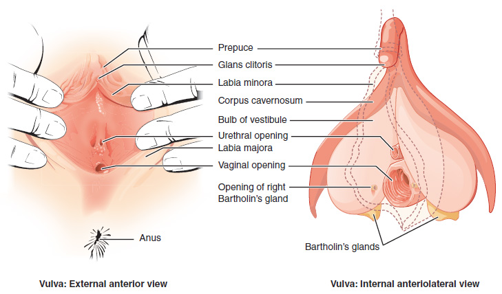
See Figure 7.1[1] for an illustration of the external female genitals. The external female reproductive structures, referred to collectively as the vulva (VŬL-vă), include the following[2]:
- The mons pubis (MŎNZ PYŪ-bĭs) is a pad of fat that is located anteriorly over the pubic bone. After puberty, it becomes covered in pubic hair.
- The labia majora (LĀ-bē-uh MĀ-jŏr-uh) are larger outer folds of hair-covered skin that begin just posterior to the mons pubis.
- The labia minora (LĀ-bē-uh mĭ-NŌR-uh) are thinner, hairless, and more pigmented folds found medially to the labia majora.
- Although they naturally vary in shape and size from woman to woman, the labia minora serve to protect the female urethra and the entrance to the female reproductive tract.
- The superior, anterior portions of the labia minora come together to encircle the clitoris (KLĬT-ŏ-rĭs). The clitoris is erectile tissue that originates from the same fetal cells as the penis and has abundant nerves that are important in sexual sensation and orgasm.
- The vestibule is the area between the labia minora and behind the clitoris that contains the urethral and vaginal openings. It is flanked by outlets to the vestibular glands, also known as Bartholin’s glands (BAR-tō-lĭns glăns), that secrete mucus to keep the vestibular area moist. Read more about the urethra in the “Anatomy of the Urinary System” section in the “Urinary System Terminology” chapter.
- The perineum (pĕr-ĭ-NĒ-um) is the area between the vaginal opening and the anus.

Internal Female Reproductive Organs
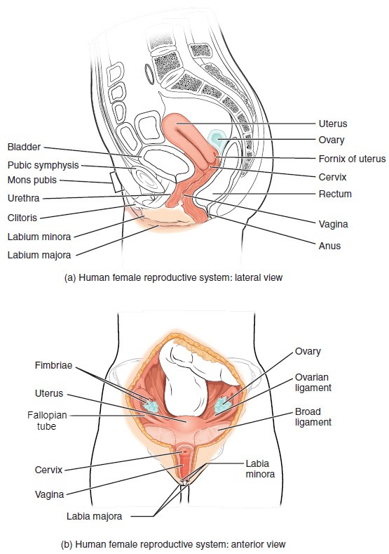
The internal female reproductive organs include the vagina, uterus, cervix, ovaries, and Fallopian tubes. See Figure 7.2[3] for an illustration of the internal and external structures of the female reproductive system.

Vagina
The vagina (vă-JĪN-uh) is a muscular canal that is approximately ten centimeters (cm) long. It serves as the entrance to the reproductive tract, as well as the exit from the uterus during menstruation and childbirth.[4]
The vagina is composed of smooth muscle that allows for expansion during intercourse and childbirth. The vagina is lined with mucous membranes that secrete mucus to keep it moist. The superior portion of the vagina meets the cervix (the opening and lower part of the uterus). The inferior portion of the vagina may have a thin, perforated hymen that partially surrounds the opening to the vagina.[5]
The vagina contains a normal population of healthy bacteria called normal flora that help protect against infection. In a healthy woman, the most common type of normal flora is lactobacillus that secretes lactic acid. The lactic acid protects the vagina by maintaining an acidic pH (below 4.5). Lactic acid, in combination with other vaginal secretions, makes the vagina a self-cleansing organ. However, douching can disrupt the normal balance of healthy microorganisms and increase a woman’s risk for infections and irritation. It is recommended that women do not douche and that they allow the vagina to maintain its normal healthy population of protective normal flora.[6]
Uterus and Cervix
The uterus (YŪ-tĕr-us) is a muscular, pear-shaped organ that is approximately five cm wide by seven cm long. It is composed of three sections[7]:
- The inferior portion, called the cervix (SĔR-vĭks), connects the uterus and the vagina.
- The middle section of the uterus is called the corpus (body of the uterus).
- The superior portion is called the fundus.
An opening in the middle of the cervix allows menstrual blood to exit and sperm to enter the uterus. The cervix dilates (opens) during childbirth to allow the baby to exit the mother’s body. The corpus of the uterus expands during pregnancy. The fundus is the location of the uterus that is commonly felt by the health care provider during prenatal exams to indirectly measure the growth of the fetus.[8]
The wall of the uterus is made up of these three layers[9]:
- Perimetrium (pār-ĭ-MĒ-trē-um): The serous membrane surrounding the uterus.
- Myometrium (my-ō-MĒ-trē-um): A thick layer of smooth muscle responsible for uterine contractions.
- Endometrium (en-dō-MĒ-trē-um): The innermost layer that provides the site for implantation of a fertilized egg or sheds during menstruation if an egg is not fertilized.
Ovaries
The ovary (Ō-văr-ē) is the female reproductive gland located in the pelvic cavity. There are two ovaries, one at the entrance to each Fallopian tube, which are attached to the uterus via the ovarian ligaments. The ovaries create oocytes (eggs) and hormones.[10]
Fallopian Tubes
The Fallopian (fă-LŌP-ē-an) tubes transport oocytes from the ovary to the uterus. Each of the Fallopian tubes is close to, but not directly connected to, the ovary, so the fimbriae (FĬM-brē-ē) catch the oocyte like a baseball in a glove. The middle region of the Fallopian tube, called the ampulla, is where fertilization often occurs. The fertilized egg then moves from the Fallopian tube into the uterus, where it implants into the endometrium.[11]
Breasts
Although the breasts are located far from the other reproductive organs, they are considered accessory organs of the female reproductive system. The function of female breasts is to supply milk to an infant in a process called lactation (lak-TĀ-shŏn).[12]
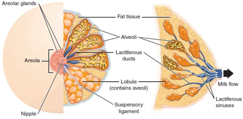
The external features of the breast include a nipple surrounded by a pigmented areola. The areolar region is characterized by small, raised areolar glands that secrete lubricating fluid during lactation to protect the nipple from chafing. When a baby nurses (i.e., draws milk from the breast), the entire areolar region is taken into their mouth.[13]
A breast is made up of three main parts: lobules, ducts, and connective tissue. The lobules are the mammary glands composed of alveoli (tiny sacs) that produce milk. The lactiferous (lak-TĬF-ĕr-us) ducts are tubes that carry milk to the nipple. The connective tissue (which consists of fibrous and fatty tissue) surrounds and holds everything together.[14]
An infant can draw milk from the lobules and through the ducts and the nipple by suckling. Alveoli are surrounded by fat tissue, which determines the size of the breast. Breast size differs between individuals and does not affect the amount of milk produced.[15] See Figure 7.3[16] for an illustration of a breast and milk flow.

Some women have fibrocystic breasts (fī-brō-SĬS-tĭk brests) with connective tissue that feels lumpy or ropelike in texture. Fibrocystic breast changes are common and no longer considered a disease, although it was formerly referred to as fibrocystic breast disease. Some women also experience breast pain, tenderness, and lumpiness, especially in the upper, outer areas of the breasts, just before menstruation.[17]
- “Vulva_Figure_28_02_02.jpg” by OpenStax College is licensed under CC BY 3.0 ↵
- Anatomy & Physiology by OpenStax is licensed under CC BY 4.0. Access for free at https://openstax.org/books/anatomy-and-physiology/pages/1-introduction ↵
- This image is derivative of “Figure_28_02_01.JPG” by OpenStax College is licensed under CC BY 3.0 ↵
- Anatomy & Physiology by OpenStax is licensed under CC BY 4.0. Access for free at https://openstax.org/books/anatomy-and-physiology/pages/1-introduction ↵
- Anatomy & Physiology by OpenStax is licensed under CC BY 4.0. Access for free at https://openstax.org/books/anatomy-and-physiology/pages/1-introduction ↵
- Anatomy & Physiology by OpenStax is licensed under CC BY 4.0. Access for free at https://openstax.org/books/anatomy-and-physiology/pages/1-introduction ↵
- Anatomy & Physiology by OpenStax is licensed under CC BY 4.0. Access for free at https://openstax.org/books/anatomy-and-physiology/pages/1-introduction ↵
- Anatomy & Physiology by OpenStax is licensed under CC BY 4.0. Access for free at https://openstax.org/books/anatomy-and-physiology/pages/1-introduction ↵
- Anatomy & Physiology by OpenStax is licensed under CC BY 4.0. Access for free at https://openstax.org/books/anatomy-and-physiology/pages/1-introduction ↵
- Anatomy & Physiology by OpenStax is licensed under CC BY 4.0. Access for free at https://openstax.org/books/anatomy-and-physiology/pages/1-introduction ↵
- Anatomy & Physiology by OpenStax is licensed under CC BY 4.0. Access for free at https://openstax.org/books/anatomy-and-physiology/pages/1-introduction ↵
- Anatomy & Physiology by OpenStax is licensed under CC BY 4.0. Access for free at https://openstax.org/books/anatomy-and-physiology/pages/1-introduction ↵
- Anatomy & Physiology by OpenStax is licensed under CC BY 4.0. Access for free at https://openstax.org/books/anatomy-and-physiology/pages/1-introduction ↵
- Centers for Disease Control and Prevention. (2023, July 25). What is breast cancer? https://www.cdc.gov/cancer/breast/basic_info/what-is-breast-cancer.htm ↵
- Anatomy & Physiology by OpenStax is licensed under CC BY 4.0. Access for free at https://openstax.org/books/anatomy-and-physiology/pages/1-introduction ↵
- “Anatomy_of_the_breast” by OpenStax is licensed under CC BY 4.0. Access for free at https://openstax.org/books/anatomy-and-physiology/pages/1-introduction ↵
- Mayo Clinic. (2023, April 4). Fibrocystic breasts. https://www.mayoclinic.org/diseases-conditions/fibrocystic-breasts/symptoms-causes/syc-20350438 ↵
