2.5 Tissue Membranes
Tissue Membranes
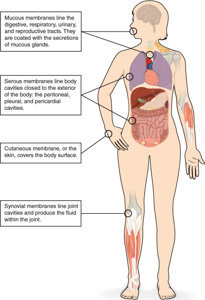
A tissue membrane is a thin layer of cells that covers the outside of the body, an organ, internal passageways that lead to the exterior of the body, or the lining of joint cavities. See Figure 2.5[1] for an illustration of tissue membranes. Consider these examples of tissue membranes:
- Synovial membranes line movable joints and produce fluid within the joint.
- Epithelial membranes can be found in the skin, which covers and protects the surface of the body.
- Mucous membranes are epithelial membranes that line and protect the digestive, respiratory, urinary, and reproductive tracts and are coated with mucous for protection.
- Serous membranes line body cavities such as the peritoneal, pleural, and pericardial cavities.

Connective Tissue Membranes
Connective tissue membranes are formed solely from connective tissue and cover organs and line movable joints.
A synovial membrane is a type of connective tissue membrane that lines the cavities of joints that hold synovial fluid. Synovial fluid lubricates joints for movement. For example, synovial membranes surround the joints of the shoulder, elbow, and knee.
Epithelial Membranes
Epithelial membranes are composed of a thin layer of protective cells and are attached to a layer of connective tissue. Skin, mucous membranes, and serous membranes are types of epithelial membranes.
Skin is an epithelial membrane, also referred to as cutaneous membrane, that rests on top of connective tissue. The outer surface of the epithelial membrane is exposed to the external environment and is covered with dead, tough cells to protect the body from invading organisms and from desiccation (i.e., drying out).
Mucous membranes are another type of epithelial membrane that line body cavities and their passageways that open to the external environment. Mucus covers the epithelial membrane and provides protection. The digestive, respiratory, excretory, and reproductive tracts contain mucous membranes.
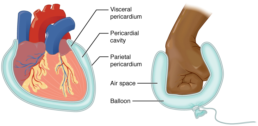
Serous membranes are a third type of epithelial membrane that are contained in body cavities and are composed of layers. A parietal layer lines the walls of a body cavity, and a visceral layer covers the organ within the body cavity. (Note that viscer is the word root for “organ.”) Between the parietal and visceral layers is a thin, fluid-filled serous space. Serous membranes provide protection to the organs they enclose by reducing friction that can lead to inflammation of the organs. There are three body cavities with serous membranes:
- Pleura: Serous membranes surround the lungs in the pleural cavity and reduce friction between the lungs and the body wall.
- Pericardium: Serous membranes surround the heart in the pericardial cavity and reduce friction between the heart and the wall of the pericardium. (Note the word components of pericardial are peri-: surrounding; cardia-: heart; and -al: pertaining to.)
- Peritoneum: Serous membranes surround several organs in the abdominopelvic cavity and reduce friction between the abdominal and pelvic organs and the body wall.
Consider the pericardium serous membranes around the heart as an example of a serous membrane. A visceral layer covers the heart, and a parietal layer lines the pericardial cavity. Between these two layers is a fluid-filled serous space that reduces friction for the beating heart. An analogy of this serous membrane is a fist surrounded by a thin, air-filled balloon to provide protection. See Figure 2.6[2] for an illustration of the visceral and parietal membranes of the pericardium compared to the analogy of a fist surrounded by a balloon.

- “413_Types_of_Membranes.jpg” by OpenStax is licensed under CC BY 3.0. Access for free at https://openstax.org/books/anatomy-and-physiology-2e/pages/1-introduction ↵
- “Serous_Membrane.jpg” by OpenStax is licensed under CC BY 3.0. Access for free at https://openstax.org/books/anatomy-and-physiology-2e/pages/1-introduction ↵

