5.7 Medical Specialists, Diagnostic Testing, and Procedures Related to the Urinary System
Medical Specialists
Nephrology (nĕf-RŎL-ŏ-jē) is the study of kidney diseases. A nephrologist (nĕf-RŎL-ŏ-jĭst) is a physician who specializes in diagnosing and treating kidney conditions. Urology (yū-RŎL-ŏ-jē) is the study of male and female urinary systems as well as the male reproductive system. A urologist (yū-RŎL-ŏ-jĭst) is a specialist who diagnoses and treats urinary system conditions, disorders, and diseases.
To learn more about urology, visit the Why Urology? by the American Urological Association.
Diagnostic Tests
Common diagnostic tests related to the urinary system are described in this section. Diagnostic tests performed for specific medical conditions are discussed in the “Diseases and Disorders of the Urinary System” section.
Blood Tests
Blood Urea Nitrogen (BUN)
Urea nitrogen is a waste product made when the liver breaks down protein. Urea is carried in the blood, filtered out by the kidneys, and removed from the body in urine. A diagnostic test called blood urea nitrogen (blŭd yū-rē-ă nī-trŏ-jĕn) (BUN) provides important information about kidney and liver function. If the liver is unhealthy, it may not break down proteins the way it should, and if the kidneys aren’t healthy, they may not correctly filter urea. Either of these problems can lead to an increased amount of urea nitrogen in the blood. Additionally, dehydration (not enough fluid in the body) can cause higher levels of urea in the blood because not much urine is being created.
Creatinine
A creatinine (krē-ĂT-ĭ-nēn) test is used to determine if they kidneys are working normally. Creatinine is a waste product made by muscles as part of regular, everyday activity. Healthy kidneys filter creatinine from the blood and eliminate it in the urine. However, if there is a problem with kidney functioning, creatinine can build up in the blood and less will be released in urine. If creatinine levels in the blood and/or urine are not in normal range, it can be a sign of kidney disease.
Glomerular Filtration Rate
Glomerular filtration rate (GFR) is one of the most common blood tests to check for kidney disease and to determine how well the nephrons are filtering creatinine. People make different amounts of creatinine, depending on their age, gender, height, weight, diet, and activity levels, so a formula is used to calculate their estimated GFR (eGFR). Lower than normal eGFR levels indicate kidney disease, and very low levels indicate kidney failure.[1]
Urine Tests
Urine Dip
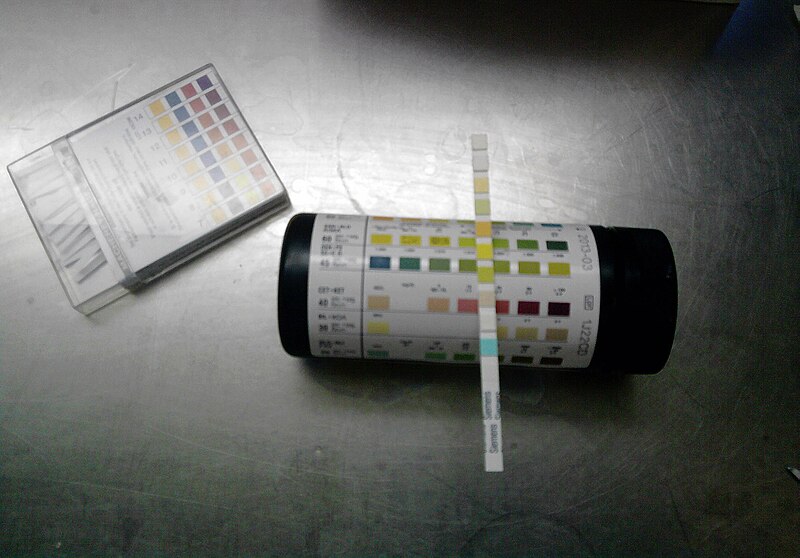
A urine dip (yū-rēn dĭp) test refers to a treated chemical strip (dipstick) being placed in a person’s urine sample. Patches on the dipstick change color to indicate the presence of substances such as white blood cells, red blood cells, protein, or glucose. See Figure 5.15[2] for an image of a urine dipstick test. Urine is collected for a urine dip test in a clean specimen container. Using the “clean catch” technique, the skin surrounding the urethra is cleaned with a special towelette before the urine is collected.
Urinalysis
A urinalysis (yūr-ĭ-NĂL-ĭ-sĭs) (UA) includes a physical, chemical, and microscopic examination of urine by a lab technician. It requires collection of a “clean catch” urine sample in a sterile container. It involves analyzing the urine with a microscope for the following[3]:
- Color
- Appearance (i.e., clear or cloudy)
- Odor
- pH level (acidity)
- Specific gravity (concentration of all particles in the urine)
- Substances not usually found in significant amounts in the urine, such as red blood cells, white blood cells, leukocyte esterase, bacteria, protein, glucose, ketones, and bilirubin
- Cells, crystals, and casts
Urine Culture and Sensitivity
Urine culture and sensitivity (yū-rēn KŬL-chŭr ănd sĕn-sĭ-TĬV-ĭ-tē) (C&S) detects and identifies the number and type of bacteria growing in urine that can indicate a urinary tract infection (UTI). If pathogenic bacteria are found, a sensitivity report is generated that lists antibiotics to which the bacteria are sensitive to for treatment. A culture that is reported as “no growth in 24 or 48 hours” usually indicates that there is no infection. If a culture shows growth of several different types of bacteria, then it is likely due to contamination of the urine sample during collection, and a new collection of urine may be required.[4]
24-Hour Urine Collection
During a 24-hour urine collection test (twehn-tee fôr hour yŭ-rēn kŏ-lĕk-shŭn tĕst), all urinary output from an individual is collected over a 24-hour period of time and analyzed for protein, creatinine, and other substances to evaluate kidney function.
Other Diagnostic Tests
Bladder Scan
A bladder scan (blăd-ĕr skăn) uses a portable, noninvasive medical device that uses sound waves to calculate the amount of urine in a patient’s bladder. Nurses use bladder scanners at patients’ bedsides to determine post-void residual urine amounts without the need to perform urinary catheterization.
CT Scan of Kidney
A computed tomography scan of the kidney (kŏm-PYŌŌ-tĕd tŏ-mŏg-ră-fē skăn ăv thĕ kĭd-nē) (CT) is a diagnostic imaging procedure that uses a combination of X-rays and computer technology to produce a variety of images. It provides detailed images of the kidney looking for disease, cancer, obstructions, and other kidney conditions.[5]
Intravenous Pyelogram (IVP)
An intravenous pyelogram (ĭn-tră-vē-nŭs PĪ-ĕ-lō-gram) (IVP) is a special X-ray exam of the kidneys, bladder, and ureters. An iodine-based contrast (dye) is administered intravenously (into a vein). A series of X-ray images are taken at different times to see how the kidneys remove the dye and collect the urine. Before the final image is taken, the patient is asked to urinate again to see how well the bladder has emptied.[6]
Kidney, Ureter, and Bladder X-ray
A kidney, ureter, and bladder X-ray, often abbreviated KUB, is used to visualize these structures in the urinary system.
Urodynamic Flow Test
Urodynamic flow testing (yū-rō-dī-NĂM-ĭk flō tĕs-tĭng) is a procedure that looks at how well the bladder, sphincters, and urethra are storing and releasing urine. Most urodynamic tests focus on the bladder’s ability to hold urine and empty steadily and completely. Urodynamic tests can also show whether the bladder is having involuntary contractions that cause urine leakage.[7]
Voiding Cystourethrogram
A voiding cystourethrogram (VOY-dĭng SĬS-tō-ūr-ĒTH-rō-gram) (VCUG) uses X-ray technology called fluoroscopy to visualize the urinary tract and bladder to examine the size, shape, and direction of urine flow. A thin, flexible tube called a catheter is inserted into the urethra and passed into the bladder. Contrast dye flows through the catheter into the bladder. X-rays are taken from various angles while the bladder is full of contrast dye, and then the catheter is removed, and X-rays are taken as the patient empties their bladder.[8]
Procedures and Surgeries
Cystoscopy
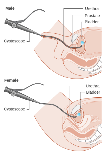
Cystoscopy (sĭs-TŎS-kŏ-pē) is a procedure performed by a urologist who uses a cystoscope to look inside the urethra and bladder. A cystoscope is a long, thin optical instrument with an eyepiece at one end, a rigid or flexible tube in the middle, and a tiny lens and light at the other end of the tube. The urologist fills the bladder with fluid and looks at detailed images of the urethra and bladder on a computer monitor. During a cystoscopy, the urologist can see kidney stones, tumors, stricture (STRIK-chŭr) or a narrowing of the urethra, and other signs of blockage, such as an enlarged prostate gland.[9] See Figure 5.16[10] for an illustration of cystoscopy.
During a cystoscopy, a urologist may also perform treatments, such as removing a stone, taking a sample of the urethra or bladder tissue for a biopsy, injecting medicines into the bladder to treat urinary leakage, or remove abnormal tissue. The urologist may also perform retrograde pyelography (rĕ-trō-grād pī-ĕ-lō-gră-fē), an X-ray procedure where a special dye is injected into the urinary tract to create images of urinary flow to show obstructions such as kidney stones and tumors.[11]
Dialysis
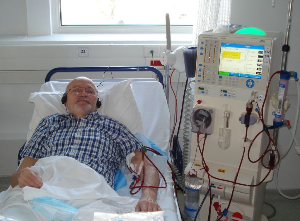
When kidney failure occurs, treatment is required to eliminate excess wastes, potassium, salt, and water. There are two types of dialysis: hemodialysis and peritoneal dialysis. Hemodialysis (hē-mō-dī-ĂL-ĭ-sĭs) uses a special machine that filters the blood. People who are not hospitalized go to a special dialysis clinic for hemodialysis treatments several times a week. Peritoneal dialysis (pĕr-ĭ-tŏ-NĒ-ăl dī-ĂL-ĭ-sĭs) uses the lining of the abdomen, called the peritoneal membrane, to filter the blood. Peritoneal dialysis can be performed in a person’s home. See Figure 5.17[12] for an image of a patient undergoing hemodialysis.
Kidney Transplantation
A kidney transplant (KĬD-nē TRĂNS-plănt), also referred to as renal transplant (RĒ-năl TRĂNS-plănt), is a surgical procedure used for some patients in end-stage renal disease. During a transplant, the surgeon places the donated kidney in the lower abdomen and connects a renal artery and renal vein. The healthy donated kidney takes over the work of the two kidneys that failed, so dialysis is no longer needed. Often, the new kidney will start making urine as soon as blood starts flowing through it, but sometimes it takes a few weeks for it to start working. People who have received a kidney transplant must take immunosuppressive medications for the rest of their lives to keep their bodies from rejecting the new kidney.[13]
Ureteroscopy
Ureteroscopy (yū-rĕ-tĕr-ŎS-kŏ-pē) is a procedure that uses a ureteroscope to look inside the ureters and kidneys. Like a cystoscope, a ureteroscope has an eyepiece at one end, a rigid or flexible tube in the middle, and a tiny lens and light at the other end of the tube. However, a ureteroscope is longer and thinner than a cystoscope so the urologist can see detailed images of the lining of the ureters and kidneys.[14]
Urinary Catheterization
Urinary catheterization (YŪR-ĭ-nĕr-ē kăth-ĭ-tĕr-ī-ZĀ-shŭn) is the insertion of a catheter tube into a person’s urethra and then into their bladder to drain urine. This procedure is commonly performed by nurses. Catheterization may include an indwelling catheter or intermittent catheterization.
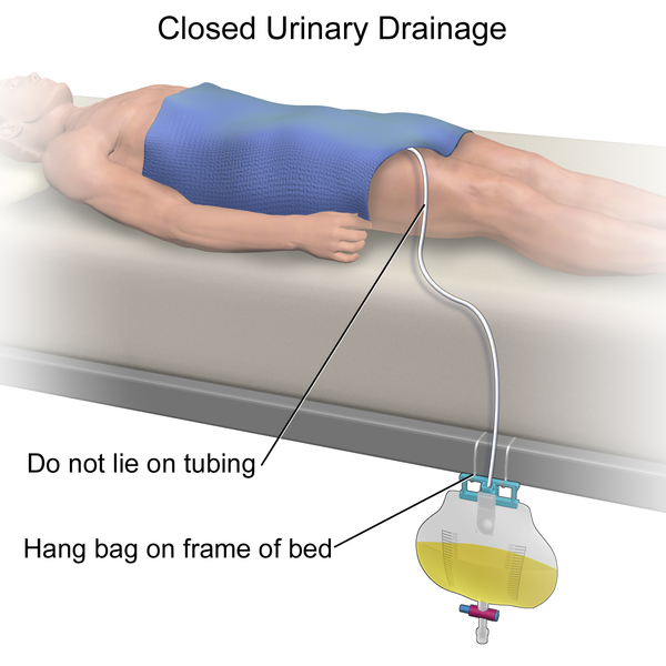
An indwelling catheter (ĭn-DWĔL-ĭng KĂTH-ĭ-tĕr), often referred to as a “Foley catheter,” refers to a urinary catheter that remains in place after insertion into the bladder for the continual collection of urine. It has a balloon on the insertion tip to maintain placement in the neck of the bladder. The other end of the catheter is attached to a drainage bag for the collection of urine. See Figure 5.18[15] for an illustration of a urine collection bag connected to an indwelling catheter as the patient is lying in bed.
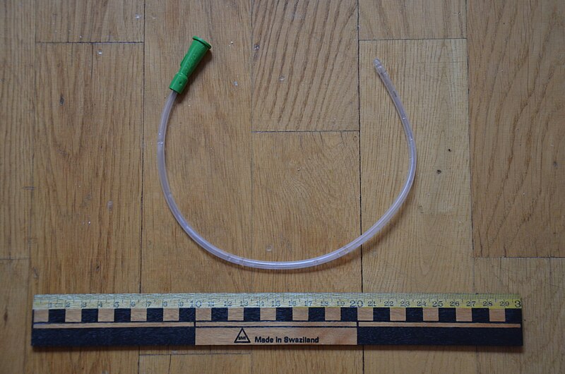
Intermittent catheterization (ĭn-tĕr-MĬT-ĕnt KĂTH-ĕ-tĕr-ī-ZĀ-shŭn) is used for the relief of urinary retention. It may be performed once, such as after surgery when a patient is experiencing urinary retention due to the effects of anesthesia or performed several times a day to manage chronic urinary retention. A straight catheter is used for intermittent urinary catheterization. The catheter is inserted into the urethra and bladder to allow for the flow of urine and then immediately removed, so a balloon is not required at the insertion tip.[16] See Figure 5.19[17] for an image of a straight catheter.
- MedlinePlus [Internet]. Bethesda (MD): National Library of Medicine (US); [updated 2023, July 6]. Glomerular Filtration Rate (GFR) Test; [cited 2023, Oct 12]. https://medlineplus.gov/lab-tests/glomerular-filtration-rate-gfr-test/ ↵
- “Chemstrip2.jpg” by J3D3 is licensed under CC BY-SA 3.0 ↵
- MedlinePlus [Internet]. Bethesda (MD): National Library of Medicine (US); [updated 2016, May 5]. Urinalysis; [cited 2023, Oct 12]. https://medlineplus.gov/urinalysis.html ↵
- LabTestsOnline.org. (2022, September 29). Urine culture test. https://labtestsonline.org/tests/urine-culture ↵
- Johns Hopkins Medicine. (n.d.). Computed tomography (CT or CT scan) of the kidney. https://www.hopkinsmedicine.org/health/treatment-tests-and-therapies/ct-scan-of-the-kidney ↵
- A.D.A.M. Medical Encyclopedia [Internet]. Atlanta (GA): A.D.A.M., Inc.; c1997-2023. Intravenous pyelogram; [reviewed 2023, Jan 1; cited 2023, Oct 12]. https://medlineplus.gov/ency/article/003782.htm ↵
- National Institute of Diabetes and Digestive and Kidney Diseases. (2021, September). Urodynamic testing. National Institutes of Health. https://www.niddk.nih.gov/health-information/diagnostic-tests/urodynamic-testing ↵
- A.D.A.M. Medical Encyclopedia [Internet]. Atlanta (GA): A.D.A.M., Inc.; c1997-2021. Voiding cystourethrogram; [updated 2023, Jan 1 cited 2023, Oct 12]. https://medlineplus.gov/ency/article/003784.htm ↵
- National Institute of Diabetes and Digestive and Kidney Disorders. (2021, July). Cystoscopy & ureteroscopy. National Institutes of Health. https://www.niddk.nih.gov/health-information/diagnostic-tests/cystoscopy-ureteroscopy ↵
- “Diagram_showing_a_cystoscopy_for_a_man_and_a_woman_CRUK_064.svg” by Cancer Research UK is licensed under CC BY-SA 4.0 ↵
- National Institute of Diabetes and Digestive and Kidney Disorders. (2021, July). Cystoscopy & ureteroscopy. National Institutes of Health. https://www.niddk.nih.gov/health-information/diagnostic-tests/cystoscopy-ureteroscopy ↵
- “Dializa-02-2021.jpg” by Mishu57 is licensed under CC BY-SA 4.0 ↵
- MedlinePlus [Internet]. Bethesda (MD): National Library of Medicine (US); [updated 2016, Apr 13]. Kidney Transplantation; [cited 2023, Oct 12]. https://medlineplus.gov/kidneytransplantation.html ↵
- National Institute of Diabetes and Digestive and Kidney Disorders. (2021, July). Cystoscopy & ureteroscopy. National Institutes of Health. https://www.niddk.nih.gov/health-information/diagnostic-tests/cystoscopy-ureteroscopy ↵
- “Closed Urinary Drainage.png” by BruceBlaus is licensed under CC BY-SA 4.0 ↵
- Centers for Disease Control and Prevention. (2015, November 15). Catheter-associated urinary tract infections (CAUTI). https://www.cdc.gov/infectioncontrol/guidelines/cauti/ ↵
- “Urinary catheter.JPG” by Bengt Oberger is licensed under CC BY-SA 3.0 ↵
