15.5 Diseases and Disorders Related to the Sensory Systems
Eye Diseases and Disorders
Astigmatism
Astigmatism (ă-STIG-mă-tizm) refers to blurry vision due to the irregular curvature of the cornea or lens. It is treated with corrective glasses or contact lenses.
Blindness
The term “blindness” covers a broad spectrum of visual impairments due to injury, disease, or a congenital condition. Total blindness (TŌ-tăl BLĪND-nĕss) is a term used to describe those who have complete lack of light perception. Low vision (LŌ VIZH-ŭn) is a term used to describe those individuals whose vision cannot be fully corrected by conventional methods such as glasses, contact lenses, medicine, surgery, magnification aids, or assistive technology. Visual impairment (VIZH-ŭ-al Im-PĀR-mĕnt) is a term used to describe decreased visual function that interferes with an individual’s ability to perform activities of daily living, such as reading, driving, and watching TV. Legal blindness (LĒ-găl BLĪND-nĕss), a term used for establishing disability benefits and rehabilitation training, refers to visual acuity (VIZH-u-ăl ă-KŪ-ĭt-ē) (VA) of 20/200 or less in the best-seeing eye after correction.[1]
Cataract
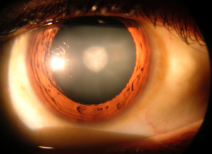
A cataract (KAT-ă-rakt) is a progressive disease of the lens that causes cloudiness and a lack of transparency. Cataracts are very common in older adults. Over half of all Americans aged 80 or older either have cataracts or have had surgery to remove cataracts. See Figure 15.7[2] for an image of a cataract. Cataracts develop slowly, and symptoms include faded colors, blurred or double vision, halos around light, and trouble seeing at night. See Figure 15.8[3] for a simulated image of a person’s vision who has cataracts. Decreased vision due to cataracts may result in trouble reading and driving and increases the risk of falling. Patients often undergo surgery for cataracts. During cataract surgery, the doctor removes the clouded lens and replaces it with a new, artificial lens.[4]

Color Blindness
Color blindness, also called color vision deficiency, makes it difficult to differentiate between certain colors. Color blindness tends to run in families as a genetic condition, but it can also occur due to damage to the eye or to the brain. There’s no cure for color blindness, but special glasses and contact lenses can help people differentiate between colors. Most people who have color blindness are able to use visual strategies related to color selection and don’t have problems participating in everyday activities.[5]
Conjunctivitis
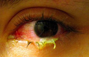
Conjunctivitis (kŏn-jŭnk-tĭ-VĪT-ĭs) is a viral or bacterial infection that causes swelling and redness in the conjunctiva and sclera. See Figure 15.9[6] for an image of conjunctivitis. The eye may feel itchy and painful with crusty yellow drainage present. Bacterial conjunctivitis is treated with antibiotic eye drops. Viral conjunctivitis is typically mild and resolves on its own.[7]
Diabetic Retinopathy
Diabetic retinopathy (ret-ĭn-OP-ă-thē) is a complication caused by diabetes mellitus. Fluid leakage from blood vessels causes the retinal tissue to swell, resulting in cloudy or blurred vision. If left untreated, diabetic retinopathy can cause blindness. There are various treatments for diabetic retinopathy, such as laser surgery to seal leaking blood vessels or the injection of medications into the eye to decrease inflammation. People with advanced cases of diabetic retinopathy may require surgery to remove and replace the gel-like fluid in the back of the eye, called the vitreous humor. Surgery may also be needed to repair a retinal detachment, a separation of the light-receiving lining in the back of the eye.[8]
Glaucoma
Glaucoma (glaw-KŌ-mă) is a disease that causes vision loss due to increased pressure inside the eye damaging the optic nerve. Treatment includes prescription eye drops to lower the pressure inside the eye and slow the progression of the disease. If not treated appropriately, glaucoma can cause blindness. Symptoms of glaucoma include gradual loss of peripheral vision. See Figure 15.10[9] for a simulated image of a person’s vision with glaucoma.[10]

Macular Degeneration
Age-related macular degeneration (MĂK-yŭ-lăr Dĕ-gĕn-ĕ-RĀ-shŭn) is a common condition that causes blurred central vision. It is the leading cause of vision loss for people 50 and older. See Figure 15.11[11] for a simulated image of a person’s vision who has macular degeneration. There are two types of macular degeneration: dry (nonexudative) and wet (exudative). During dry macular degeneration, cellular debris called drusen accumulates and scars the retina. In the wet (exudative) form, which is more severe, blood vessels grow behind the retina and leak exudate fluid, causing hemorrhaging and scarring. There is no treatment for dry macular degeneration, but laser therapy can be used to help treat wet (exudative) macular degeneration.[12]

Myopia
Myopia (mī-Ō-pē-ă), also known as nearsightedness, means normal near vision but far-away objects appear blurry. Myopia occurs when the eyeball grows too long from front to back or when there are problems with the shape of the cornea or the lens. These problems make light focus in front of the retina, instead of on it, causing blurriness. See Figure 15.12[13] for a simulated image of a person’s vision who has myopia. Nearsightedness usually becomes apparent between ages 6 and 14. It is corrected with glasses, contacts, or LASIK surgery.[14]

Night Blindness (Nyctalopia)
Nyctalopia (nik-ta-LŌ-pē-ă) refers to poor vision at night or in dim light. It is also associated with an inability to quickly adapt from a well-illuminated to a poorly illuminated environment. Nyctalopia is not a disease in itself, but rather a symptom of an underlying eye problem, usually a retinal problem.[15]
Nystagmus
Nystagmus (nĭs-TĂG-mŭs) is a disorder that causes involuntary, rhythmic eye movements. Most nystagmus disorders are caused by an underlying medical condition. The repetitive movements can make it impossible for someone to keep their eyes fixed and focused on any given object. Individuals can be born with congenital nystagmus, or it can occur as a result of another condition or a side effect of medication.[16]
Hyperopia and Presbyopia
Hyperopia (hī-pĕr-Ō-pē-ă) refers to impaired near vision, commonly referred to as “farsightedness” because distance vision is normal but near vision is impaired. When it occurs in middle-aged and older adults, it is referred to as presbyopia (prez-bī-Ō-pē-ă). As people age, the lens in the eye gets harder and less flexible and stops focusing light correctly on the retina. Presbyopia can be corrected with glasses and/or contacts.[17] See Figure 15.13[18] for a simulated image of a person’s vision who has presbyopia.

Retinal Detachment
Retinal detachment occurs when the retina is pulled away or separated from its normal position. Sudden flashing lights, floaters, and the appearance of a dark shadow in the center of one’s vision are symptoms of a retinal tear. A retinal tear is a medical emergency because it can lead to a retinal detachment. If left untreated, retinal detachment can lead to blindness. Ophthalmologists repair the retinal detachment using laser surgery, freezing treatment, and/or other types of surgery to fix the retinal tear and reattach the retina to the back of the eye.[19]
Strabismus
Usually, the eyes move together as a pair, but with strabismus (stră-BĬZ-mŭs), the eyes don’t move together in coordination. One eye might drift in, out, up, or down. If not managed, amblyopia can develop. Amblyopia (am-blē-Ō-pē-ă), commonly known as lazy eye, develops when there is a breakdown in how the brain and the eye work together. Over time, the brain relies more and more on the other, stronger eye, while vision in the weaker eye gets worse.[20]
Stye
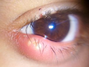
A stye (stī) is a bacterial infection of an oil gland of the eyelid, causing a red, tender bump. See Figure 15.14[21] for an image of a stye. Treatment includes applying warm compresses to the eyelid and prescription eyedrops.[22]
Ear Diseases and Disorders
Hearing Loss
Hearing loss is classified as conductive hearing loss or sensorineural hearing loss. Conductive hearing loss (kŏn-DUC-tĭv HĒR-ing LŎS) occurs when something in the external or middle ear is obstructing the transmission of sound. For example, cerumen impaction or a perforated tympanic membrane can cause conductive hearing loss. Sensorineural hearing loss (sĕn-sō-rē-NOOR-ăl HĒR-ing LŎS) is caused by pathology of the inner ear, nerve damage, or damage to the auditory areas of the brain. Presbycusis (prĕz-bī-KŪ-sĭs) is sensorineural hearing loss that occurs with aging due to gradual nerve degeneration.[23]
Cerumen Impaction
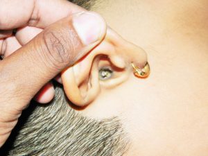
Cerumen impaction (sĕ-ROO-mĕn Im-PĂK-shŭn) is earwax buildup that blocks the ear canal and can cause symptoms of hearing loss. See Figure 15.15 for an image of cerumen impaction.[24] Cerumen can be removed via irrigation of the ear canal, ear drops to dissolve the wax, or manual removal.[25] See Figure 15.15 for an image of an ear irrigation procedure, commonly performed by nurses in a clinic setting.[26]

Otitis Externa
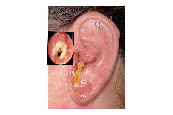
Otitis externa (ō-TĪ-tĭs eks-TUR-nă) is an external ear inflammation and/or infection. See Figure 15.17[27] for an image of otitis externa. It is also known as “swimmer’s ear” because it commonly occurs in swimmers due to the continuous exposure to water that washes away the protective wax in their ear canals. Otitis externa causes a reddened and swollen ear canal with associated yellow, white, or grey debris. Patients often report itching in the ear canal with pain that is worsened by pulling upwards and outwards on the auricle. Otitis externa is treated with antibiotic drops placed in the affected ear canal.[28]
Otitis Media
Otitis media (ō-TĪ-tĭs MĒ-dē-ă) refers to a middle ear infection. Ear infections are common in children between the ages of 6 months and 2 years because of the size and shape of their Eustachian tubes. Acute otitis media typically occurs after an upper respiratory infection when the Eustachian tube becomes inflamed and the middle ear fills with fluid, causing ear pain and irritability. This fluid can become infected, causing purulent fluid and low-grade fever. Otitis media is diagnosed by a health care provider using an otoscope (Ō-tō-skōp) to examine the tympanic membrane for bulging and purulent fluid. If not treated, acute otitis media can potentially cause perforation of the tympanic membrane. Treating early acute otitis media with antibiotics is controversial in the United States due to the effort to prevent antibiotic resistance. However, the treatment goals are to control pain and treat infection with antibiotics if a bacterial infection is present.[29]
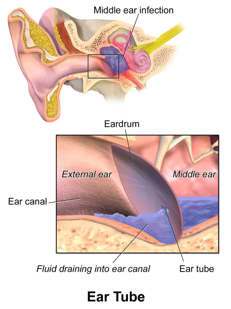
Some children develop recurrent ear infections that cause intermittent hearing loss affecting their language development. For children experiencing recurring infections, a surgery called myringotomy (mĭr-ĭn-GŎT-ō-mē) is performed by an otolaryngologist. During myringotomy surgery, a tympanostomy tube is placed in the tympanic membrane to drain fluid from the middle ear and prevent infection from developing.[30] See Figure 15.18[31] for an image of a tympanostomy tube in the ear.
Otosclerosis
Otosclerosis (ō-tō-sklĕ-RŌ-sĭs) is abnormal bone formation of the middle ear that can cause hearing loss. Treatment for mild otosclerosis is use of a hearing aid, while more severe otosclerosis is treated with surgery called stapedectomy (stā-pĕ-DEK-tŏ-mē).[32]
Presbycusis
Presbycusis (prez-bĭ-KŪ-sĭs) refers to hearing impairment that occurs with aging. One in three people in the U.S. between the ages of 65 and 74 has hearing loss, and nearly half of those older than 75 have difficulty hearing. Having trouble hearing can make it hard to understand and follow a doctor’s advice; respond to warnings; and hear phones, doorbells, and smoke alarms. As we age, changes in the inner ear, middle ear, and nerve pathways from the ear to the brain are common. Long-term exposure to noise and some medical conditions can also play a role in hearing loss. Recent research suggests that certain genes make some people more susceptible to hearing loss as they age. Hearing loss may be treated with hearing aids or cochlear implants.[33]
Tinnitus
Tinnitus (tĭ-NĪ-tŭs) is a ringing, buzzing, roaring, hissing, or whistling sound in the ears. The noise may be intermittent or continuous. Tinnitus can be caused by cerumen impaction, noise trauma, or ototoxic medications, such as diuretics or high doses of aspirin. Military personnel have a high incidence of tinnitus due to noise trauma from loud explosions and gunfire. There are no medications to treat tinnitus, but patients can be referred to an otolaryngologist for treatment such as cognitive therapy or noise masking.[34]
Vertigo
Vertigo (VUR-tĭ-gō) is a type of dizziness that is often described by patients as, “the room feels as if it is spinning.” Benign positional vertigo (BPV) is a common condition caused by crystals becoming lodged in the semicircular canals in the vestibule of the inner ear that send false signals of movement to the brain. BPV can be treated by trained professionals using a specific set of maneuvers that guide the crystals back to the chamber where they are supposed to be in the inner ear.[35]
Olfactory Disorders
Anosmia
Trauma to the face and certain medications can damage the olfactory nerve and cause the loss of smell, known as anosmia (a-NOZ-mē-ă). There are also temporary causes of anosmia, such as the inflammatory response related to upper respiratory infections or allergies. The sense of smell is important because it is often the first warning signal alerting us to the smoke of a fire, spoiled food, or the odor of a natural gas leak or dangerous fumes. Anosmia can also result in the loss of taste. A person with an impaired sense of taste may require additional spice and seasonings on their food, and in severe cases, cause the loss of their desire to eat.[36]
- This work is a derivative of StatPearls by Lee & Mesfin and is licensed under CC BY 4.0 ↵
- “Cataract in human eye.png” by Rakesh Ajuja, MD is licensed under CC BY-SA 3.0 ↵
- “Eye disease simulation, cataract.jpg” by National Eye Institute, National Institutes of Health is in the Public Domain. ↵
- National Eye Institute. (2023, November 15). Cataracts. National Institutes of Health. https://www.nei.nih.gov/learn-about-eye-health/eye-conditions-and-diseases/cataracts ↵
- National Eye Institute. (2023, November 15). Color blindness. National Institutes of Health. https://www.nei.nih.gov/learn-about-eye-health/eye-conditions-and-diseases/color-blindness ↵
- “Swollen eye with conjunctivitis.jpg” by Tanalai at English Wikipedia is licensed under CC BY 3.0 ↵
- National Eye Institute. (2019, July 8). Causes of pink eye. National Institutes of Health. https://www.nei.nih.gov/learn-about-eye-health/eye-conditions-and-diseases/pink-eye/causes-pink-eye ↵
- American Optometric Association. (n.d.). Diabetic retinopathy. https://www.aoa.org/healthy-eyes/eye-and-vision-conditions/diabetic-retinopathy?sso=y ↵
- “Eye disease simulation, glaucoma.jpg” by National Eye Institute, National Institutes of Health is in the Public Domain. ↵
- National Eye Institute. (2023, November 15). Glaucoma. National Institutes of Health. https://www.nei.nih.gov/learn-about-eye-health/eye-conditions-and-diseases/glaucoma ↵
- “Eye disease simulation, age-related macular degeneration.jpg” by National Eye Institute, National Institutes of Health is in the Public Domain. ↵
- National Eye Institute. (2021, June 22). Age-related macular degeneration. National Institutes of Health. https://www.nei.nih.gov/learn-about-eye-health/eye-conditions-and-diseases/age-related-macular-degeneration ↵
- “Eye disease simulation, myopia.jpg” by National Eye Institute, National Institutes of Health is in the Public Domain. ↵
- National Eye Institute. (2020, August 28). Types of refractive errors. National Institutes of Health. https://www.nei.nih.gov/learn-about-eye-health/eye-conditions-and-diseases/refractive-errors/types-refractive-errors#section-id-6802 ↵
- Cleveland Clinic. (2020, December 1). Night blindness (nyctalopia). https://my.clevelandclinic.org/health/symptoms/10118-night-blindness-nyctalopia ↵
- All About Vision. (2019, March 4). Nystagmus. https://www.allaboutvision.com/conditions/nystagmus.htm ↵
- National Eye Institute. (2020, August 28). Types of refractive errors. National Institutes of Health. https://www.nei.nih.gov/learn-about-eye-health/eye-conditions-and-diseases/refractive-errors/types-refractive-errors#section-id-6802 ↵
- “Pesto ingredients - blurred.jpg” by Colin is licensed under CC BY-SA 4.0 ↵
- National Eye Institute. (2023, November 15). Retinal detachment. National Institutes of Health. https://www.nei.nih.gov/learn-about-eye-health/eye-conditions-and-diseases/retinal-detachment ↵
- National Eye Institute. (2022, September 22). Amblyopia (lazy eye). National Institutes of Health. https://www.nei.nih.gov/learn-about-eye-health/eye-conditions-and-diseases/amblyopia-lazy-eye ↵
- “External hordeolum.jpg” by Inrankabirhossain is licensed under CC BY-SA 4.0 ↵
- National Eye Institute. (2023, November 15). Blepharitis. National Institutes of Health. https://www.nei.nih.gov/learn-about-eye-health/eye-conditions-and-diseases/blepharitis ↵
- This work is a derivative of Open RN Nursing Skills 2e and is licensed by CC BY 4.0 ↵
- “Ear Wax.JPG” by Anand2202 is licensed under CC BY-SA 4.0 ↵
- This work is a derivative of StatPearls by Mankowski & Raggio and is licensed under CC BY 4.0 ↵
- “150915-F-GO352-025.jpg” by Staff Sgt. Jason Huddleston for U.S. Air Force is licensed under CC0. Access for free at https://www.59mdw.af.mil/News/Article-Display/Article/647342/photo-essay-559th-medical-group-at-a-glance/ ↵
- “Otitis externa.gif” by S. Bhjimji MD is licensed under CC BY 4.0. Access for free at https://www.ncbi.nlm.nih.gov/books/NBK556055/ ↵
- This work is a derivative of StatPearls by Medina-Blasini & Sharman and is licensed under CC BY 4.0 ↵
- This work is a derivative of StatPearls by Danishyar & Ashurst and is licensed under CC BY 4.0 ↵
- This work is a derivative of StatPearls by Danishyar & Ashurst and is licensed under CC BY 4.0 ↵
- “Ear Tube.png” by BruceBlaus is licensed under CC BY-SA 4.0 ↵
- National Institute on Deafness and Other Communication Disorders. (2022, March 16). Otosclerosis. National Institutes of Health. https://www.nidcd.nih.gov/health/otosclerosis ↵
- National Institute on Deafness and Other Disorders. (2023, March 17). Age-related hearing loss (presbycusis). National Institutes of Health. https://www.nidcd.nih.gov/health/age-related-hearing-loss ↵
- This work is a derivative of StatPearls by Grossan & Peterson and is licensed under CC BY 4.0 ↵
- Woodhouse, S., & Doriangricchia, J. P. (2022, November). Benign paraoxysmal positional vertigo (BPPV). Vestibular Disorders Association. https://vestibular.org/article/diagnosis-treatment/types-of-vestibular-disorders/benign-paroxysmal-positional-vertigo-bppv/ ↵
- National Institute on Deafness and Other Communication Disorders. (2023, July 31). Smell disorders. National Institutes of Health. https://www.nidcd.nih.gov/health/smell-disorders ↵
