15.4 Anatomy and Physiology of the Sensory Systems
Vision
Vision is the special sense of sight based on the transduction of light stimuli received through the eyes. See Figure 15.1[1] for an illustration of the eye. The bony orbits surround the eyeballs, protecting them and anchoring the soft tissues of the eye. The eyelashes and eyelids help protect the eye by blocking particles from landing on the surface of the eye.[2]
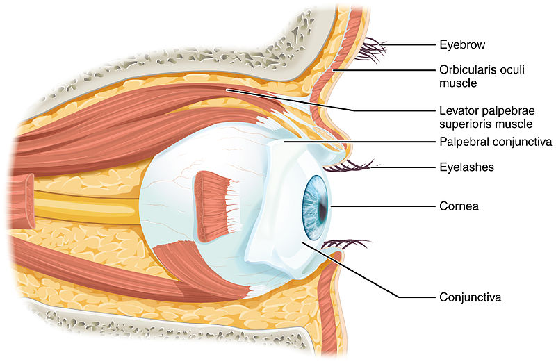
The inner surface of each lid is a thin membrane known as the conjunctiva (kŏn-jŭnk-TI-va). The conjunctiva extends over the white areas of the eye called the sclera (sklĕr-ă), connecting the eyelids to the eyeball. The iris (ir-ĭs) is the colored part of the eye. The iris is a smooth muscle that opens and closes the pupil (pū-pĭl), the hole at the center of the eye that allows light to enter. The iris constricts the pupil in response to bright light and dilates the pupil in response to dim light. The cornea (KOR-nē-ă) is the transparent front part of the eye that covers the iris, pupil, and anterior chamber. The cornea, with the anterior chamber and lens, refracts light and contributes to vision. The innermost layer of the eye is the retina (RĔT-ĭ-nă). The retina contains the nervous tissue and specialized cells called photoreceptors for the initial processing of visual stimuli. These nerve cells of the retina leave the eye and enter the brain via the optic nerve (OP-tik nerve). See Figure 15.2[3] for an illustration of these structures of the eye.[4]
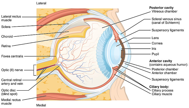
Tears are produced by the lacrimal gland (LAK-rĭ-măl gland) that is located beneath the lateral edges of the nose. Tears are continuously produced by the lacrimal duct and secreted through a duct onto the surface of the eye to wash away foreign particles. Movement of the eye occurs by the contraction of six voluntary extraocular extrinsic muscles that originate from the bones of the orbit and insert into the surface of the eyeball.[5]
View a supplementary YouTube video[6] from Crash Course on vision: Vision: Crash Course Anatomy & Physiology #18
Auditory
Hearing is a special sense based on the transduction of sound waves into a neural signal that is made possible by the structures of the ear. See Figure 15.3[7] for an illustration of the ear structures.[8]
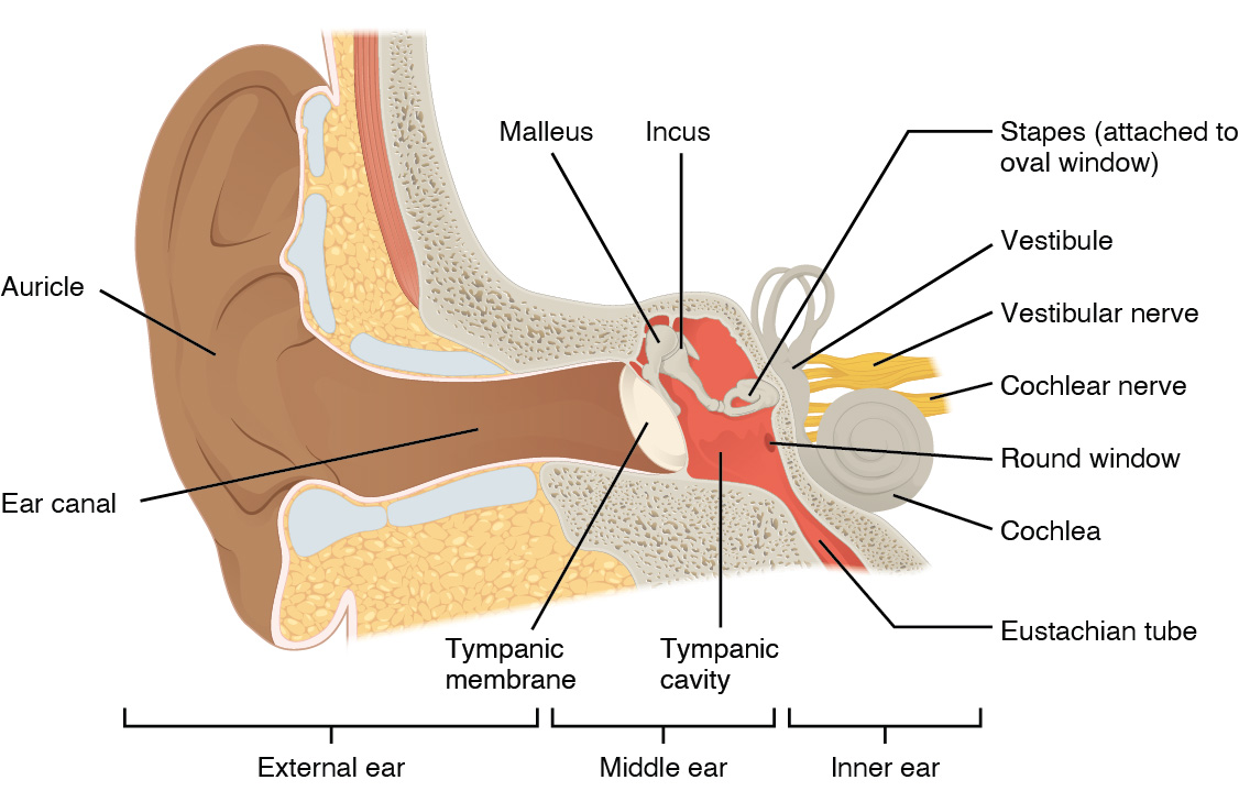
The large, fleshy structure on the lateral aspect of the head is the auricle (AW-rĭ-kl), also known as the pinna (PĬN-ă). The C-shaped curves of the auricle direct sound waves toward the ear canal. At the end of the ear canal is the tympanic membrane (tĭm-păn-ik mem-brān), commonly referred to as the eardrum, that vibrates from sound waves. The auricle, ear canal, and tympanic membrane are referred to as the external ear.[9]
The middle ear consists of a space with three small bones called the malleus, incus, and stapes, the Latin names that roughly translate to “hammer,” “anvil,” and “stirrup.” The malleus (MĂL-ē-ŭs) is attached to the tympanic membrane and articulates with the incus. The incus (ĬN-kŭs), in turn, articulates with the stapes. The stapes (stā-pēz) is attached to the inner ear, where the sound waves are converted into a neural signal. The middle ear is also connected to the pharynx through the Eustachian tube (yōō-STĀ-shən tūb) that helps equalize air pressure across the tympanic membrane. The Eustachian tube is normally closed but will pop open when the muscles of the pharynx contract during swallowing or yawning.[10]
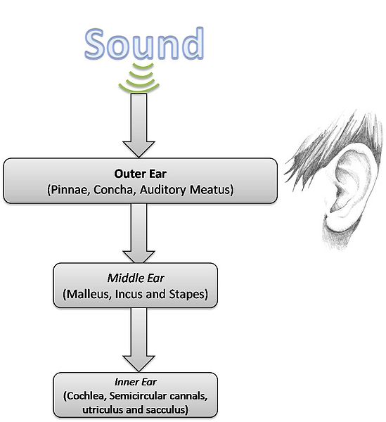
The inner ear (IN-er ĒR) is often described as a bony labyrinth because it is composed of a series of semicircular canals. The semicircular canals have two separate regions called the cochlea (KŎK-lē-ă) and the vestibule (ves-tĭ-būl) that are responsible for hearing and balance. The neural signals from these two regions travel together from the inner ear to the brain via the vestibulocochlear nerve (ves-tĭ-būl-ō-KŌ-klē-ar nerve). See Figure 15.4[11] for an illustration of the transmission of sound from the outer ear to the middle ear and to the inner ear.[12]
Hearing
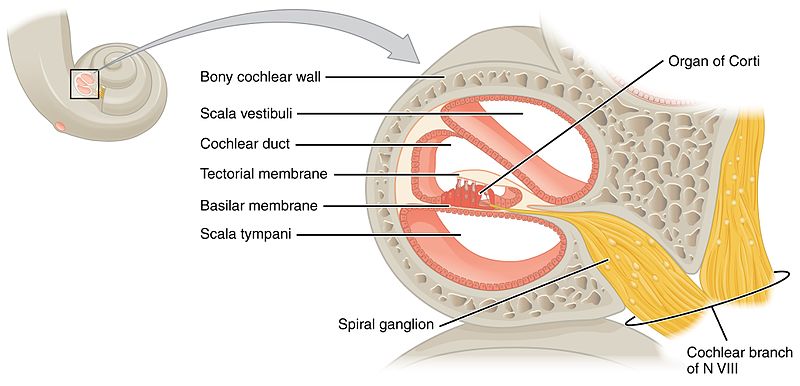
The cochlea encodes auditory stimuli for frequencies between 20 and 20,000 Hz, which is the range of sound that human ears can detect. The hair cells along the length of the cochlear duct, which are each sensitive to a particular frequency, allow the cochlea to separate auditory stimuli by frequency, just as a prism separates visible light into its component colors. See Figure 15.5[13] for an illustration of the cochlea.[14]
Along with hearing, the inner ear is also responsible for the sense of balance. There are three semicircular canals (superior, posterior, and lateral) extending from the vestibule filled with fluid that work to maintain balance. Hair cells within the vestibule sense head position, head movement, and body motion. Medical conditions affecting the semicircular canals cause incorrect balance signals to be sent to the brain, resulting in a spinning type of dizziness called vertigo (VUR-tĭ-gō).[15]
View a supplementary YouTube video[16] from Crash Course on hearing and balance: Hearing & Balance: Crash Course Anatomy & Physiology #17
Taste
Taste is the special sense associated with the tongue. The surface of the tongue contains raised bumps called papillae that contain the structures for taste transmission. Within the structure of the papillae are taste buds that contain specialized receptor cells for the transduction of taste stimuli. These receptor cells are sensitive to the chemicals contained within foods that are ingested, and they release neurotransmitters based on the amount of the chemical in the food. Until recently, only four tastes were recognized: sweet, salty, sour, and bitter. Recent research has suggested that there may also be additional tastes for fats and glutamates (tomatoes, cheese, and mushrooms).[17]
Smell
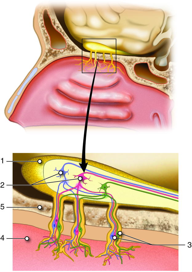
Olfaction (smell) is a special sense based on receptors in a small region of the nasal cavity that are responsive to chemical stimuli. Scent receptor messages travel to the brain, where smells are interpreted and can even become associated with long-term memories and emotional responses.[18] See Figure 15.6[19] for an illustration of olfaction. In this illustration, the olfactory bulb (1) contains mitral cells (2) that receive information from the olfactory cells (3). The olfactory cells are found within the nasal epithelium (4) and pass their information through the cribriform plate (5) of the ethmoid bone.
View a supplementary YouTube video[20] from Crash Course on taste and smell: Taste & Smell: Crash Course Anatomy & Physiology #16
Touch
Touch is considered a general sense, as opposed to the special senses that were previously discussed in this section. Many of the receptors for touch are located in the skin, but receptors are also found in muscles, tendons, joint capsules, ligaments, and in the walls of visceral organs. These receptors detect sensations such as pressure, vibration, light touch, tickle, itch, temperature, pain, and proprioception (i.e., the sense of location, movement, and action of our body parts).[21]
- “1411_Eye_in_The_Orbit.jpg” by OpenStax College is licensed under CC BY 3.0 ↵
- This work is a derivative of Anatomy & Physiology by OpenStax and is licensed under CC BY 4.0. Access for free at https://openstax.org/details/books/anatomy-and-physiology-2e ↵
- “1413_Structure_of_the_Eye.jpg” by OpenStax College is licensed under CC BY 3.0 ↵
- This work is a derivative of Anatomy & Physiology by OpenStax and is licensed under CC BY 4.0. Access for free at https://openstax.org/details/books/anatomy-and-physiology-2e ↵
- This work is a derivative of Anatomy & Physiology by OpenStax and is licensed under CC BY 4.0. Access for free at https://openstax.org/details/books/anatomy-and-physiology-2e ↵
- CrashCourse. (2015, May 11). Vision: Crash Course Anatomy & Physiology #18 [Video]. YouTube. All rights reserved. https://www.youtube.com/watch?v=o0DYP-u1rNM ↵
- “5dfc7e834c03e3e7dbbf82de10413b92379a1a57.png” by Clark, et al., is licensed under CC BY 4.0. Access for free at https://openstax.org/books/biology-2e/pages/1-introduction. ↵
- This work is a derivative of Anatomy & Physiology by OpenStax and is licensed under CC BY 4.0. Access for free at https://openstax.org/details/books/anatomy-and-physiology-2e ↵
- This work is a derivative of Anatomy & Physiology by OpenStax and is licensed under CC BY 4.0. Access for free at https://openstax.org/details/books/anatomy-and-physiology-2e ↵
- This work is a derivative of Anatomy & Physiology by OpenStax and is licensed under CC BY 4.0. Access for free at https://openstax.org/details/books/anatomy-and-physiology-2e ↵
- “Outer_Ear.jpg" by Nzachariah3 and is licensed under CC BY 3.0 ↵
- This work is a derivative of Anatomy & Physiology by OpenStax and is licensed under CC BY 4.0. Access for free at https://openstax.org/details/books/anatomy-and-physiology-2e ↵
- “1406_Cochlea.jpg” by OpenStax College is licensed under CC BY 4.0 ↵
- This work is a derivative of Anatomy & Physiology by OpenStax and is licensed under CC BY 4.0. Access for free at https://openstax.org/details/books/anatomy-and-physiology-2e ↵
- This work is a derivative of Anatomy & Physiology by OpenStax and is licensed under CC BY 4.0. Access for free at https://openstax.org/details/books/anatomy-and-physiology-2e ↵
- CrashCourse. (2015, May 4). Hearing & balance: Crash Course Anatomy & Physiology #17 [Video]. YouTube. All rights reserved. https://www.youtube.com/watch?v=Ie2j7GpC4JU ↵
- This work is a derivative of Anatomy & Physiology by OpenStax and is licensed under CC BY 4.0. Access for free at https://openstax.org/details/books/anatomy-and-physiology-2e ↵
- This work is a derivative of Anatomy & Physiology by OpenStax and is licensed under CC BY 4.0. Access for free at https://openstax.org/details/books/anatomy-and-physiology-2e ↵
- “Cenveo - Drawing Anatomy of the Structures Involved in Smell (Olfaction) - Numbered labels” by Cenveo is licensed under CC BY 4.0 ↵
- CrashCourse. (2015, April 27). Taste & smell: Crash Course Anatomy & Physiology #16 [Video]. YouTube. All rights reserved. https://www.youtube.com/watch?v=mFm3yA1nslE ↵
- This work is a derivative of Anatomy and Physiology by OpenStax licensed under CC BY 4.0. Access for free at https://openstax.org/books/anatomy-and-physiology/pages/1-introduction ↵
