9.5 Physiology of the Cardiovascular System
Pulmonary and Systemic Circulation
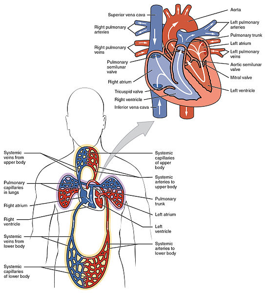
There are two distinct but linked circuits in the human circulation called pulmonary and systemic circuits.
The pulmonary circuit transports oxygenated blood from the right ventricle to the lungs, where carbon dioxide is delivered for exhalation, oxygen is picked up, and this newly oxygenated blood returns to the left atrium of the heart.[1]
The systemic circuit transports oxygenated blood from the left ventricle of the heart, through the aorta, and into the systemic arteries throughout the body where it diffuses into the tissues at the capillaries. Deoxygenated blood travels back to the heart by entering small veins that merge and eventually drain into the superior and inferior vena cava and into the right atrium of the heart. See Figure 9.7[2] for an illustration of blood flow through the pulmonary and systemic circuits.[3]

Transportation
The systemic and pulmonary circuits transport blood and its components for physiological processes that occur throughout the body[4]:
- The right ventricle pumps deoxygenated blood through the pulmonary arteries away from the heart to the lungs. (Note this is the only place in the body where arteries carry deoxygenated blood.) Oxygen from air inhaled into the lungs diffuses into the pulmonary capillaries through the thin walls of the alveoli. At the same time, carbon dioxide diffuses out of the pulmonary capillaries into the alveoli and then exhaled out of the body. The pulmonary veins return oxygenated blood to the left atria of the heart, which moves into the left ventricle where it is pumped out to the rest of the body via the aorta to the systemic arteries.
- Nutrients from the foods eaten are absorbed in the small intestine, where they diffuse into the systemic circulation and are transported to the liver through the hepatic portal vein and then throughout the rest of the body.
- Systemic arteries carry oxygenated blood to the liver where it mixes with the nutrient-rich blood coming in from the hepatic portal vein. Wastes and toxins are filtered out of the blood, bile is produced to help remove wastes and break down fats, medications are metabolized, and nutrients are broken down.
- Systemic arteries carry blood to the kidneys, where wastes are filtered out and urine is created.
- Endocrine glands scattered throughout the body release hormones into the bloodstream where they are transported to distant target cells.
Heart Rate
Heart rate (HART RĀT) (HR) is the number of heart beats per minute (bpm). HR can vary considerably among people based on their age, as well as their fitness level. For an adult, normal resting HR is 60–100 bpm. Although similar to the pulse, the heart rate is auscultated directly using a stethoscope and is a more accurate measurement than the pulse.[5]
Abnormal heart rates are referred to as bradycardia and tachycardia. Bradycardia (brād-i-KARD-ē-ă) refers to a slow heart rate, less than 60 bpm in an adult. Tachycardia (tak-ē-KARD-ē-ă) refers to a fast heart rate, greater than 100 bpm in an adult. Read more about bradycardia and tachycardia in the “Diseases and Disorders of the Cardiovascular System” section.
Pulse
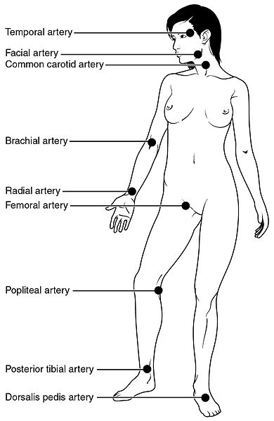
Each time the heart pumps, ejecting blood forcefully into the circulation, the arteries expand and recoil to accommodate the surge of blood moving through them. This expansion and recoiling of the arterial wall are called the pulse (PULS) and allow for the measurement of heart rate. The pulse can be palpated manually by placing the tips of the fingers across an artery that runs close to the body surface, such as the radial artery (on the thumb side of the wrist) or the common carotid artery (in the neck).[6] These sites and other pulse sites are shown in Figure 9.8[7] below.

Both the rate and the strength of the pulse are important clinically. A high pulse rate can be temporarily caused by physical activity, but an extended fast or irregular pulse may indicate a heart condition. The pulse strength indicates the strength of ventricular contraction, cardiac output, and perfusion. Cardiac output (KAR-dē-ăk OUT-put) is the amount of blood pumped by the heart per minute. Perfusion (pĕr-FŪ-zhŏn) is the passage of blood through the blood vessels.[8]
If the pulse is strong, then cardiac output is high and perfusion to that site is good. If the pulse is weak, cardiac output is low or perfusion is impaired, and medical intervention may be warranted.
Cardiac Cycle
The period of time that begins with contraction of the atria, continues through ventricular contraction, and ends with ventricular relaxation is known as the cardiac cycle. The phase of the cardiac cycle when ventricles contract and eject blood is called systole (SIS-tŏ-lē). The phase of the cardiac cycle where the heart muscles relax, allowing the chambers to fill with blood, is called diastole (dī-AS-tŏ-lē).[9]
Blood Pressure
Blood pressure (BLŬD PRESH-ŭr) (BP) is the force of the blood against the vessel walls. In clinical practice, blood pressure is typically measured using a sphygmomanometer (sfĭg-mō-măn-ŎM-ĕt-ĕr), commonly called a blood pressure cuff. The cuff is placed over the brachial artery of the patient’s upper arm and inflated with air to temporarily occlude (ŏ-KLOOD) (block) blood flow and measure blood pressure. Blood pressure is recorded as a ratio of two numbers expressed as systolic pressure over diastolic pressure, measured in millimeters of mercury (mm Hg). For example, 120/80 mm Hg is a normal adult blood pressure. The systolic pressure is the higher value and reflects the arterial pressure resulting from the ejection of blood during ventricular contraction called systole. The diastolic pressure is the lower value and represents the arterial pressure of blood during ventricular relaxation called diastole.[10]
Low blood pressure, called hypotension (hī-pō-TEN-shŏn), has several causes, such as dehydration, vomiting, diarrhea, or medications used to treat hypertension. Treatment for hypotension typically includes fluid replacement. High blood pressure is called hypertension (hī-pĕr-TEN-shŏn) (HTN). Hypertension has a variety of causes, with risk factors such as obesity, lack of exercise, smoking, high salt diet, diabetes, and kidney disease.
- This work is a derivative of Anatomy and Physiology by OpenStax licensed under CC BY 4.0. Access for free at https://openstax.org/books/anatomy-and-physiology/pages/1-introduction ↵
- “2003_Dual_System_of_Human_Circulation.jpg” by OpenStax College is licensed under CC BY 3.0 ↵
- This work is a derivative of Anatomy and Physiology by OpenStax licensed under CC BY 4.0. Access for free at https://openstax.org/books/anatomy-and-physiology/pages/1-introduction ↵
- This work is a derivative of Anatomy and Physiology by OpenStax licensed under CC BY 4.0. Access for free at https://openstax.org/books/anatomy-and-physiology/pages/1-introduction ↵
- This work is a derivative of Anatomy and Physiology by OpenStax licensed under CC BY 4.0. Access for free at https://openstax.org/books/anatomy-and-physiology/pages/1-introduction ↵
- This work is a derivative of Anatomy and Physiology by OpenStax licensed under CC BY 4.0. Access for free at https://openstax.org/books/anatomy-and-physiology/pages/1-introduction ↵
- “2110_Pulse_Sites.jpg” by OpenStax College is licensed under CC BY 3.0 ↵
- This work is a derivative of Anatomy and Physiology by OpenStax licensed under CC BY 4.0. Access for free at https://openstax.org/books/anatomy-and-physiology/pages/1-introduction ↵
- This work is a derivative of Anatomy and Physiology by OpenStax licensed under CC BY 4.0. Access for free at https://openstax.org/books/anatomy-and-physiology/pages/1-introduction ↵
- This work is a derivative of Anatomy and Physiology by OpenStax licensed under CC BY 4.0. Access for free at https://openstax.org/books/anatomy-and-physiology/pages/1-introduction ↵

