9.4 Anatomy of the Cardiovascular System
The Heart
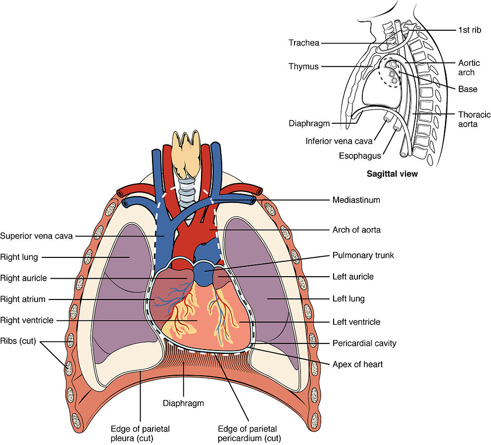
The heart is located within the thoracic cavity, medially between the lungs, and in the space known as the mediastinum. The great veins (the superior and inferior venae cavae) and the great arteries (the aorta and pulmonary artery) are attached to the superior surface of the heart called the base. See Figure 9.1[1] for an illustration of the heart in the thoracic cavity.[2] The inferior tip of the heart, the apex (Ā-peks), lies just to the left of the sternum between the junction of the fourth and fifth ribs. Health care professionals must know the position of the heart when placing a stethoscope (STETH-ŏ-skōp) to listen to heart and lung sounds, referred to as auscultation (os-kŭl-TĀ-shŏn).[3]
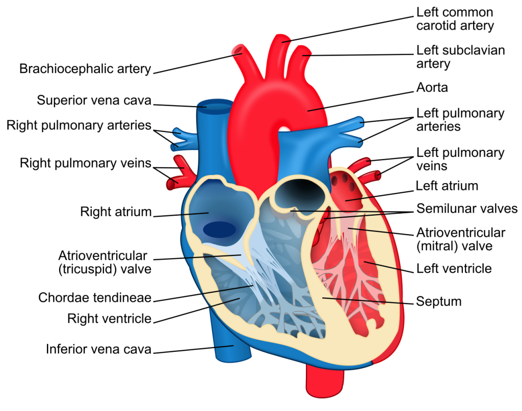
See Figure 9.2[4] for an illustration of the heart. The heart consists of four chambers: two atria and two ventricles. The right atrium receives deoxygenated blood from the body via the inferior and superior vena cava. The right ventricle pumps the deoxygenated blood to the lungs. The left atrium receives oxygenated blood from the lungs and propels it into the left ventricle. The left ventricle pumps blood to the rest of the body.[5]
Blood Vessels
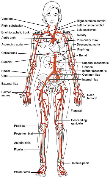
After blood is pumped out of the left ventricle through the aorta, it is carried throughout the body via systemic arteries. An artery (AR-tĕr-ē) is a blood vessel that carries blood away from the heart. Arteries branch into ever-smaller vessels called arterioles (ar-TĒR-ē-ōlz) and eventually into tiny vessels called capillaries. See Figure 9.3[6] for an illustration of the systemic arteries that carry oxygenated blood throughout the body to organs and tissues, as indicated by the red color.
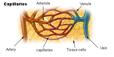
Oxygen and nutrients are exchanged with cells at the capillary (KAP-ĭ-lār-ē) level. A capillary is a microscopic channel that supplies blood to the tissue cells. Capillaries connect arterioles and venules (VEN-yoolz), small veins (VĀNZ). See Figure 9.4[7] for an illustration of capillaries supplying blood to tissue cells.[8]
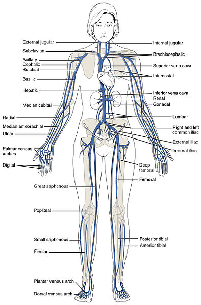
Veins return blood to the heart. Two large veins, the inferior vena cava and superior vena cava (VĒ-nă KĀ-vă), connect to the heart. See Figure 9.5[9] for an illustration of the systemic veins that carry deoxygenated blood back to the heart, indicated in blue. Medications may be administered by health care professionals into veins, referred to intravenous (ĭn-tră-VĒ-nŭs) (IV) medications.[10]
View the following YouTube video[11] on blood vessels: Blood Vessels, Part 1 – Form and Function: Crash Course Anatomy & Physiology #27
Coronary Arteries and Veins
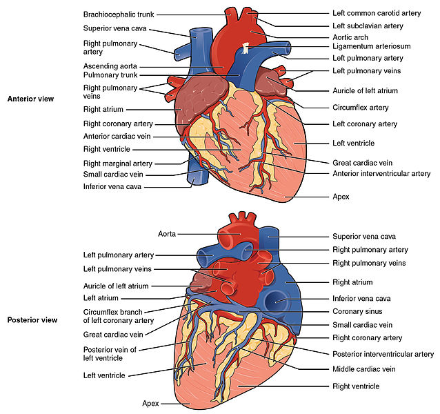
Coronary arteries are arteries that branch off the aorta and carry oxygenated blood to the heart muscle itself. Coronary veins bring deoxygenated blood back to the right atrium. Blood circulates through the coronary arteries and veins with each heartbeat. See Figure 9.6[12] for an illustration of the coronary arteries and veins.
When a person has a myocardial infarction (mī-ŏ-kar′dē-ăl in-FARK-shŏn) (MI), a blockage in a coronary artery causes a lack of oxygenated blood flow to the heart muscle. If a significant area of the heart’s muscle tissue dies from lack of oxygenation, the heart is no longer able to pump.
- “2001_Heart_Position_in_ThoraxN.jpg” by OpenStax College is licensed under CC BY 3.0 ↵
- “Position of the Heart in the Thorax” by OpenStax College is licensed under CC BY 4.0. Access for free at https://openstax.org/books/anatomy-and-physiology/pages/19-1-heart-anatomy ↵
- This work is a derivative of Anatomy and Physiology by OpenStax licensed under CC BY 4.0. Access for free at https://openstax.org/books/anatomy-and-physiology/pages/1-introduction ↵
- “Heart_diagram-en.svg.png” by ZooFari is licensed under CC BY-SA 3.0 ↵
- This work is a derivative of Anatomy and Physiology by OpenStax licensed under CC BY 4.0. Access for free at https://openstax.org/books/anatomy-and-physiology/pages/1-introduction ↵
- “2120_Major_Systemic_Artery.jpg” by OpenStax College is licensed under CC BY 3.0 ↵
- “Illu_capillary_en.jpg” by unknown author is licensed in the Public Domain. ↵
- This work is a derivative of Anatomy and Physiology by OpenStax licensed under CC BY 4.0. Access for free at https://openstax.org/books/anatomy-and-physiology/pages/1-introduction ↵
- “2131_Major_Systematic_Veins.jpg” by OpenStax College is licensed under CC BY 3.0 ↵
- This work is a derivative of Anatomy and Physiology by OpenStax licensed under CC BY 4.0. Access for free at https://openstax.org/books/anatomy-and-physiology/pages/1-introduction ↵
- CrashCourse. (2015, July 20). Blood Vessels, Part 1 - Form and function: Crash Course Anatomy & Physiology #27. [Video]. YouTube. All rights reserved. https://youtu.be/v43ej5lCeBo ↵
- “Surface Anatomy of the Heart” by OpenStax College is licensed under CC BY 4.0 ↵

