7.7 Medical Specialists, Diagnostic Testing, and Procedures Related to the Female Reproductive System
Medical Specialists
Gynecology (gī-nĕ-KOL-ŏ-jē) refers to the study of the female reproductive system. A gynecologist (gīn-ĕ-KOL-ŏ-jĭst) (GYN) is a physician who specializes in the diagnosis and treatment of diseases and disorders of the female reproductive system. Obstetrics (ŏb-STĔT-rĭks) is a specialty regarding pregnancy, labor, and delivery of a baby. An obstetrician (OB) is a physician who specializes in providing medical care through pregnancy, labor, and delivery of a baby. Other subspecialties in women’s health include contraception, reproductive endocrinology, infertility, adolescent gynecology, endoscopy, and gynecological oncology.[1]
To learn more about obstetrics or gynecology, visit the specialty and subspecialty page of the American Board of Obstetrics and Gynecology.
Common Diagnostic Tests and Procedures
Many diagnostic tests and procedures are discussed as they apply to female reproductive system disorders in the “Diseases and Disorders of the Female Reproductive System” section. Common diagnostic tests and procedures used for multiple disorders are described below.
Dilation & Curettage (D&C)
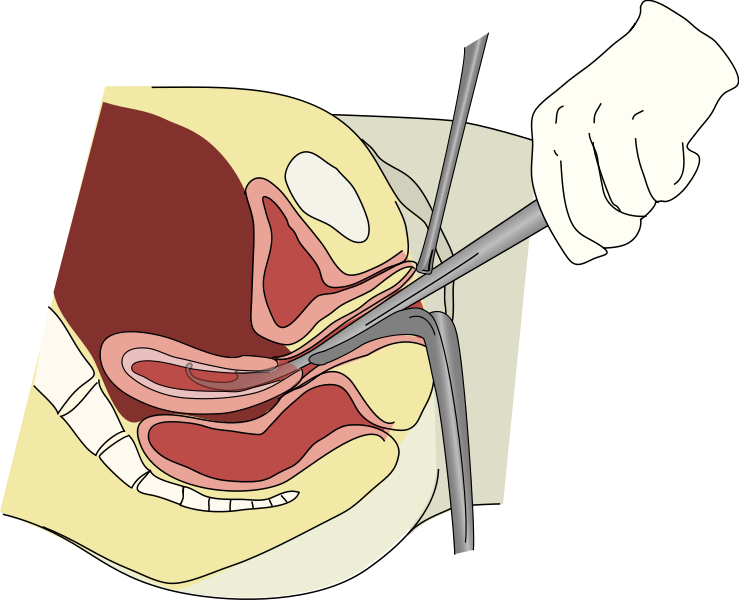
Dilation & curettage (dī-LĀ-shŏn and kūr-ĕ-täj) (D&C) is a procedure performed by a physician in which the opening of the cervix is stretched so that a surgical tool can be inserted into the uterus to scrape away the excess endometrial lining. The removed lining is then biopsied for abnormal tissue. A D&C is performed to determine the cause of abnormal vaginal bleeding, as well as to treat it.[2] See Figure 7.10[3] for an illustration of a D&C.
Hysterectomy, Oophorectomy, and Salpingectomy
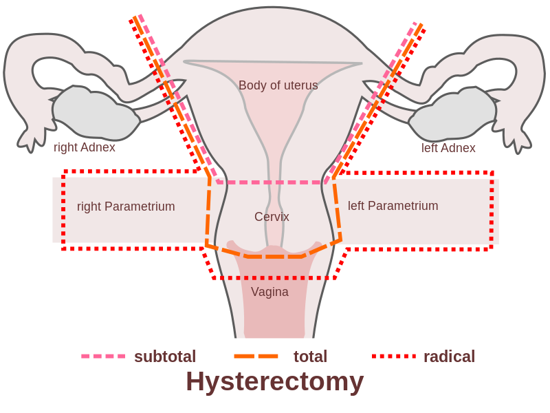
A hysterectomy (his-tĕ-REK-tŏ-mē) is a surgery used to treat a variety of disorders, such as dysfunctional uterine bleeding, fibroids, severe endometriosis, endometrial cancer, and ovarian cancer. See Figure 7.11[4] for an illustration of different types of hysterectomies. There are several types of hysterectomies[5]:
- Total hysterectomy (tō-tăl his-tĕ-REK-tŏ-mē): Surgery to remove the uterus, including the cervix. If the uterus and cervix are taken out through the vagina, the operation is called a vaginal hysterectomy (vă-jĭ-năl his-tĕ-REK-tŏ-mē). If the uterus and cervix are taken out through a large incision in the abdomen, the operation is called a total abdominal hysterectomy (tō-tăl ăb-DŎM-ĭ-năl his-tĕ-REK-tŏ-mē). If the uterus and cervix are taken out through a small incision in the abdomen using a laparoscope, the operation is called a total laparoscopic hysterectomy (tō-tăl lăp-ăr-ŏ-SKŌP-ĭk his-tĕ-REK-tŏ-mē). A subtotal hysterectomy (sŭb-tō-tăl his-tĕ-REK-tŏ-mē) removes part of the uterus but leaves the cervix intact.
- Radical hysterectomy (răd-ĭ-kăl his-tĕ-REK-tŏ-mē): Surgery to remove the uterus, cervix, and part of the vagina. The ovaries, Fallopian tubes, or nearby lymph nodes may also be removed during this procedure, referred to as a total hysterectomy/bilateral salpingectomy oophorectomy (tō-tăl his-tĕ-REK-tŏ-mē / bī-LĂT-ĕr-ăl săl-pĭn-JEK-tŏ-mē ō-ŏ-fŏ-REK-tŏ-mē) (TAH/BSO). An oophorectomy (ō-ŏ-fŏ-REK-tŏ-mē) is the surgical removal of one ovary. A bilateral oophorectomy (bī-LĂT-ĕr-ăl ō-ŏ-fŏ-REK-tŏ-mē) is the removal of both ovaries. A salpingectomy (sal-pĭn-JEK-tŏ-mē) is the removal of a Fallopian tube. A salpingo-oophorectomy (săl-pĭn-jō-ō-ŏf-ō-RĔK-tō-mē) is removal of one Fallopian tube and the associated ovary. Bilateral salpingo-oophorectomy (bī-LĂT-ĕr-ăl săl-pĭng-gō-ō-ŏ-fŏ-REK-tŏ-mē) means removal of both ovaries and Fallopian tubes.
Hysterosalpingogram
A hysterosalpingogram (hĭs-tĕ-RO-săl-pĭnj-ō-gram) is a common imaging procedure performed to assess for potential causes of infertility in women. During the procedure, radiopaque dye is injected into the uterus and fills the uterine cavity, continues into the Fallopian tubes, and eventually reaches fimbriated ends next to the ovaries. Structures are visualized with an X-ray.
Hysteroscopy
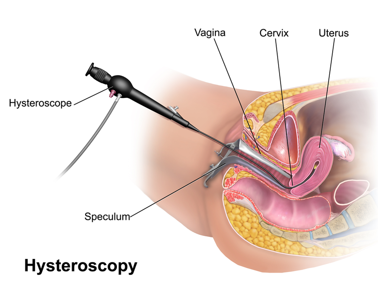
A hysteroscopy (his-tĕ-ROS-kŏ-pē) is a procedure to visually examine the uterus for abnormal areas. A hysteroscope is a thin, tube-like instrument with a light and a lens for viewing and is inserted through the vagina and cervix into the uterus. It may also have a tool to remove tissue samples, which are examined under a microscope for signs of cancer.[6] See Figure 7.12[7] for an illustration of hysteroscopy.
Mammogram
A mammogram (MĂM-ō-grăm) is a radiographic image of breast tissue to detect signs of breast cancer. See Figure 7.13[8] for an image of a women undergoing a mammogram.

Pap Smear
A Papanicolaou smear (păp-ă-NĒ-kă-low smēr) or Pap smear is a diagnostic test that screens for suspicious changes in cells of the cervix. During a Pap smear, a health care provider inserts a speculum (SPEK-yŭ-lŭm) into the vagina to allow visualization of the cervix. Cells are collected from the cervix using sterile swabs and/or cytobrushes and sent to a lab for analysis for abnormal cells or cancer. See Figure 7.14[9] for an image of a cytobrush used to collect cervical cell samples during a PAP smear.

Transvaginal Ultrasound
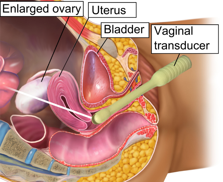
A transvaginal ultrasound (trăns-vă-jĭ-năl ŬL-tră-sound) is a procedure used to examine the vagina, uterus, Fallopian tubes, and bladder. An ultrasound transducer (probe) is inserted into the vagina and used to bounce high-energy sound waves (ultrasound) off internal tissues or organs and make echoes. The echoes form a picture of body tissues called a sonogram. See Figure 7.15[10] for an illustration of the ultrasound transducer placement during a transvaginal ultrasound.
Tubal Ligation
Tubal ligation (TOO-băl lī-GĀ-shŏn) is surgical procedure performed to prevent pregnancy and is also called female sterilization and commonly referred to as “getting your tubes tied.” “Tubal” refers to the Fallopian tubes, and “ligation” means to tie off. During this surgery, both Fallopian tubes are blocked or cut to prevent fertilization of the egg by sperm. The procedure is usually performed in the hospital or in an outpatient surgical clinic. After the procedure, the woman still has a monthly menstrual cycle and menstrual flow.[11]
- American Board of Medical Specialties. (n.d.). American board of obstetrics and gynecology specialties & subspecialties. https://www.abms.org/board/american-board-of-obstetrics-gynecology/ ↵
- Family Doctor. (2023, June 8). Abnormal vaginal bleeding. American Academy of Family Physicians. https://familydoctor.org/condition/abnormal-uterine-bleeding ↵
- “Dilation_%26_curettage.svg.png” by Andrew c is licensed under CC BY-SA 3.0 ↵
- “me_hysterectomy-en.svg” by Hic et nunc is licensed in the Public Domain. ↵
- National Cancer Institute. (2020, November 13). Endometrial cancer treatment (PDQ) - Patient version. https://www.cancer.gov/types/uterine/patient/endometrial-treatment-pdq#top ↵
- National Cancer Institute. (2023, June 26). Endometrial cancer screening (PDQ) - Patient version. https://www.cancer.gov/types/uterine/patient/endometrial-screening-pdq ↵
- “Hysteroscopy.png” by BruceBlaus is licensed under CC BY-SA 4.0 ↵
- “Woman_receives_mammogram.jpg” by Rhoda Baer and is licensed in the Public Domain. ↵
- “Cytobrush_for_sampling_endocervical_cells.jpg” by Whispyhistory is licensed under CC BY-SA 4.0 ↵
- “Vaginal_ultrasonography_in_OHSS_-_coronal.png” by BruceBlaus CC BY 3.0 ↵
- Johns Hopkins Medicine. (n.d.). Tubal ligation. https://www.hopkinsmedicine.org/health/treatment-tests-and-therapies/tubal-ligation ↵

