2.4 Body Regions
Body Regions
Body regions are used to specifically identify a body area, such as the abdominopelvic region and the spinal region.
Abdominopelvic Region
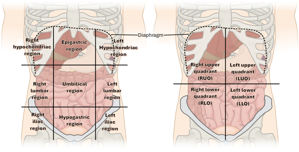
The abdominopelvic region can be divided into nine anatomic regions to assess and precisely communicate abdominal and pelvic locations. These regions include three across the top, three across the middle, and three across the bottom. See Figure 2.4[1] for an illustration of the abdominal region.

The center portion of the nine squares is the umbilical region. Directly above the umbilical region is the epigastric region. Directly below the umbilical region is the hypogastric region. On either side of the epigastric region are the right and left hypochondriac regions. On either side of the hypogastric region are the right and left iliac regions. For example, a nurse may document that an individual has “pain in the epigastric region after eating spicy meals.”
As illustrated in Figure 2.4, the abdominopelvic area can also be divided into four abdominal quadrants:
- Right upper quadrant (RUQ): Contains the gallbladder, the right lobe of the liver, and parts of the small and large intestines.
- Left upper quadrant (LUQ): Contains the stomach, pancreas, spleen, left lobe of the liver, and parts of the small and large intestines.
- Right lower quadrant (RLQ): Contains the appendix, right ureter, right ovary and Fallopian tube, and parts of the small and large intestines.
- Left lower quadrant (LLQ): Contains the left ureter, left ovary and Fallopian tube, and parts of the small and large intestines.
For example, a nurse may document that a patient with severe constipation “has a firm mass in the left lower quadrant.” Keep in mind that “right” and “left” refer to the patient’s right and left side, not the examiner’s view of the individual.
View the following YouTube video[2] on abdominopelvic quadrants and organs contained in each quadrant:
Vertebral Cavity Regions
The vertebral (i.e., spinal) cavity is divided into five regions. From the top, near the head, to the bottom, near the tailbone, these regions are as follows[3]:
- Cervical region: The neck region with seven cervical vertebrae from C1 to C7.
- Thoracic region: The chest region, including the thoracic vertebrae from T1 to T12. Each vertebra in this region is joined to a rib.
- Lumbar region: The region between the ribs and the hip bones, including lumbar vertebrae L1 to L5.
- Sacral region: Five sacral vertebrae, S1 to S5, that are fused to form the sacrum.
- Coccygeal region: The coccyx (i.e., tailbone) that is composed of several fused bones.
For example, a physician may document that a patient has “chronic pain in the L4 region.”
Vertebrae are further discussed in the “Anatomy of the Skeletal System” section.
- This image is a derivative of “Abdominal_Quadrant_Regions.jpg” by OpenStax is licensed under CC BY 3.0. ↵
- RegisteredNurseRN. (2019, May 23). Four abdominal quadrants and nice abdominal regions - Anatomy and physiology [Video]. YouTube. All rights reserved. Reused with permission. https://www.youtube.com/watch?v=ELRzBa-eAA ↵
- This work is a derivative of StatPearls by Peabody, Black, & Das and is licensed under CC BY 4.0 ↵

