17.4 Anatomy of the Endocrine System
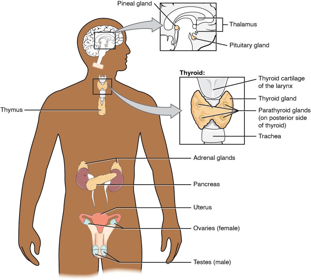
The endocrine system includes the pineal, pituitary, thyroid, parathyroid, and adrenal glands, as well as the pancreas, ovaries, and testes. See Figure 17.1[1] for an illustration of the endocrine system.[2]

Endocrine glands secrete hormones (HŌR-mōnz) as chemical messengers. Hormones are transported via the bloodstream throughout the body, where they bind to receptors on target cells, triggering a characteristic response. This long-distance communication is the fundamental function of the endocrine system.[3]
Pineal Gland
The pineal gland (PĪ-nē-ăl gland) is a small cone-shaped structure that extends from a ventricle of the brain. The pineal gland produces the hormone melatonin (MĔL-ă-tō-nĭn). Melatonin affects reproductive development and daily physiological cycles.[4]
Pituitary Gland
The pituitary gland (pĭ-TŪ-ĭ-tĕr-ē gland) is about the size of a pea. It is a protrusion off the bottom of the hypothalamus at the base of the brain. There are two parts of the pituitary gland called the anterior and the posterior pituitary.
Anterior Pituitary Gland
The anterior pituitary gland secretes several hormones that stimulate the other endocrine glands. These hormones include human growth hormone (HYOO-măn GRŌTH HŌR-mōn) (HGH), thyroid-stimulating hormone (THĪ-rŏid-STĬM-yū-lāt-ing HŌR-mōn) (TSH), adrenocorticotropic hormone (ă-drē-nō-kôr-tĭ-kō-TRŌP-ik HŌR-mōn) (ACTH), follicle-stimulating hormone (FŎL-ĭ-kŭl STĬM-yū-lāt-ing HŌR-mōn) (FSH), luteinizing hormone (LŪ-tē-ĭ-nīz-ing HŌR-mōn) (LH), beta endorphin (BĀ-tă ĕn-DŌR-fĭn), and prolactin (prō-LĂK-tĭn).[5]
Hypopituitarism (hī-pō-pĭ-TŪ-ĭt-ă-rizm) refers to deficient pituitary gland activity. Human growth hormone (HGH) deficiency, also known as dwarfism (DWÔR-fĭz-əm), is a condition caused by insufficient amounts of human growth hormone in the body. In contrast, gigantism (jī-GĂN-tĭz-əm) is caused by excessive human growth hormone in childhood that causes excessive growth and height.
Posterior Pituitary Gland
The posterior pituitary secretes antidiuretic hormone (ăn-tī-dī-yū-RĔT-ik HŌR-mōn) (ADH) that acts on the kidneys. Its effect is to regulate water reabsorption and control fluid balance. For example, if a person becomes dehydrated, the posterior pituitary releases ADH to stimulate additional water reabsorption by the kidneys and return more water to the bloodstream. In contrast, if a person becomes overhydrated from drinking too much water without other substances, the posterior pituitary decreases ADH release. In response, the kidneys decrease water reabsorption, and the excessive water is eliminated in urine output. The posterior pituitary gland also secretes the hormone oxytocin. Oxytocin (ŏk-sē-TŌ-sĭn) is a hormone that stimulates labor contractions and lactation after delivery.[6]
Thyroid Gland
The thyroid gland (THĪ-rŏid gland) is a butterfly-shaped endocrine gland located in the neck, just below the larynx. It helps to regulate metabolic processes in the body by producing and releasing thyroid hormones called thyroxine (thī-RŌK-sĭn) (T4) and triiodothyronine (trī-ī-ō-dō-THĪ-rō-nēn) (T3). These hormones are critical for regulating the basal metabolic rate (BĀ-săl MĔT-ă-bŏl-ĭk RĀT) (BMR), the rate at which the body burns energy while at rest. T3 and T4 control how the body uses energy and oxygen, impacting processes such as digestion, heart rate, and temperature regulation. The thyroid also secretes calcitonin (kăl-sĭ-TŌ-nĭn), a hormone that lowers blood calcium levels.[7]
Euthyroid (yū-THĪ-rŏid) refers to normal thyroid gland functioning with the production of the correct amount of thyroid hormones. Hypothyroidism (hī-pō-THĪ-rŏid-ĭzm) refers to deficient thyroid gland activity. Hyperthyroidism (hī-pĕr-THĪ-rŏid-ĭzm) refers to excessive thyroid gland activity. Goiter (GOI-tĕr) is the abnormal enlargement of the thyroid gland that is a symptom of either hypothyroidism or hyperthyroidism. Read more information about hypothyroidism and hyperthyroidism in the “Diseases and Disorders of the Endocrine System” section.
Parathyroid Glands
Four small masses of tissue are embedded on the surface of the thyroid gland called parathyroid glands (PĂR-ă-THĪ-rŏid glandz). Parathyroid glands secrete parathyroid hormone (PĂR-ă-THĪ-rŏid HŌR-mōn) (PTH) that increases blood calcium levels when necessary to maintain homeostatic balance.[8]
Adrenal Glands
The adrenal glands (ă-DRĒ-nal glandz) are small glands located on top of each kidney. There are two parts to each adrenal gland called the adrenal cortex and the adrenal medulla.
The adrenal cortex (ă-DRĒ-nal KŌR-tĕks), the outer part of the adrenal gland, consists of three different regions, with each region producing different types of hormones called mineralocorticoids, glucocorticoids, and androgens. The principal mineralocorticoid is aldosterone (ăl-DŌS-tĕ-rōn), which acts on the kidneys to reabsorb sodium and water and return these substances to the bloodstream. The principal glucocorticoid is cortisol (KŌR-tĭ-sŏl). Cortisol helps control blood glucose, blood pressure, and metabolism. Androgens (ĂN-drō-jĕnz) contribute to the development and maintenance of male characteristics. Androgens are secreted in minimal amounts in both sexes by the adrenal cortex, but their effects in females are typically masked by hormones secreted by the ovaries.[9]
The adrenal medulla, the inner part of the adrenal gland, secretes two hormones, epinephrine (ĕp-ĭ-NĔF-rĭn) and norepinephrine (nôr-ĕp-ĭ-NĔF-rĭn), also referred to as catecholamines (kăt-ĕ-KŌL-ă-mēns). These hormones are secreted by the nervous system in response to stress.[10]
Pancreas
The pancreas (PĂN-krē-ăs) is a long, flat gland that lies behind the stomach. The pancreas serves two functions called exocrine and endocrine roles. The exocrine (ĔK-sō-krīn) role refers to the release of digestive enzymes, including amylase and lipase that help to digest food. The endocrine (ĔN-dō-krīn) role refers to the production of hormones called glucagon and insulin that regulate blood glucose levels.[11]
Glucose (GLŪ-kōs) is the preferred fuel for all body cells. The digestive system breaks down carbohydrate-containing foods and fluids into glucose, where it is absorbed into the bloodstream. Glucose is taken up by cells from the bloodstream for fuel.[12]
Blood glucose levels are maintained by healthy individuals’ bodies between 70 mg/dL and 110 mg/dL. Hyperglycemia (hī-pĕr-glī-SĒ-mē-ă) refers to excessively high blood glucose. Hypoglycemia (hī-pō-glī-SĒ-mē-ă) refers to abnormally low blood glucose. Receptors located in the pancreas sense blood glucose levels. Specialized cells called islet cells secrete glucagon or insulin to maintain normal blood glucose levels. For example, if blood glucose levels rise above normal range, insulin (ĬN-sū-lĭn) is released, which facilitates the uptake of glucose into cells. However, if blood glucose levels drop below normal range, glucagon (GLŪ-kă-gŏn) is released, which stimulates the liver cells to release more glucose into the bloodstream.[13]
Ovaries and Testes
The ovaries and testes are responsible for producing ova and sperm and also secrete hormones.
In females, ovaries secrete estrogen (ĕs-trō-jĕn) and progesterone (prō-JĔS-tĕ-rōn). At the onset of puberty, estrogen promotes the development of breasts and the maturation of the uterus. Progesterone causes the uterine lining to thicken in preparation for pregnancy. Together, progesterone and estrogen are responsible for the changes that occur in the uterus during the menstrual cycle.[14]
In males, testes secrete testosterone (tĕs-TŌS-tĕ-rōn). At the onset of puberty, testosterone is responsible for the following actions[15]:
- Growth and development of the male reproductive structures
- Increased skeletal and muscular growth
- Enlargement of the larynx accompanied by a deepening voice
- Growth and distribution of body hair
- “e73bab44e2c1a6d18058e8ac13c76807add6c209.jpg” by Betts et al., is licensed under CC BY 4.0. Access for free at https://openstax.org/books/anatomy-and-physiology/pages/17-1-an-overview-of-the-endocrine-system ↵
- This work is a derivative of Anatomy and Physiology by OpenStax licensed under CC BY 4.0. Access for free at https://openstax.org/books/anatomy-and-physiology/pages/1-introduction ↵
- This work is a derivative of Anatomy and Physiology by OpenStax licensed under CC BY 4.0. Access for free at https://openstax.org/books/anatomy-and-physiology/pages/1-introduction ↵
- This work is a derivative of Anatomy and Physiology by OpenStax licensed under CC BY 4.0. Access for free at https://openstax.org/books/anatomy-and-physiology/pages/1-introduction ↵
- National Cancer Institute. (n.d.). Endocrine glands & their function. National Institutes of Health. https://training.seer.cancer.gov/anatomy/endocrine/glands/ ↵
- This work is a derivative of Anatomy and Physiology by OpenStax licensed under CC BY 4.0. Access for free at https://openstax.org/books/anatomy-and-physiology/pages/1-introduction ↵
- This work is a derivative of Anatomy and Physiology by OpenStax licensed under CC BY 4.0. Access for free at https://openstax.org/books/anatomy-and-physiology/pages/1-introduction ↵
- National Cancer Institute. (n.d.). Endocrine glands & their function. National Institutes of Health. https://training.seer.cancer.gov/anatomy/endocrine/glands/ ↵
- National Cancer Institute. (n.d.). Endocrine glands & their function. National Institutes of Health. https://training.seer.cancer.gov/anatomy/endocrine/glands/ ↵
- National Cancer Institute. (n.d.). Endocrine glands & their function. National Institutes of Health. https://training.seer.cancer.gov/anatomy/endocrine/glands/ ↵
- This work is a derivative of Anatomy and Physiology by OpenStax licensed under CC BY 4.0. Access for free at https://openstax.org/books/anatomy-and-physiology/pages/1-introduction ↵
- This work is a derivative of Anatomy and Physiology by OpenStax licensed under CC BY 4.0. Access for free at https://openstax.org/books/anatomy-and-physiology/pages/1-introduction ↵
- This work is a derivative of Anatomy and Physiology by OpenStax licensed under CC BY 4.0. Access for free at https://openstax.org/books/anatomy-and-physiology/pages/1-introduction ↵
- National Cancer Institute. (n.d.). Gonads. National Institutes of Health. https://training.seer.cancer.gov/anatomy/endocrine/glands/gonads.html ↵
- National Cancer Institute. (n.d.). Gonads. National Institutes of Health. https://training.seer.cancer.gov/anatomy/endocrine/glands/gonads.html ↵

