16.4 Anatomy of the Nervous System
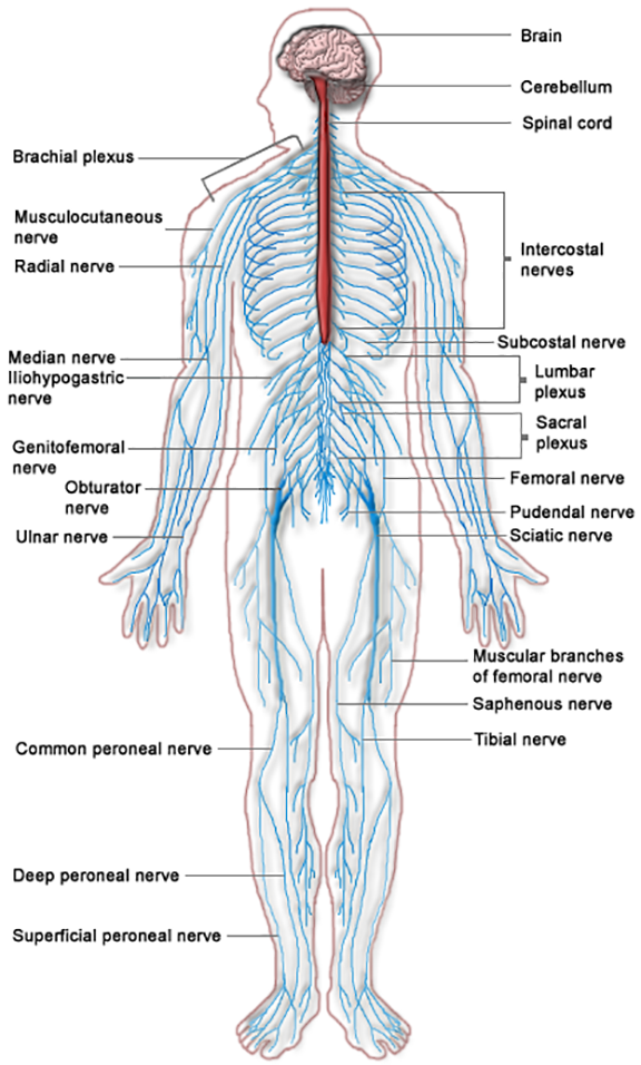
The nervous system is divided into two main parts called the central nervous system and the peripheral nervous system. See Figure 16.1[1] for an illustration of the nervous system.
The Central Nervous System
The central nervous system (SEN-trăl NŬR-vŭs SĬS-tĕm) (CNS) includes the brain and the spinal cord. The brain can be described as the interpretation center, and the spinal cord can be described as the transmission pathway.[2]
Brain
The major regions of the brain are the cerebrum, cerebral cortex, hypothalamus, thalamus, brain stem, and the cerebellum.
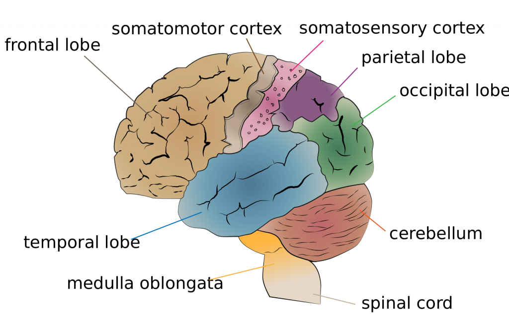
The largest portion of our brain is called the cerebrum (sĕ-rē-brŭm). The cerebrum is covered by a wrinkled outer layer of gray matter called the cerebral cortex (sĕ-rē-brăl kôr-tĕks). The cerebral cortex is involved in complex brain functions including memory, attention, perceptual awareness, thought, language, and consciousness. The cerebral cortex is divided into four lobes named the frontal, parietal, occipital, and temporal lobes. Each lobe has the following specific functions[3]:
- Frontal Lobe (FRŎN-tăl lōb): The frontal lobe is associated with movement. It contains neurons that instruct cells in the spinal cord to move skeletal muscles. The anterior portion of the frontal lobe is called the prefrontal cortex (prē-FRŎN-tăl KÔR-tĕks). The prefrontal cortex provides cognitive functions such as planning and problem-solving that are the basis of our personality, short-term memory, and consciousness. Broca’s area (brō-kăz ăr-ē-ă) is located in the frontal lobe of the dominant hemisphere and is responsible for the production of language and controlling movements responsible for speech.
- Parietal Lobe (pă-rī-ĕ-tăl lōb): The parietal lobe processes general sensations, including touch, pressure, tickle, pain, itch, and vibration.
- Temporal Lobe (tĕm-pŏ-răl lōb): The temporal lobe processes auditory information. Wernicke’s area (wĕr-nĭk-kēz ăr-ē-ă) is located in the temporal lobe of the dominant hemisphere and is responsible for the comprehension of written and spoken language. Because regions of the temporal lobe are part of the limbic system, memory is also an important function associated with the temporal lobe. The limbic system is involved with our behavioral and emotional responses needed for survival, such as feeding, reproduction, and the fight-or-flight responses.
- Occipital Lobe (ŏk-SĬP-ĭ-tăl lōb): The occipital lobe primarily processes visual information.
The cerebellum (sĕr-ĕ-bĕl-ŭm) is the posterior part of the brain that controls fine motor skills. See Figure 16.2[4] for an illustration of the cerebellum and the lobes of the cerebral cortex.
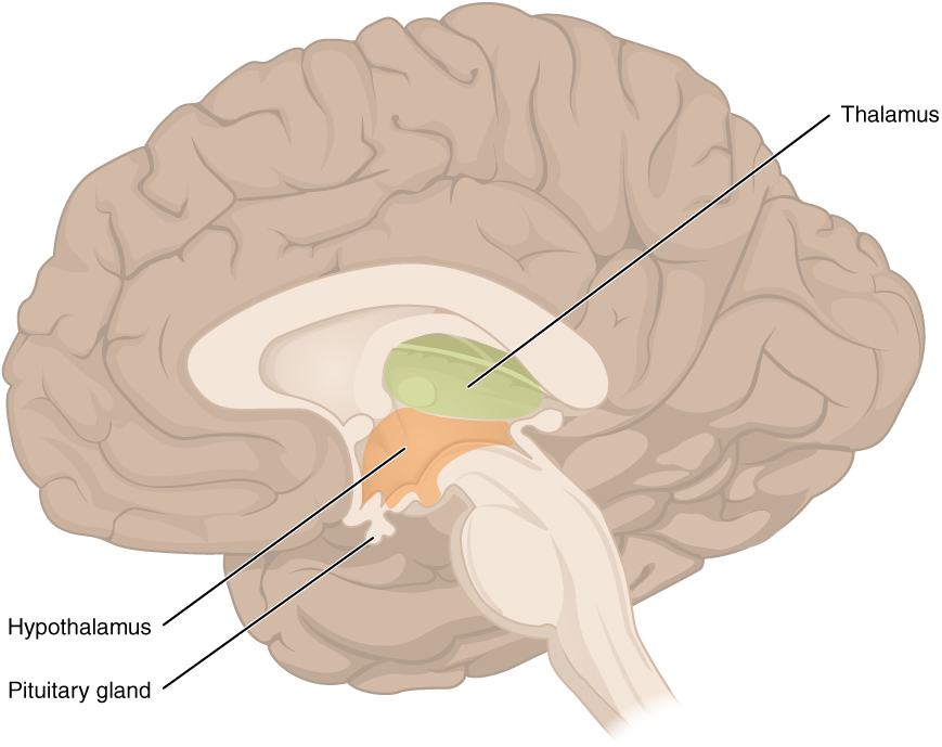
Deep within the cerebrum are the hypothalamus (hī-pō-THAL-ă-mŭs) and the thalamus (thăl-ă-mŭs). The hypothalamus coordinates the autonomic nervous system and the activity of the pituitary, controlling body temperature, thirst, hunger, and other homeostatic systems. The thalamus is the relay center for sensory and motor signals to the cerebral cortex.[5] See Figure 16.3[6] for an illustration of the hypothalamus and the thalamus.
The ventricles (vĕn-trĭ-kŭls) are a group of interconnected, fluid-filled cavities within the brain. The meninges (mĕn-ĭn-jēz) are three layered membranes that cover the brain and spinal cord. The dura mater (dŏŏ-ră mā-tĕr) is the tough outermost membrane enveloping the brain and spinal cord. The arachnoid layer is the web-like middle layer, and the pia mater is the delicate innermost layer of the meninges. The brain stem (brān stĕm) connects the spinal cord with the brain. It is composed of three parts, the midbrain, pons, and medulla oblongata. The brain stem regulates several crucial autonomic functions in the body, including involuntary functions in the cardiovascular and respiratory systems and reflexes like vomiting, coughing, sneezing, and swallowing.[7]
Spinal Cord
The spinal cord (spī-năl kôrd) is a bundle of nerve fibers enclosed in the spine that connects nearly all parts of the body to the brain. It is a continuation of the brain stem that transmits sensory information to the brain and motor impulses to the muscles.[8]
Peripheral Nervous System
The peripheral nervous system (pĕ-rĬF-ĕr-ăl NŬR-vŭs SĬS-tĕm) (PNS) consists of the remaining parts of the nervous system outside of the brain and spinal cord, including the cranial nerves that branch out from the brain and the spinal nerves that branch out from the spinal cord. The peripheral nervous system is the communication network between the brain and the body’s parts.[9]
Cranial nerves (krā-nē-ăl nĕrvz) are directly connected from the brain to the periphery. They are primarily responsible for the sensory and motor functions of the head and neck. The vagus nerve (vā-gŭs nĕrv) is a cranial nerve that innervates the heart and digestive system and is involved in regulating many critical bodily functions.[10]
Spinal nerves (spī-năl nĕrvz) are named based on the level of the spinal cord where they emerge. See Figure 16.4[11] for an illustration of spinal nerves. There are eight pairs of cervical nerves designated C1 to C8, twelve thoracic nerves designated T1 to T12, five pairs of lumbar nerves designated L1 to L5, five pairs of sacral nerves designated S1 to S5, and one pair of coccygeal nerves. All spinal nerves are mixed nerves, meaning they can carry both sensory and motor information. Spinal nerves extend outward from the vertebral column to innervate the periphery while also transmitting sensory information back to the brain.[12]
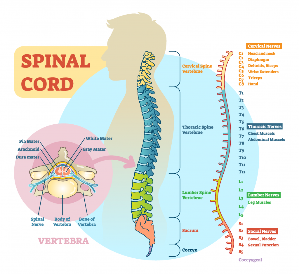
Each spinal nerve innervates a specific region of the body[13]:
- C1 provides motor innervation to muscles at the base of the skull.
- C2 and C3 provide both sensory and motor control to the back of the head and behind the ears.
- The phrenic nerve from C3, C4, and C5 innervates the diaphragm to enable breathing. This is vital because if a patient has an injury where the spinal cord is cut above C3, then spontaneous breathing is not possible.
- C5 through C8 and T1 combine to form the brachial plexus, a tangled array of nerves that serve the upper limbs and upper back.
- The lumbar plexus arises from L1-L5 and innervates the pelvic region and the anterior legs.
- The sacral plexus comes from the lower lumbar nerves L4 and L5 and the sacral nerves S1 to S4. The sciatic nerve is a part of the sacral plexus. If the sciatic nerve becomes compressed due to degeneration of an intervertebral disc, a medical condition called sciatica (sī-ăt-ĭ-kă) occurs that causes pain in the back, hip, and outer side of the leg.
If a patient experiences a spinal cord injury, the degree of paralysis can be predicted by the location of the spinal cord injury. The higher up on the spinal cord an injury occurs, the greater loss of function. For example, a spinal cord injury higher on the spinal cord can cause paralysis in most of the body and affect all limbs (tetraplegia or quadriplegia). An injury that occurs lower on the spinal cord may only affect a person’s lower body and legs (paraplegia). Review information about paralysis in “Diseases and Disorders of the Musculoskeletal System” section of the Musculoskeletal System chapter.
It is also important to remember when a patient has a spinal cord injury and motor nerves are damaged, their sensory nerves may still be intact. If this occurs, the patient can still feel sensation even if they can’t move the extremity.
Neurons
Nervous tissues present in both the CNS and PNS contain two basic types of cells: neurons and neuroglia (glial cells). Neurons (nŭr-ŏns) are responsible for the communication that the nervous system provides. They are electrically active and release chemical signals called neurotransmitters. Neuroglia plays a supporting role for nervous tissue.[14]
Neurons provide electrical signals that communicate information about sensations and, in response, stimulate movements. The three-dimensional shape of neurons makes massive numbers of connections within the nervous system. The main part of a neuron is the cell body (sĕl bŏd-ē). A fiber that emerges from the cell body and projects to target cells is called the axon (ăk-sŏn). A single axon can branch repeatedly to communicate with many target cells by sending nerve impulses. Dendrites (dĕn-drītz) conduct information received from other neurons at contact areas called a synapse (sĭn-ăps). A synapse is a junction between two nerve cells, consisting of a tiny gap through which neurotransmitters pass.[15] See Figure 16.5[16] for an illustration of a neuron.
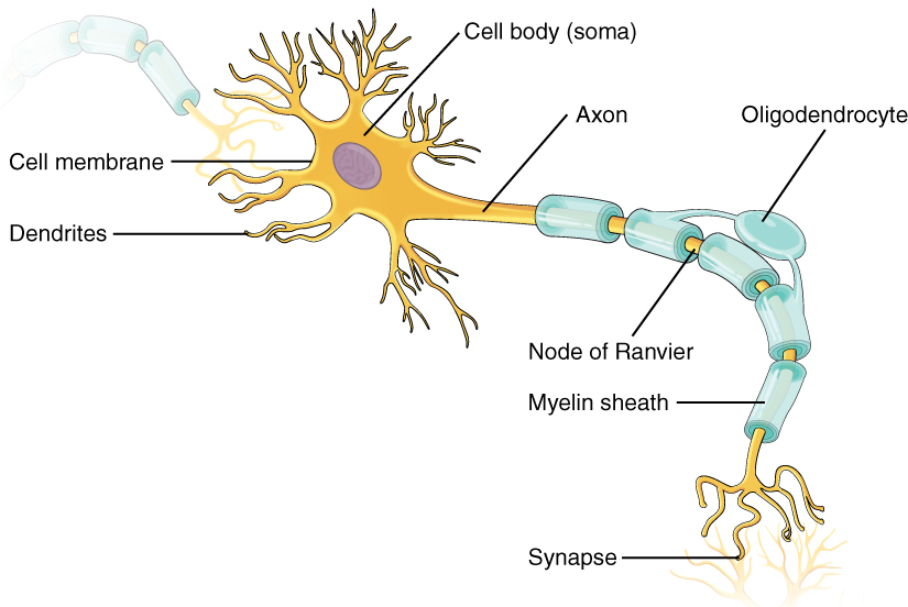
Many axons are wrapped by an insulating substance called myelin (mī-ĕ-lĭn), which is made from neuroglia. Myelin acts as insulation much like the plastic or rubber that is used to insulate electrical wires.[17]
Neurotransmitters
Electrical impulses from neurons signal the release of a neurotransmitter (nŭr-ō-trăns-mĭt-ĕr) into a synapse. Neurotransmitters allow the impulse to be transferred to a neuroreceptor on another neuron, muscle fiber, or other structure. Neuroreceptors are specific for certain neurotransmitters, and the two fit together like a key and lock.[18] See Figure 16.6[19] for an illustration of neuron communication.
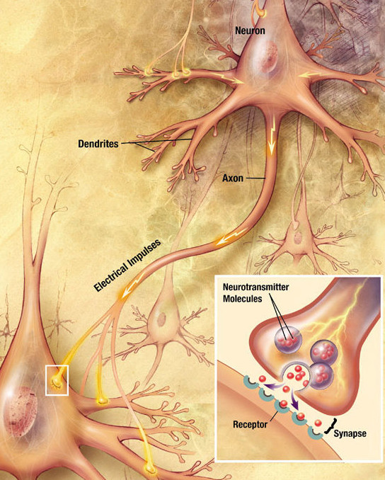
Many nervous system diseases and mental health disorders are related to abnormal impulse transmission and/or imbalanced levels of neurotransmitters. For example, dopamine is a neurotransmitter that influences movement and cognition. Dopamine imbalances are associated with Parkinson’s disease, schizophrenia, and substance use disorders. Serotonin is another type of neurotransmitter whose imbalance affects moods and feelings of depression. Medications used to treat many mental health disorders help balance neurotransmitter levels.[20]
- “Nervous system diagram.png” by unknown is licensed under CC BY-NC-SA 3.0. Access for free at https://med.libretexts.org/Bookshelves/Nursing/Book%3A_Clinical_Procedures_for_Safer_Patient_Care_(Doyle_and_McCutcheon)/02%3A_Patient_Assessment/2.07%3A_Focused_Assessments ↵
- This work is a derivative of Anatomy & Physiology by OpenStax and is licensed under CC BY 4.0. Access for free at https://openstax.org/books/anatomy-and-physiology/pages/1-introduction ↵
- This work is a derivative of Anatomy and Physiology by OpenStax licensed under CC BY 4.0. Access for free at https://openstax.org/books/anatomy-and-physiology/pages/1-introduction ↵
- “Cerebrum lobes.svg” by Jkwchui is licensed under CC BY-SA 3.0 ↵
- This work is a derivative of Anatomy and Physiology by OpenStax licensed under CC BY 4.0. Access for free at https://openstax.org/books/anatomy-and-physiology/pages/1-introduction ↵
- “1310_Diencephalon.jpg” by OpenStax is licensed under CC BY 4.0 ↵
- This work is a derivative of Anatomy and Physiology by OpenStax licensed under CC BY 4.0. Access for free at https://openstax.org/books/anatomy-and-physiology/pages/1-introduction ↵
- This work is a derivative of Anatomy and Physiology by OpenStax licensed under CC BY 4.0. Access for free at https://openstax.org/books/anatomy-and-physiology/pages/1-introduction ↵
- This work is a derivative of Anatomy & Physiology by OpenStax and is licensed under CC BY 4.0. Access for free at https://openstax.org/books/anatomy-and-physiology/pages/1-introduction ↵
- This work is a derivative of Anatomy and Physiology by OpenStax licensed under CC BY 4.0. Access for free at https://openstax.org/books/anatomy-and-physiology/pages/1-introduction ↵
- “1008694237-vector.png” by VectorMine on Shutterstock. All rights reserved. Image used with purchased permission. ↵
- This work is a derivative of Anatomy and Physiology by OpenStax licensed under CC BY 4.0. Access for free at https://openstax.org/books/anatomy-and-physiology/pages/1-introduction ↵
- This work is a derivative of Anatomy and Physiology by Boundless.com and is licensed under CC BY-SA 4.0 ↵
- This work is a derivative of Anatomy & Physiology by OpenStax and is licensed under CC BY 4.0. Access for free at https://openstax.org/books/anatomy-and-physiology/pages/1-introduction ↵
- This work is a derivative of Anatomy and Physiology by OpenStax licensed under CC BY 4.0. Access for free at https://openstax.org/books/anatomy-and-physiology/pages/1-introduction ↵
- “1206_The_Neuron-1.jpg” by OpenStax is licensed under CC BY 4.0 ↵
- This work is a derivative of Anatomy and Physiology by OpenStax licensed under CC BY 4.0. Access for free at https://openstax.org/books/anatomy-and-physiology/pages/1-introduction ↵
- This work is a derivative of Anatomy and Physiology by OpenStax licensed under CC BY 4.0. Access for free at https://openstax.org/books/anatomy-and-physiology/pages/1-introduction ↵
- “Chemical synapse schema cropped.jpg” by Looie496 is licensed under Public Domain. Access for free at https://med.libretexts.org/Bookshelves/Anatomy_and_Physiology/Book%3A_Anatomy_and_Physiology_(Boundless)/10%3A_Overview_of_the_Nervous_System/10.1%3A_Introduction_to_the_Nervous_System/10.1A%3A_Organization_of_the_Nervous_System ↵
- This work is a derivative of Open RN Nursing Pharmacology 2e and Open RN Nursing Skills 2e both licensed under CC BY 4.0 ↵

