14.4 Anatomy of the Muscular System
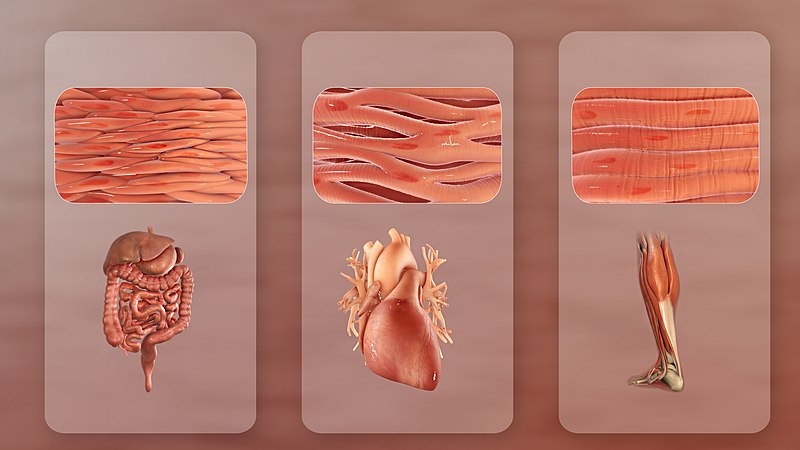
There are three major types of muscle tissue categorized as smooth, cardiac, and skeletal muscle. See Figure 14.1[1] for an illustration of these three types of muscle. Cardiac and skeletal muscles are striated (STRĪ-ā-tĕd), meaning they contain functional units called sarcomeres. Sarcomeres are composed of two protein filaments called actin (ĂK-tin) and myosin (MĪ-ŏ-sin) that are responsible for muscular contraction.[2]

Smooth Muscle
Smooth muscle (SMŎŎTH MŬS-ăl) is responsible for involuntary muscle movement. Smooth muscle is present in the following areas[3]:
- Walls of hollow organs, like the urinary bladder, uterus, stomach, and intestines, where muscle contractions cause the movement of fluids and other substances
- Walls of passageways, such as the arteries and veins of the circulatory system, where it causes vasodilation and vasoconstriction
- Tracts of the respiratory, urinary, and reproductive systems, where contraction and relaxation affect the movement of air, urine, and reproductive fluids
- Eyes, where it functions to change the size of the pupil
- Skin, where it causes hair to stand erect in response to cold temperature or fear, commonly called goose bumps
Cardiac Muscle
Cardiac muscle (KÄR-dē-ăk MŬS-ăl) is only found in the heart. Highly coordinated contractions of cardiac muscle pump blood throughout the circulatory system. Cardiac muscle fiber cells are extensively branched and connected to one another at their ends to allow the heart to contract in a wavelike pattern and work as a pump.[4]
Skeletal Muscle
Skeletal muscles (SKĔL-ĕ-tăl MŬS-ălz) are located throughout the body. They are under voluntary control and primarily produce movement of the arms, legs, back, and neck and maintain posture by resisting gravity. Small, constant adjustments of the skeletal muscles are needed to hold the body upright or balanced in any position.[5]
Skeletal muscles also have several additional functions. Ring-shaped skeletal muscles called sphincters are located throughout the body at the openings of internal tracts to control the movement of substances. These skeletal muscles allow voluntary control of functions such as swallowing, defecation, and urination in the digestive and urinary systems.[6]
Skeletal muscles, such as the rectus abdominus, protect internal organs (particularly abdominal and pelvic organs) by acting as an external barrier against trauma and supporting the weight of the organs.[7]
Skeletal muscles also contribute to maintaining homeostasis by generating heat. This heat generation is very noticeable during exercise, when sustained muscle movement causes a person’s body temperature to rise, or conversely during cold environmental temperatures when shivering produces random skeletal muscle contractions to generate heat.[8]
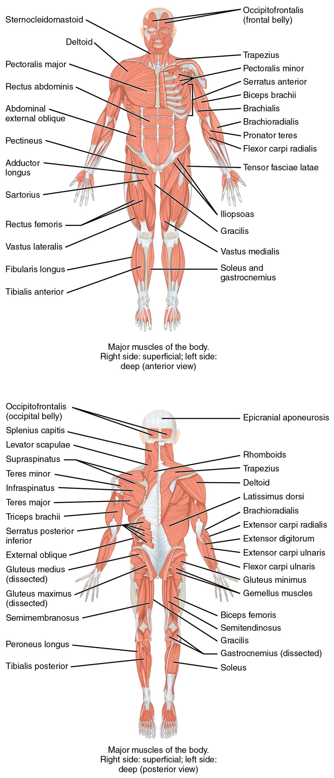
See Figure 14.2[9] for an illustration of the major skeletal muscles of the body. For the anterior and posterior views in this figure, superficial muscles are shown on the right side of the body, and deep muscles are shown on the left side of the body. For the legs, superficial muscles are shown in the anterior view while the posterior view shows both superficial and deep muscles.[10]

Muscles are named based on various characteristics, such as the following[11]:
- Body location: The area of the body, for example, biceps, triceps, and quadriceps
- Size: The size of the muscle, such as maximus (largest) and minimus (smallest)
- Shape: The shape of the muscle, such as deltoid (triangular) or trapezius (trapezoid)
- Action: The action of the muscle, such as flexor (i.e., to flex) or adductor (i.e., towards midline of body)
- Fiber direction: The direction of the muscle fibers (such as external oblique)
Major Skeletal Muscles
Major skeletal muscles include the following[12]:
- Biceps brachii (BĪ-sĕps bră-ki): Muscle on the anterior upper arm.
- Biceps brachialis (bră-kē-ĂL-ĭs): Muscle located in the upper and lower arm that flexes the elbow joint and rotates the forearm.
- Deltoid (DĔL-toid): A large triangular muscle covering the shoulder joint.
- Gastrocnemius (găs-trŏk-NĒ-mē-ŭs): The chief muscle of the calf of the leg.
- Gluteus maximus (glŪ-tē-ŭs MĂK-sĭ-mŭs): The largest and outermost of the three gluteal muscles in the buttocks.
- Latissimus dorsi (lă-tĭs-Ĭ-mŭs DŌR-sī): A large muscle in the back.
- Pectoralis major (pĕk-tŏr-ĂL-ĭs MĀ-jŏr): A thick, fan-shaped muscle situated on the chest.
- Quadriceps (kwŎD-rĭ-sĕps): A large muscle group on the front of the thigh.
- Rectus abdominis (RĔK-tŭs ăb-DŎM-ĭ-nĭs): A paired muscle running vertically on each side of the anterior wall of the abdomen.
- Triceps brachii (TRĪ-sĕps bră-ki): Muscle on the posterior of the upper arm.
Tendons
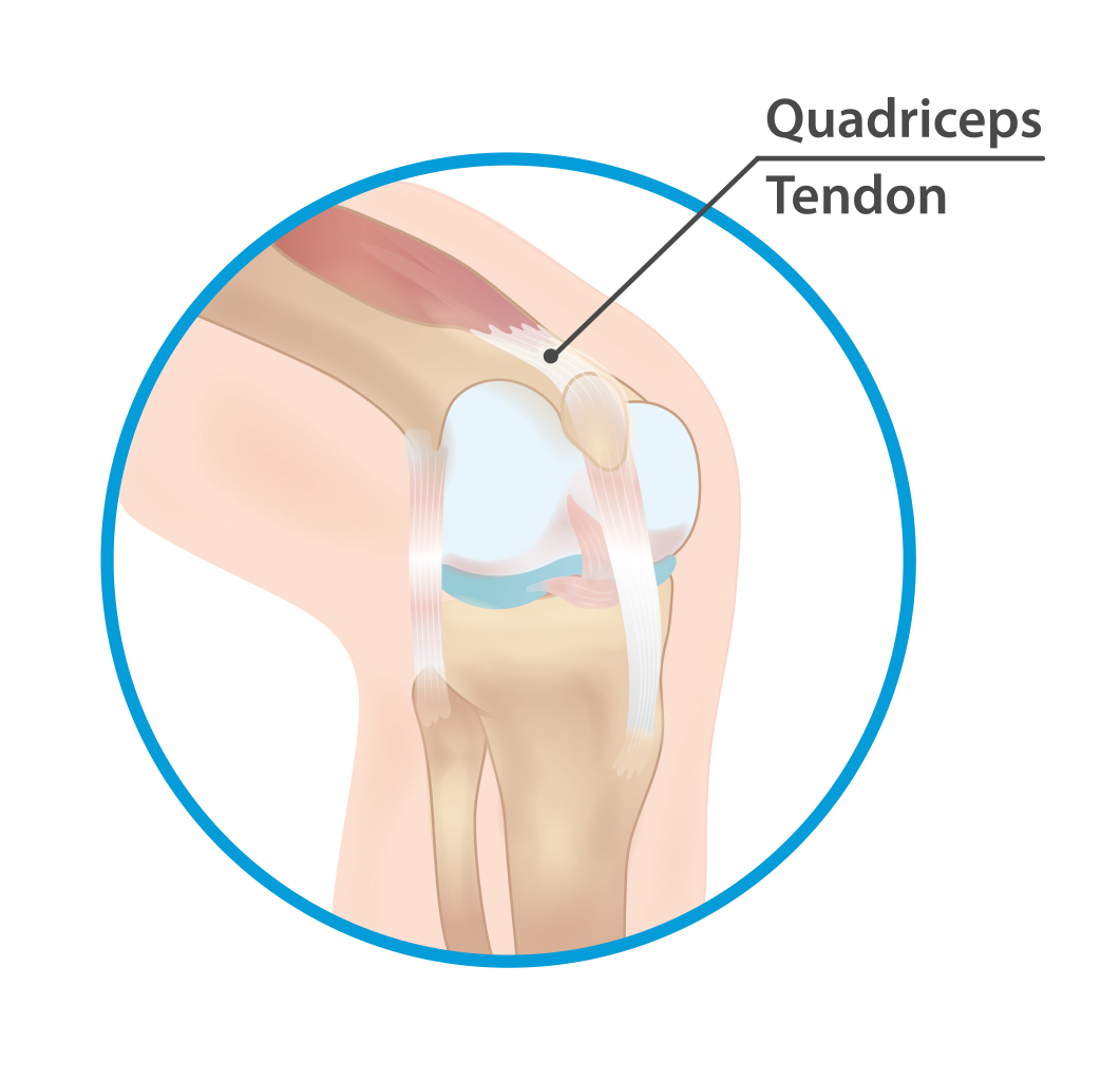
Muscles attach to bones via tendons (TĔN-dŏnz). A tendon is a flexible but inelastic cord of strong fibrous collagen tissue. For example, consider the Achilles tendon, hamstring, and rotator cuff. The Achilles tendon (ə-KĬL-ēz TĔN-dŏn) attaches the calf muscles to the heel bone. The hamstring (HĂM-strĭng) tendons refer to five tendons at the back of a person’s knee that connect a group of three hamstring muscles to bones in the pelvis, knee, and lower leg. The rotator cuff (rō-TĀ-tŏr KŬF) is a group of muscles and tendons that stabilize the shoulder.[13] See Figure 14.3[14] for an illustration of the tendon that attaches the quadriceps muscle to the top of the patella.

- “Types_Of_Muscle.jpg” by www.scientificanimations.com is licensed under CC BY-SA 4.0 ↵
- This work is a derivative of Anatomy and Physiology by OpenStax licensed under CC BY 4.0. Access for free at https://openstax.org/books/anatomy-and-physiology/pages/1-introduction ↵
- This work is a derivative of Anatomy & Physiology by OpenStax and is licensed under CC BY 4.0. Access for free at https://openstax.org/details/books/anatomy-and-physiology-2e ↵
- This work is a derivative of Anatomy and Physiology by OpenStax licensed under CC BY 4.0. Access for free at https://openstax.org/books/anatomy-and-physiology/pages/1-introduction ↵
- This work is a derivative of Anatomy and Physiology by OpenStax licensed under CC BY 4.0. Access for free at https://openstax.org/books/anatomy-and-physiology/pages/1-introduction ↵
- This work is a derivative of Anatomy and Physiology by OpenStax licensed under CC BY 4.0. Access for free at https://openstax.org/books/anatomy-and-physiology/pages/1-introduction ↵
- This work is a derivative of Anatomy and Physiology by OpenStax licensed under CC BY 4.0. Access for free at https://openstax.org/books/anatomy-and-physiology/pages/1-introduction ↵
- This work is a derivative of Anatomy and Physiology by OpenStax licensed under CC BY 4.0. Access for free at https://openstax.org/books/anatomy-and-physiology/pages/1-introduction ↵
- “1105_Anterior_and_Posterior_Views_of_Muscles.jpg” by OpenStax is licensed under CC BY 4.0 ↵
- This work is a derivative of Anatomy and Physiology by OpenStax licensed under CC BY 4.0. Access for free at https://openstax.org/books/anatomy-and-physiology/pages/1-introduction ↵
- This work is a derivative of Anatomy and Physiology by OpenStax licensed under CC BY 4.0. Access for free at https://openstax.org/books/anatomy-and-physiology/pages/1-introduction ↵
- This work is a derivative of Anatomy and Physiology by OpenStax licensed under CC BY 4.0. Access for free at https://openstax.org/books/anatomy-and-physiology/pages/1-introduction ↵
- This work is a derivative of Anatomy and Physiology by OpenStax licensed under CC BY 4.0. Access for free at https://openstax.org/books/anatomy-and-physiology/pages/1-introduction ↵
- “Quadriceps_tendon.svg” by InjuryMap is licensed under CC BY-SA 4.0 ↵

