11.10 Respiratory System
Most humans cannot survive without breathing for more than three minutes. If you experiment by trying to hold your breath longer, your autonomic nervous system will take over and resume a normal breathing pattern. This is because cells need to maintain a delicate balance of using oxygen for functioning and releasing carbon dioxide as a waste product.[1]
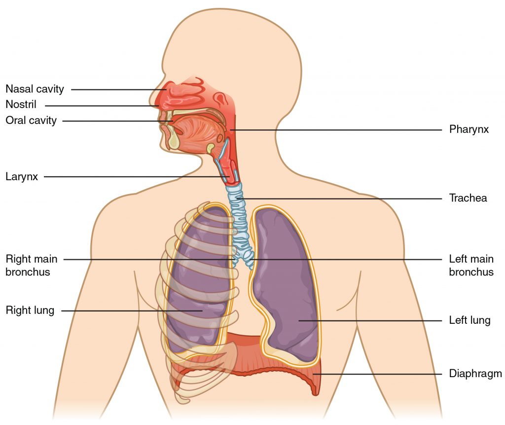
Although oxygen is critically needed for cell functioning, it is the accumulation of carbon dioxide that primarily drives the need to breathe. The major organs of the respiratory system function to provide oxygen to body tissues for cellular respiration, remove the waste product carbon dioxide, and help maintain an acid-base balance. Portions of the respiratory system are also used for nonvital functions, such as sensing odors, producing speech, and for straining, such as during childbirth or coughing. The respiratory system includes muscles that move air into and out of the lungs, the passageways through which air moves, and alveoli. Alveoli are microscopic gas exchange surfaces covered by capillaries. See Figure 11.14[2] for an image of the anatomy of the respiratory system.[3]

Capillaries carry deoxygenated blood to the alveoli, where carbon dioxide is released and oxygen is picked up by hemoglobin. The cardiovascular system transports oxygenated blood from the lungs to the tissues throughout the body where it picks up carbon dioxide and carries it back to the alveoli.[4]
A variety of diseases can affect the respiratory system, such as asthma, chronic obstructive pulmonary disorder (COPD), pneumonia, and lung cancer. These conditions affect the gas exchange process and can result in decreased oxygen saturation levels, increased respiratory rates, labored breathing, and other respiratory difficulties.[5]
Anatomy of the Lungs
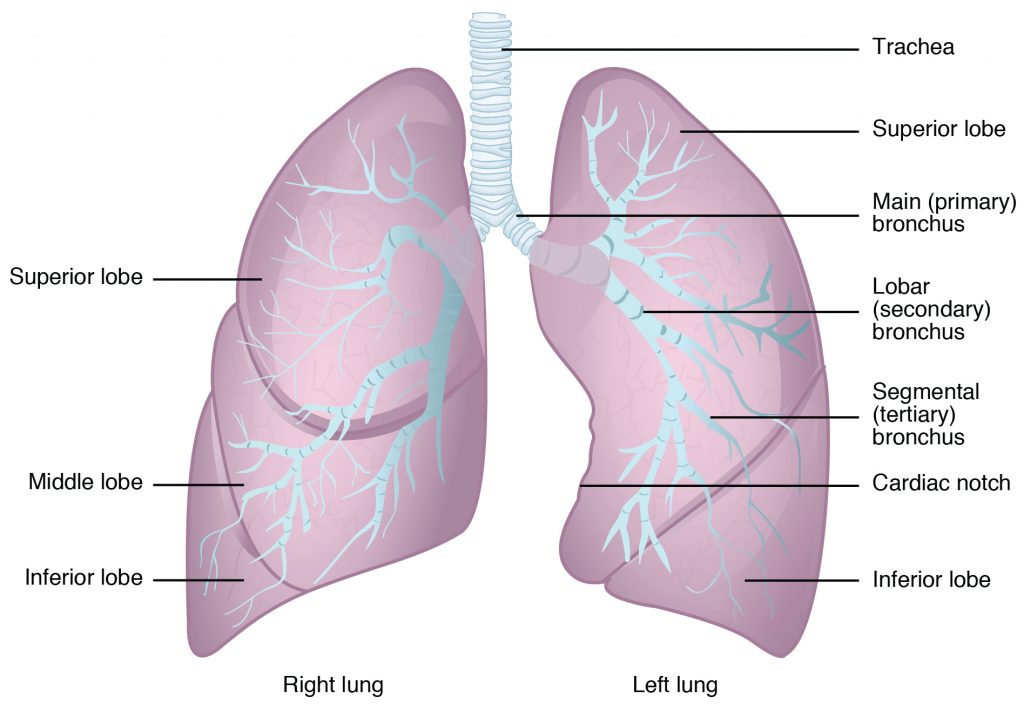
The lungs are pyramid-shaped, paired organs that are connected to the trachea by the right and left bronchi. Below the lungs is the diaphragm, a flat, dome-shaped muscle located at the base of the lungs. Each lung is composed of smaller units called lobes. The right lung consists of three lobes and the left lung consists of two lobes to accommodate the positioning of the heart. As discussed previously, the respiratory system works in conjunction with the cardiovascular system to transport oxygen and nutrients to cells and remove waste from cells and out of the body during exhalation. See Figure 11.15[6] for an image of the anatomy of the lungs.[7]

As the body ages, chest muscles lose their strength, and the smaller airways of the respiratory system, called the bronchioles, lose their elasticity and can collapse. These age-related changes decrease the ability of an older adult to inhale oxygen, resulting in fatigue and increased difficulty completing activities of daily living. Although some respiratory function can be maintained or restored through exercise, the aging respiratory system leaves older adults more susceptible to acute respiratory illnesses such as influenza, pneumonia, and COVID-19.[8]
Because the respiratory system is necessary for life, if a nursing assistant notices any issues with breathing or a blocked airway, these concerns should be reported immediately to the nurse. See the section “Choking and Airway Clearance” in Chapter 3 for a review of interventions for a blocked airway.
Because the cardiovascular system transports oxygenated blood, some symptoms of respiratory distress are similar to cardiovascular complications such as cyanosis, fatigue, dizziness, and shortness of breath. Exercising and avoiding smoking are vital for maintaining or improving respiratory function. Table 11.10 outlines common respiratory diagnoses in the aging population, symptoms to report, and associated interventions provided by nursing assistants.
Table 11.10 Common Respiratory Conditions and Related Nursing Assistant Interventions[9]
| Condition | Definition | Symptoms to Report | Nursing Assistant Interventions |
|---|---|---|---|
| Bronchitis | Acute or chronic inflammation of the bronchioles (i.e., small airways) | Cough that produces mucus, fatigue, shortness of breath, and chest discomfort | Encourage fluids to thin the mucous, segment ADLs, and position the patient for breathing comfort. |
| Asthma | A chronic condition in which irritants cause inflammation and narrowing of the respiratory tract | Coughing, wheezing, chest tightness, difficulty breathing, and shortness of breath | Avoid exposure to known triggers (such as smoke, pet dander, pollen, mold, etc.). During an asthma attack, try to keep the patient calm and immediately report breathing difficulties. |
| Emphysema | A chronic condition in which alveoli hyperinflate, causing reduced air exchange and resulting in a lower amount of oxygenated blood in circulation | Shortness of breath that becomes increasingly more difficult as the disease progresses | Encourage healthy weight and activity as tolerated, segment ADLs, and encourage the use of supplemental oxygen if it is prescribed. |
| Chronic Obstructive Pulmonary Disease (COPD) | Chronic inflammation of the lungs, often caused by smoking, chronic bronchitis, asthma, or emphysema | Shortness of breath, labored breathing, dizziness, disorientation, and cyanosis | Encourage healthy weight, activity as tolerated, and smoking cessation. |
| Lung Cancer | Cancer in the lungs | Chronic cough, coughing up blood, shortness of breath, and chest pain | Encourage smoking cessation, healthy weight and activity as tolerated; segment ADLs; and position the patient for breathing comfort. |
| Bacterial Pneumonia | Bacterial infection of the lungs | Fever; chills; shortness of breath; productive cough with green, yellow, or bloody mucous; sharp chest pain; fatigue; and confusion or disorientation | Encourage healthy diet, activity as tolerated, rest, and taking antibiotics as prescribed. |
View the following YouTube video[10] to learn more about the respiratory system: Respiratory System, Part 1: Crash Course Anatomy & Physiology #31.
- Human Nutrition by University of Hawai‘i at Mānoa Food Science and Human Nutrition Program is licensed under CC BY 4.0 ↵
- "image1-2-1024x861.jpg" by unknown author is licensed under CC BY-NC-SA 4.0. Access for free at https://pressbooks.oer.hawaii.edu/humannutrition/chapter/the-respiratory-system/. ↵
- Human Nutrition by University of Hawai‘i at Mānoa Food Science and Human Nutrition Program is licensed under CC BY 4.0 ↵
- Human Nutrition by University of Hawai‘i at Mānoa Food Science and Human Nutrition Program is licensed under CC BY 4.0 ↵
- Human Nutrition by University of Hawai‘i at Mānoa Food Science and Human Nutrition Program is licensed under CC BY 4.0 ↵
- "image22-1024x704.jpg" by unknown author is licensed under CC BY-NC-SA 4.0. Access for free at https://pressbooks.oer.hawaii.edu/humannutrition/chapter/the-respiratory-system/. ↵
- Human Nutrition by University of Hawai‘i at Mānoa Food Science and Human Nutrition Program is licensed under CC BY 4.0 ↵
- Human Nutrition by University of Hawai‘i at Mānoa Food Science and Human Nutrition Program is licensed under CC BY 4.0 ↵
- Human Nutrition by University of Hawai‘i at Mānoa Food Science and Human Nutrition Program is licensed under CC BY 4.0 ↵
- CrashCourse. (2015, August 24). Respiratory System, Part 1: Crash Course Anatomy & Physiology #31. [Video]. YouTube. All rights reserved. https://youtu.be/bHZsvBdUC2I ↵

