5.2 Basic Concepts of the Respiratory System
Review of the Anatomy and Physiology of the Respiratory System
The purpose of the respiratory system is to perform gas exchange. Pulmonary ventilation provides air to the alveoli for this gas exchange process. At the respiratory membrane where the alveolar and capillary walls meet, gases move across the membranes, with oxygen entering the bloodstream and carbon dioxide exiting. It is through this mechanism that blood is oxygenated and carbon dioxide, the waste product of cellular respiration, is removed from the body.
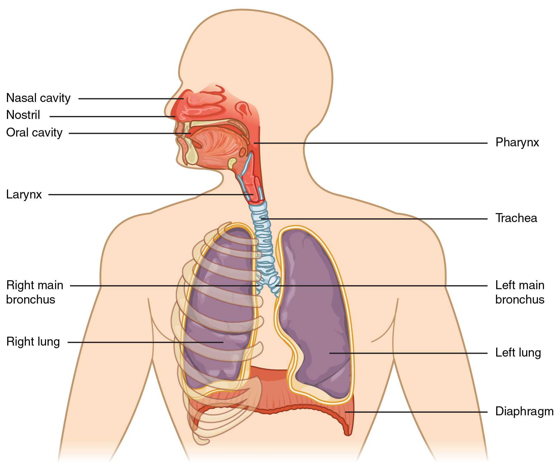
The major organs of the respiratory system function primarily to provide oxygen to body tissues for cellular respiration, remove the waste product carbon dioxide, and help maintain acid-base balance. Portions of the respiratory system are also used for nonvital functions, such as sensing odors, speech production, and for straining, such as during childbirth or coughing.[1]
See Figure 5.1[2] illustrating major respiratory structures.

Functionally, the respiratory system can be divided into a conducting zone and a respiratory zone. The conducting zone of the respiratory system includes the organs and structures not directly involved in gas exchange. The gas exchange occurs in the respiratory zone.
Conducting Zone
The major functions of the conducting zone are to provide a route for incoming and outgoing air, remove debris and pathogens from the incoming air, and warm and humidify the incoming air. Several structures within the conducting zone perform other functions as well. The epithelium of the nasal passages, for example, is essential to sensing odors, and the bronchial epithelium that lines the lungs can metabolize some airborne carcinogens.
The cilia of the respiratory epithelium help remove the mucus and debris from the nasal cavity with a constant beating motion, thus sweeping materials toward the throat to be swallowed. Interestingly, cold air slows the movement of the cilia, resulting in the accumulation of mucus that may, in turn, lead to a runny nose during cold weather. This moist epithelium functions to warm and humidify incoming air. Capillaries located just beneath the nasal epithelium warm the air by convection.
Bronchial Tree
The trachea branches into the right and left primary bronchi at the carina. A bronchial tree (or respiratory tree) is the collective term used for these multiple-branched bronchi. The main function of the bronchi, like other conducting zone structures, is to provide a passageway for air to move into and out of each lung. In addition, the mucous membrane traps debris and pathogens.
A bronchiole branches from the tertiary bronchi. Bronchioles, which are about 1 mm in diameter, further branch until they become the tiny terminal bronchioles, which lead to alveoli, where gas exchange occurs. There are more than 1,000 terminal bronchioles in each lung. The muscular walls of the bronchioles do not contain cartilage like those of the bronchi. This muscular wall can change the size of the bronchial tubing to increase or decrease airflow through the tube.
Respiratory Zone
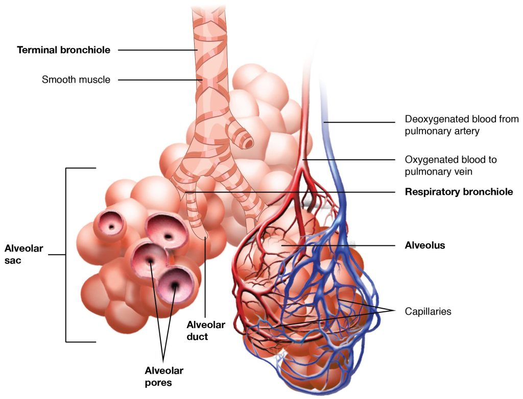
In contrast to the conducting zone, the respiratory zone includes structures that are directly involved in gas exchange. See Figure 5.2[3] for an illustration of the respiratory zone. The respiratory zone begins where the terminal bronchioles join a respiratory bronchiole, the smallest type of bronchiole, which then leads to an alveolar duct, opening into a cluster of alveoli.

Alveoli
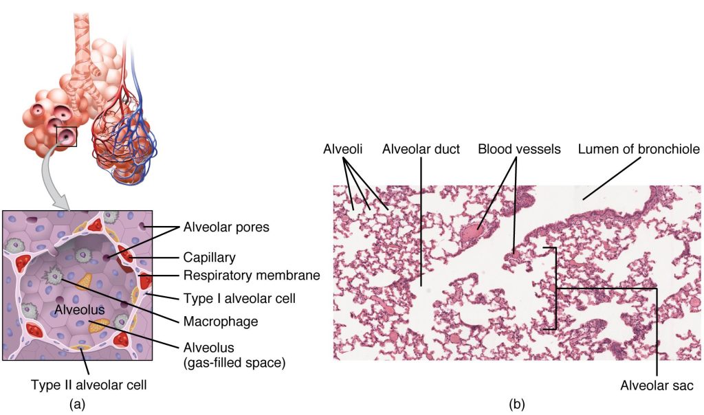
An alveolar duct is a tube composed of smooth muscle and connective tissue, which opens into a cluster of alveoli. An alveolus is one of the many small, grapelike sacs that are attached to the alveolar ducts.
An alveolar sac is a cluster of many individual alveoli that are responsible for gas exchange. See Figure 5.3[4] for an illustration of the structures of the respiratory zone.

Respiratory Rate and Control of Ventilation
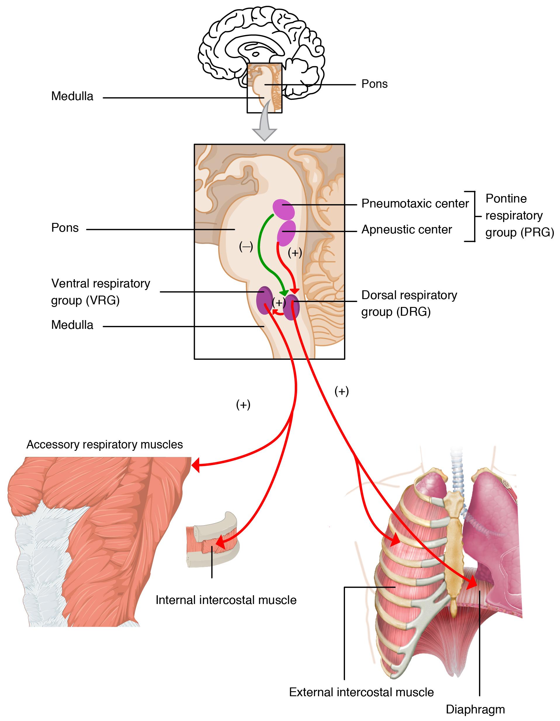
Breathing usually occurs without thought, although at times you can consciously control it, such as when you swim under water, sing a song, or blow bubbles. The respiratory rate is the total number of breaths, or respiratory cycles, that occur each minute. Respiratory rate can be an important indicator of disease, as the rate may increase or decrease during an illness. The respiratory rate is controlled by the respiratory center located within the medulla oblongata in the brain, which responds primarily to changes in carbon dioxide, oxygen, and pH levels in the blood.
The normal respiratory rate of a child decreases from birth to adolescence. A child under 1 year of age has a normal respiratory rate between 30 and 60 breaths per minute, but by the time a child is about 10 years old, the normal rate is closer to 18 to 30. By adolescence, the normal respiratory rate is similar to that of adults, 12 to 18 breaths per minute.
Neurons that stimulate the muscles of the respiratory system are responsible for controlling and regulating pulmonary ventilation. The major brain centers involved in pulmonary ventilation are the medulla oblongata and the pontine respiratory group. (See Figure 5.4[5] for an illustration of the respiratory centers of the brain.)

See the supplementary YouTube videos below related to respiratory anatomy and physiology.
Anatomy of Respiratory System[6]
Inhalation and Exhalation[7]
Carbon Dioxide Transport[8]
Surface Tension[9]
- This work is a derivative of Anatomy and Physiology by OpenStax licensed under CC BY 4.0. Access for free at https://openstax.org/books/anatomy-and-physiology/pages/1-introduction ↵
- “2301 Major Respiratory Organs.jpg” by OpenStax College is licensed under CC BY 4.0. Access for free at https://openstax.org/books/anatomy-and-physiology/pages/22-1-organs-and-structures-of-the-respiratory-system ↵
- “2309 The Respiratory Zone.jpg” by OpenStax College is licensed under CC BY 3.0. Access for free at https://openstax.org/books/anatomy-and-physiology/pages/22-1-organs-and-structures-of-the-respiratory-system ↵
- “2310 Structures of the Respiratory Zone.jpg” by OpenStax College is licensed under CC BY 3.0. Access for free at https://openstax.org/books/anatomy-and-physiology/pages/22-1-organs-and-structures-of-the-respiratory-system ↵
- “2327 Respiratory Centers of the Brain.jpg” by OpenStax College is licensed under CC BY 3.0. Access for free at https://openstax.org/books/anatomy-and-physiology/pages/22-3-the-process-of-breathing ↵
- Forciea, B. (2015, May 13). Respiratory system anatomy (v2.0) [Video]. YouTube. All rights reserved. Video used with permission. https://youtu.be/aqTwrdMS6CE ↵
- Forciea, B. (2015, May 12). Anatomy and physiology: Respiratory system: Breathing mechanics (v2.0) [Video]. YouTube. All rights reserved. Video used with permission. https://youtu.be/X-J5Xgg3l6s ↵
- Forciea, B. (2015, May 12). Respiratory system: C02 transport (v2.0) [Video]. YouTube. All rights reserved. Video used with permission. https://youtu.be/BmrvqZoxHYI ↵
- Forciea, B. (2015, May 13). Anatomy and physiology: Respiratory system: Surface tension (v2.0) [Video]. YouTube. All rights reserved. Video used with permission. https://youtu.be/YHTAausYA94 ↵
The process at the alveoli level where blood is oxygenated and carbon dioxide, the waste product of cellular respiration, is removed from the body.
The total number of breaths, or respiratory cycles, that occur each minute. A child under 1 year of age has a normal respiratory rate between 30 and 60 breaths per minute, but by the time a child is about 10 years old, the normal rate is closer to 18 to 30. By adolescence, the normal respiratory rate is similar to that of adults, 12 to 18 breaths per minute.

