10.2 Basic Concepts of Analgesics and the Musculoskeletal System
Before we discuss medications used to treat pain and musculoskeletal conditions, let’s review the physiology of pain, as well as the anatomy and physiology of the musculoskeletal system.
Review of the Physiology of Pain
Knowledge about the transmission and the processing of pain has greatly expanded in recent years due to a multidisciplinary approach. Although pain is considered something to be avoided, pain impulses are necessary for maintaining the integrity of our bodies and survival. Interactions between the nervous and immune systems are closely linked through cellular interactions in processing and transmitting pain sensation. However, prolonged or chronic pain can cause secondary symptoms, such as anxiety and depression, and can decrease an individual’s overall quality of life.[1]
The transmission of pain is linked to nociceptors, specialized sensory neurons in the central nervous system (CNS) that respond to painful stimuli. Nociceptors respond to harmful or potential tissue-damaging stimuli and transmit stimuli from the skin, muscles, joints, and viscera. The most nociceptor-rich tissue is the skin, which contains several different types of nociceptors. Nociceptors are further divided according to the type of stimuli they respond to (e.g., mechanical, chemical, thermal, or noxious stimuli).[2]
Nociceptor activation is determined by the pain stimulus and depends on the site of generation and mode of activation. The site of the stimulus is important because it can influence the intensity of the nociceptor response. An interesting example is corneal nociceptors, which are activated by weaker stimuli than skin nociceptors. The nature of the stimulus is also important. Stimuli brought about by cutting or crushing, for example, activate most skin nociceptors but do not activate nociceptor in the joints, muscles, or viscera, which instead quickly respond to other types of mechanical forces, such as rotation and distention. In addition to cutting and crushing injuries, harmful stimuli that are able to activate nociceptors in the skin also include chemical, thermal, and mechanical damage.[3]
A property of nociceptors is their ability to cause sensitization, a process that reduces the threshold of activation and an increased response rate to stimulation. Sensitization typically results from tissue injury and inflammation and can result in chronic pain.[4]
The pain sensation, transmitted by neurons in the central nervous system, is influenced by the immune system through the release of molecular mediators. These substances activate pain receptors, increase the sensitivity of pain receptors, and stimulate the release of inflammatory substances called prostaglandins.[5]
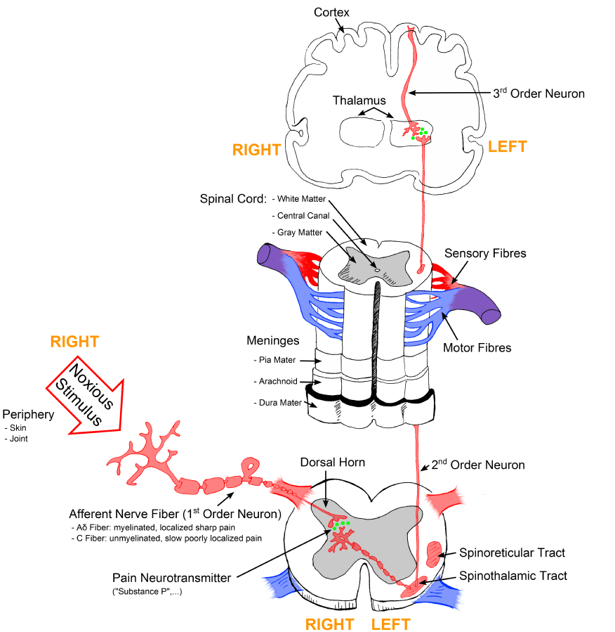
For a person to feel pain, the signal from the nociceptors in peripheral tissues must be transmitted to the spinal cord and then to the hypothalamus and cerebral cortex of the brain. The signal is transmitted to the brain by two types of nerve cells (A-delta and C fibers). The dorsal horn of the spinal cord is the relay station for information from these fibers. In the brain the thalamus is the relay station for incoming sensory stimuli, including pain. From the thalamus the pain messages are relayed to the cerebral cortex where they are perceived.[6] See Figure 10.1[7] for an illustration of how the pain signal is transmitted from peripheral tissues to the spinal cord and then to the brain.

Endogenous Pain Relief
The CNS has an endogenous (i.e., internal) system for relieving pain. The CNS can suppress pain signals from the peripheral nerves by using endogenous opioid peptides that interact with opioid receptors to inhibit perception and transmission of pain signals. These endogenous opioid peptides are endorphins, enkephalins, and dynorphins.[8]
View the supplementary YouTube video below for more information about how pain relievers work.
Review of Anatomy and Physiology of the Musculoskeletal System
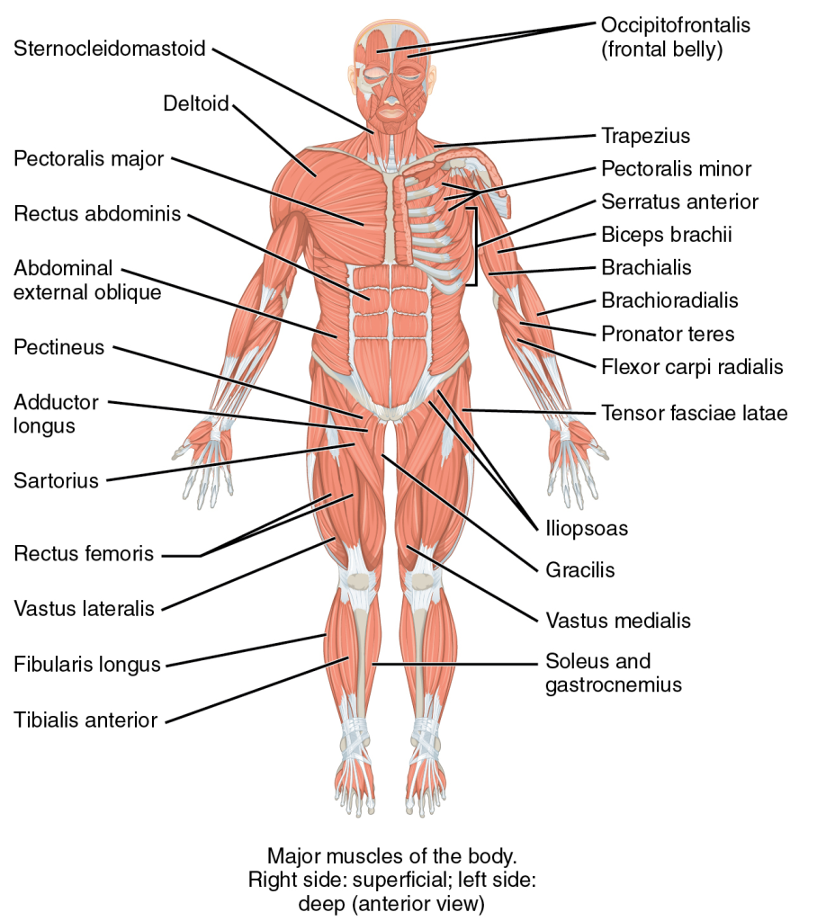
In the musculoskeletal system, the muscular and skeletal systems work together to support and move the body. The bones of the skeletal system serve to protect the body’s organs, support the weight of the body, and give the body shape. The muscles of the muscular system attach to these bones, pulling on them to allow for movement of the body.[10] See Figure 10.2[11] for an illustration of the musculoskeletal system.

Muscles
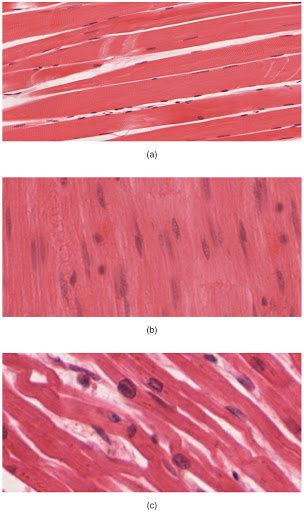
The body contains three types of muscle tissue: skeletal muscle, smooth muscle, and cardiac muscle. See Figure 10.3[12] for images of different types of muscle.

Skeletal muscle is voluntary and striated. These are the muscles that attach to bones and control conscious movement. Smooth muscle is involuntary and nonstriated. It is found in the hollow organs of the body, such as the stomach, intestines, and around blood vessels. Cardiac muscle is involuntary and striated. It is found only in the heart and is specialized to help pump blood throughout the body.[13]
When a muscle fiber receives a signal from the nervous system, myosin filaments are stimulated, pulling actin filaments closer together. This shortens sarcomeres within a fiber, causing it to contract.[14]
- This work is a derivative of Mechanisms of Transmissions and Processing of Pain: A Narrative Review by Di Maio, et. al. and is licensed under CC BY 4.0 ↵
- This work is a derivative of Mechanisms of Transmissions and Processing of Pain: A Narrative Review by Di Maio, et. al. and is licensed under CC BY 4.0 ↵
- This work is a derivative of Mechanisms of Transmissions and Processing of Pain: A Narrative Review by Di Maio, et. al. and is licensed under CC BY 4.0 ↵
- This work is a derivative of Mechanisms of Transmissions and Processing of Pain: A Narrative Review by Di Maio, et. al. and is licensed under CC BY 4.0 ↵
- This work is a derivative of Mechanisms of Transmissions and Processing of Pain: A Narrative Review by Di Maio, et. al. and is licensed under CC BY 4.0 ↵
- . Frandsen, G., & Pennington, S. (2018). Abrams’ clinical drug: Rationales for nursing practice (11th ed.). pp. 305, 310, 952-953, 959-960. Wolters Kluwer. ↵
- “Sketch colored final.png” by Bettina Guebeli is licensed under CC BY-SA 4.0 ↵
- . Frandsen, G., & Pennington, S. (2018). Abrams’ clinical drug: Rationales for nursing practice (11th ed.). pp. 305, 310, 952-953, 959-960. Wolters Kluwer. ↵
- Ted-Ed. (2012, June 26). How do pain relievers work? - George Zaidan [Video]. YouTube. All rights reserved. https://youtu.be/9mcuIc5O-DE ↵
- Khan Academy. (n.d.). The musculoskeletal system review. https://www.khanacademy.org/science/high-school-biology/hs-human-body-systems/hs-the-musculoskeletal-system/a/hs-the-musculoskeletal-system-review ↵
- This image is a derivative of “1105 Anterior and Posterior Views of Muscles.jpg” by CFCF licensed under CC BY 4.0 ↵
- “414 Skeletal Smooth Cardiac.jpg” by OpenStax College is licensed under CC BY 4.0 ↵
- Khan Academy. (n.d.). The musculoskeletal system review. https://www.khanacademy.org/science/high-school-biology/hs-human-body-systems/hs-the-musculoskeletal-system/a/hs-the-musculoskeletal-system-review ↵
- Khan Academy. (n.d.). The musculoskeletal system review. https://www.khanacademy.org/science/high-school-biology/hs-human-body-systems/hs-the-musculoskeletal-system/a/hs-the-musculoskeletal-system-review ↵
Nerve endings that selectively respond to painful stimuli and send pain signals to the brain and spinal cord.
Produced in nearly all cells and are part of the body’s way of dealing with injury and illness. Prostaglandins act as signals to control several different processes depending on the part of the body in which they are made. Prostaglandins are made at the sites of tissue damage or infection, where they cause inflammation, pain, and fever as part of the healing process.

