7.6 Checklist: Initiate Telemetry
*Disclaimer: Always follow agency policy and manufacturer recommendations
Checklist: Initiate Telemetry[1],[2]
- Verify the provider’s order.
- Gather supplies: electrodes.
- Introduce yourself and your role to the client.
- Perform hand hygiene.
- Identify the client using two identifiers and check allergies.
- Provide privacy.
- Explain the procedure to the client regarding the purpose of telemetry, what to expect with lead placement, and telemetry monitoring.
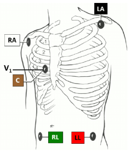
- Prior to applying the electrodes, assess the skin and ensure it is free of excess hair and sweat. Apply the electrodes to clean, dry skin, ensuring good adherence according to manufacturer instructions. The electrodes must make contact with the skin for a clear picture of the heart’s electrical activity on the monitor.[3] Apply the electrodes to the skin. See Figure 7.34[4] for an illustration of electrode placement.[5]
- White – Right arm (RA): Infraclavicular fossa close to the right shoulder
- Black – Left arm (LA): Infraclavicular fossa close to the left shoulder
- Red – Left leg (LL): Below the rib cage on the left upper quadrant of the abdomen
- Green – Right leg (RL): Below the rib cage on the right upper quadrant of the abdomen
- Brown – (V1): Fourth intercostal space on the right sternal border
- Attach the lead wires to the electrodes.
- Observe the rhythm and print a six-second strip.
- Interpret the rhythm.

View a YouTube video[6] showing an instructor demonstration of this skill:
- Clinical skills: Essentials collection (1st ed.). (2021). Elsevier. ↵
- Lippincott procedures. http://procedures.lww.com ↵
- Drew, B. J., Califf, R. M., Funk, M., Kaufman, E. S., Krucoff, M. W., Laks, M. M., Macfarlane, P. W., Sommargren, C., Swiryn, S., & Van Hare, G. F. (2004). Practice standards for electrocardiographic monitoring in hospital settings. Circulation, 110(17), 2721-2746. https://www.ahajournals.org/doi/10.1161/01.cir.0000145144.56673.59 ↵
- This image is a derivative of “Patient-monitoring-ECG-lead-configuration-A-5-electrode-lead-configuration-was-used-in.png” by Barbara Drew et al., and is licensed under CC BY 4.0. Access for free at https://www.researchgate.net/publication/267740230_Insights_into_the_Problem_of_Alarm_Fatigue_with_Physiologic_Monitor_Devices_A_Comprehensive_Observational_Study_of_Consecutive_Intensive_Care_Unit_Patients ↵
- Drew, B. J., Califf, R. M., Funk, M., Kaufman, E. S., Krucoff, M. W., Laks, M. M., Macfarlane, P. W., Sommargren, C., Swiryn, S., & Van Hare, G. F. (2004). Practice standards for electrocardiographic monitoring in hospital settings. Circulation, 110(17), 2721-2746. https://www.ahajournals.org/doi/10.1161/01.cir.0000145144.56673.59 ↵
- Chippewa Valley Technical College. (2023, January 5). Initiating telemetry [Video]. YouTube. Video licensed under CC BY 4.0. https://youtu.be/LvE4NrlcuE0 ↵

