16.2 Review of Anatomy & Physiology
This chapter discusses a wide range of common pediatric disorders that affect many body systems, including gastrointestinal, integumentary, neurological, cardiovascular, and musculoskeletal, as well as the eyes and ears. A review of the anatomy and physiology of these body systems, as well as pediatric life span considerations, is discussed in the following subsections.
Gastrointestinal System
Table 16.2a provides an overview of the major components of the gastrointestinal system and their associated functions, and these components are illustrated in Figure 16.1.[1]
Table 16.2a. Components and Functions of the Gastrointestinal System[2]
| Component | Major Functions | Other Functions |
|---|---|---|
| Mouth |
|
|
| Pharynx |
|
|
| Esophagus |
|
|
| Stomach |
|
|
| Small Intestine |
|
|
| Large Intestine |
|
|
| Accessory Organs |
|
|
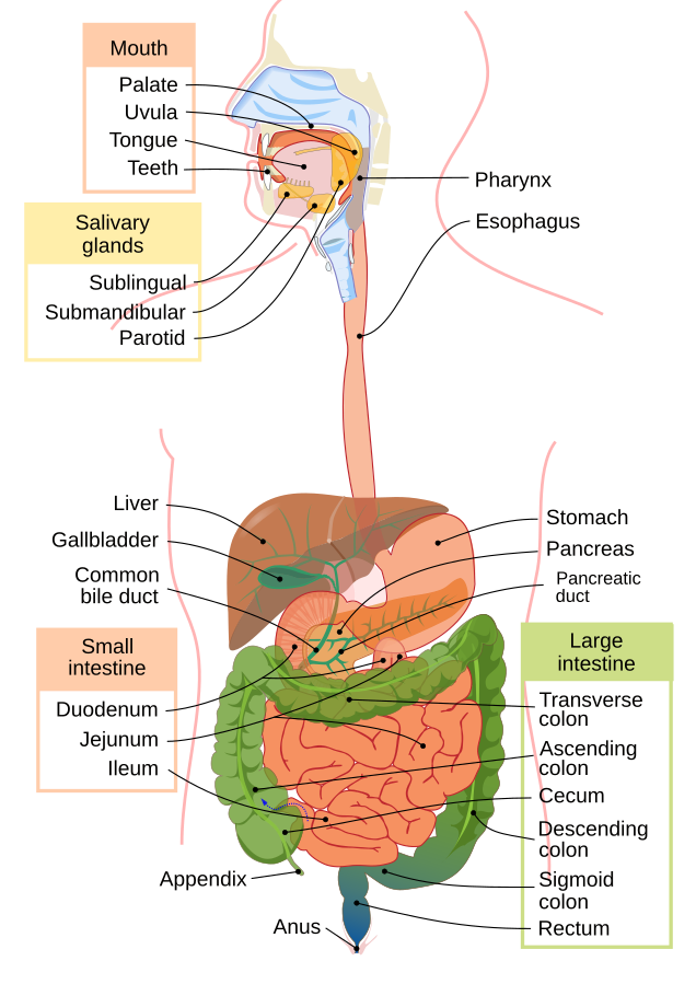
For a more detailed review of the anatomy and physiology of the gastrointestinal system, visit the “Review of Anatomy & Physiology of the Gastrointestinal System” section of the “Gastrointestinal System Alterations” chapter of Open RN Health Alterations.[3]
Pediatric Differences
Although the gastrointestinal systems of adults and children have the same components and functions, there are some pediatric differences that must be considered[4],[5],[6]:
- The exocrine functions of the pancreas are not mature until one year of age.
- Infants have quicker gastric emptying times when compared to adults.
- The small intestine is shorter in children than adults, providing less surface area for absorption of nutrients.
- The production of gastric acid is decreased at birth when compared to adults.
- Children do not gain control of bowel movements until around two years of age.
Integumentary System
Table 16.2b provides an overview of the major components of the integumentary system and their associated functions, and Figure 16.2[7] illustrates the anatomy of the skin.
Table 16.2b. Components of the Integumentary System[8]
| Integumentary System Components | Major Functions |
|---|---|
| Skin: Composed of the epidermis (outermost layer), the dermis (the layer underneath the epidermis), and the hypodermis (the layer underneath the dermis also referred to as subcutaneous tissue) |
|
| Hair |
|
| Nails |
|
| Sweat glands |
|
| Sebaceous glands |
|
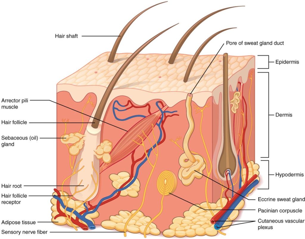
For a more detailed review of the anatomy and physiology of the integumentary system, view the “Integumentary System” chapter of OpenStax Anatomy and Physiology, 2e.[9]
Pediatric Differences
Adults and children have the same integumentary system components, but there are some pediatric differences that must be considered[10],[11],[12]:
- Sebaceous glands are present in pediatric clients, but do not become active until puberty.
- Infants have thinner and more fragile skin when compared to adults.
- The skin of infants is more permeable to water when compared to adults.
- The skin of children is more susceptible to damage from ultraviolet rays.
Neurological System
The neurological system is a complex system that is divided into two main areas called the central nervous system and the peripheral nervous system. The central nervous system consists of the brain and spinal cord. The peripheral nervous system is made up of nerves and ganglia. Ganglia are large clusters of nerve cells. See Figure 16.3[13] for an illustration of the central and peripheral nervous systems.
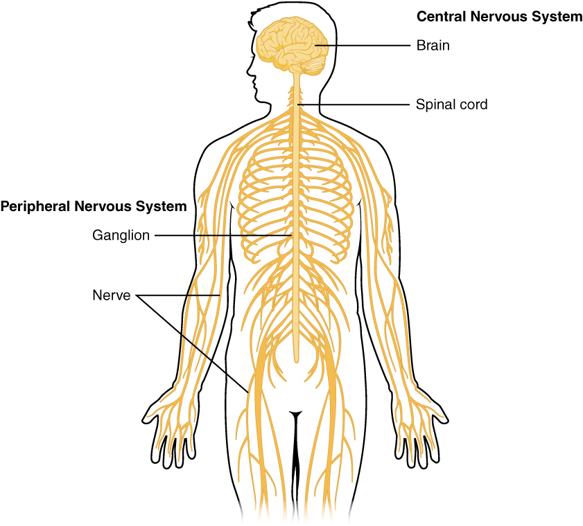
There are three basic functions of the nervous system, including sensation, response, and integration. Sensation involves the receival of information from the world around us. Response is when the nervous system acts based on the information it has received, including voluntary or involuntary responses. Integration refers to the processing of sensory information via the nervous system.[14]
For a detailed review of the anatomy and physiology of the neurological system, view “The Nervous System and Nervous Tissue” section of OpenStax Anatomy and Physiology, 2e.[15]
Pediatric Differences
Although the neurological systems of adults and children have the same components and functions, there are pediatric differences that must be considered[16],[17]:
- Newborns and infants have underdeveloped temperature regulation that can result in hypothermia if their heads are left exposed.
- Children have increased fall risk due to incomplete motor development.
- Children are more susceptible to head injuries due to their larger head in relationship to their body size.
- The cranial bones of pediatric clients are thin and leave brain tissue less protected, resulting in hemorrhages, head trauma, and diffuse brain injury as a result of accidents or injuries.
- The brain of infant clients develops rapidly, requiring an adequate supply of oxygen and glucose.
Cardiovascular System
Review Table 16.2c for an overview of the major components of the cardiovascular system and their associated functions. See Figure 16.4[18] for an illustration of the heart.
Table 16.2c. Overview of the Anatomy and Physiology of the Cardiovascular System[19]
| Cardiovascular System Component | Major Functions |
|---|---|
| Blood |
|
| Heart |
|
| Atrioventricular valves (tricuspid valve on the right side of the heart and bicuspid or mitral valve on the left side) |
|
| Semilunar valves (pulmonic on the right side of the heart and aortic valves on the left side) |
|
| Coronary arteries |
|
| Cardiac conduction system |
|
| Pulmonary arteries |
|
| Pulmonary veins |
|
| Aorta |
|
| Superior and inferior vena cava |
|
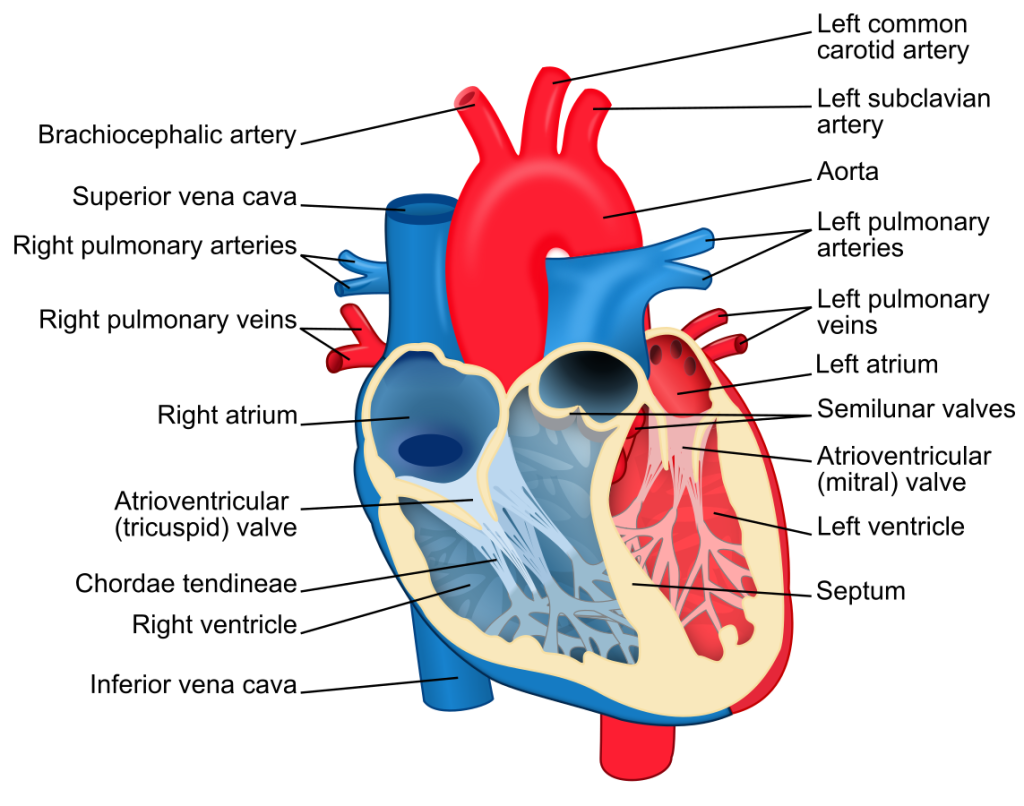
For a detailed review of the anatomy and physiology of the cardiovascular system, view the “Review of Anatomy and Physiology of the Cardiovascular System” section of the “Cardiovascular Alterations” chapter of the Open RN Health Alterations.
Pediatric Differences
Although the cardiovascular systems of adults and children have the same components and functions, there are some pediatric differences that must be considered[20],[21],[22]:
- Many profound changes occur in a newborn’s cardiovascular anatomy and physiology over the first few days and weeks following birth.
- Newborns have incomplete sympathetic innervation of the heart, predisposing them to bradycardia.
- A small volume of blood loss in small children can be significant. For example, 100 mL of blood loss in an infant weighing 5 kg is 10% of their total blood volume.
- Establishing IV access in infants and children can be challenging due to their small veins and increased subcutaneous tissue.
- Children have a higher metabolic rate and, therefore, an increased workload on their heart.
Musculoskeletal System
Table 16.2d provides an overview of the musculoskeletal system. See Figures 16.5[23] and 16.6[24] for images depicting the skeleton and muscles.
Table 16.2d. Overview of the Anatomy and Physiology of the Musculoskeletal System[25],[26]
| Body Structure | Major Functions |
|---|---|
| Bone and cartilage |
|
| Joints |
|
| Skeletal muscle |
|
| Cardiac muscle |
|
| Smooth muscle |
|
| Tendons |
|
| Ligaments |
|
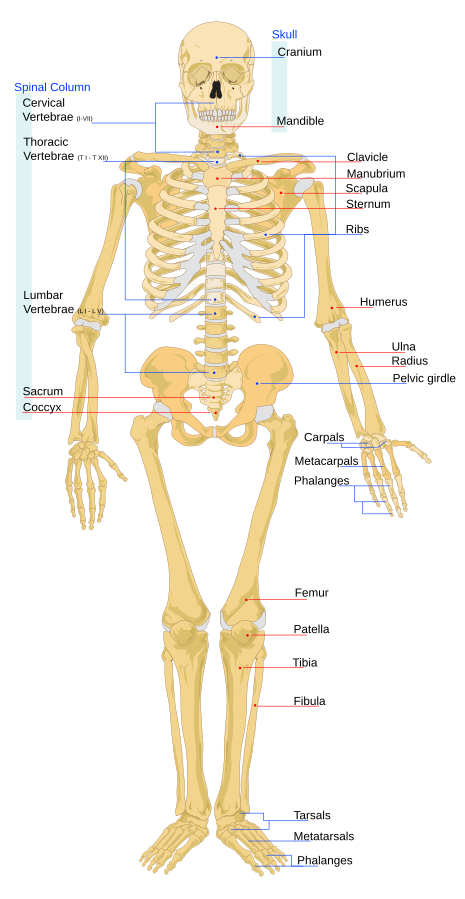

For a more detailed review of the anatomy and physiology of the musculoskeletal system, visit the “Support and Movement” chapter of OpenStax Anatomy and Physiology, 2e.[27]
Pediatric Differences
Here are some pediatric considerations in regards to the anatomy and physiology of the musculoskeletal system[28]:
- Children have more bones than adults, but as they grow up, some of their bones fuse together.
- When compared to older children, infants lack muscle tone and coordination so they require support while being held.
- The bones of children are softer than adults, meaning they will bend before breaking. Because they are more flexible, they can have severe injuries without the presence of a fracture.
- Children have growth plates in their long bones. Fractures to this area can affect their growth.
Eyes & Ears
Table 16.2e provides an overview of the eyes and ears and their associated functions. See Figures 16.7[29] and 16.8[30] for illustrations of the eyes and ears.
Table 16.2e. Overview of the Anatomy and Physiology of the Eyes and Ears[31]
| Body Structure | Major Functions |
|---|---|
| Eyes |
|
| Ears |
|
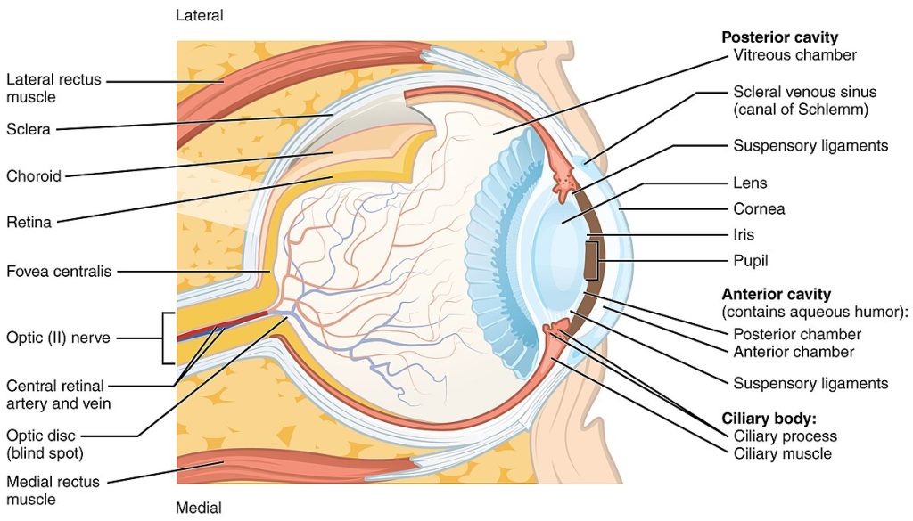
Figure 16.7 Anatomy of the Eye
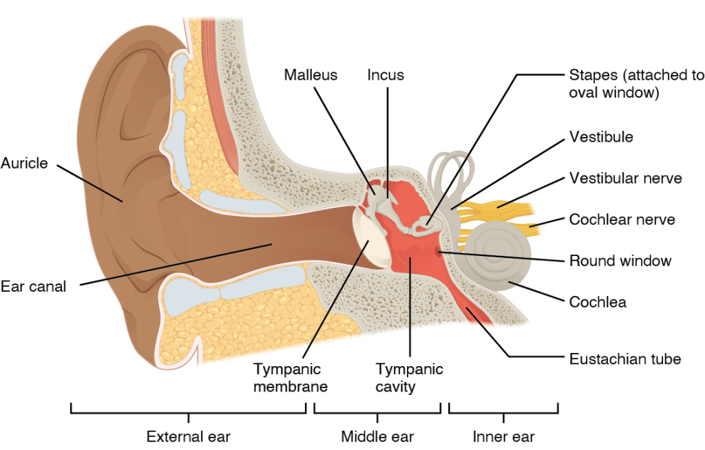
For a detailed review of the anatomy and physiology of the eyes and ears, visit the “Sensory Perception” chapter of OpenStax Anatomy and Physiology, 2e.
Pediatric Differences
The eyes and ears of adults and children have similar components and functions, but there are pediatric differences that must be considered[32],[33]:
- Children do not reach the full level of visual acuity of adults until seven years old.
- The eyes of children nine years old and younger are more sensitive to the sun when compared to adults.
- The eustachian tubes of children are smaller and positioned differently when compared to adults. This makes fluid drainage difficult and ear infections more likely.
- “Digestive_system_diagram_en” by Mariana Ruiz, Jmarchn is in the Public Domain. ↵
- Ernstmeyer, K., & Christman, E. (Eds.). (2024). Health alterations. https://wtcs.pressbooks.pub/healthalts/ ↵
- Ernstmeyer, K., & Christman, E. (Eds.). (2024). Health alterations. https://wtcs.pressbooks.pub/healthalts/ ↵
- Ernstmeyer, K., & Christman, E. (Eds.). (2024). Health alterations. https://wtcs.pressbooks.pub/healthalts/ ↵
- Indrio, F., Neu, J., Pettoello-Mantovani, M., Marchese, F., Martini, S., Salatto, A., & Aceti, A. (2022). Development of the gastrointestinal tract in newborns as a challenge for an appropriate nutrition: A narrative review. Nutrients, 14(7), 1405. https://www.ncbi.nlm.nih.gov/pmc/articles/PMC9002905/ ↵
- Johns Hopkins Medicine. (n.d.). Toilet training. https://www.hopkinsmedicine.org/health/wellness-and-prevention/toilettraining ↵
- “501_Structure_of_the_skin.jpg” by J. Gordon Betts, et al., for OpenStax is licensed under CC BY 4.0 ↵
- Betts, J. G., Young, K. A., Wise, J. A., Johnson, E., Poe, B., Kruse, D. H., Korol, O., Johnson, J. E., Womble, M., & DeSaix, P. (2022). Anatomy and physiology 2e. OpenStax. https://openstax.org/books/anatomy-and-physiology-2e/pages/1-introduction ↵
- Betts, J. G., Young, K. A., Wise, J. A., Johnson, E., Poe, B., Kruse, D. H., Korol, O., Johnson, J. E., Womble, M., & DeSaix, P. (2022). Anatomy and physiology 2e. OpenStax. https://openstax.org/books/anatomy-and-physiology-2e/pages/1-introduction ↵
- Betts, J. G., Young, K. A., Wise, J. A., Johnson, E., Poe, B., Kruse, D. H., Korol, O., Johnson, J. E., Womble, M., & DeSaix, P. (2022). Anatomy and physiology 2e. OpenStax. https://openstax.org/books/anatomy-and-physiology-2e/pages/1-introduction ↵
- Leo, P. (n.d.). How does infant skin differ from adult skin? https://www.medscape.org/viewarticle/743529 ↵
- King, A., Balaji, S., & Keswani, S. G. (2013). Biology and function of fetal and pediatric skin. Facial Plastic Surgery Clinics of North America, 21(1), 1–6. https://www.ncbi.nlm.nih.gov/pmc/articles/PMC3654382/ ↵
- “1201_Overview_of_Nervous_System” by OpenStax is licensed under CC BY 4.0 ↵
- Betts, J. G., Young, K. A., Wise, J. A., Johnson, E., Poe, B., Kruse, D. H., Korol, O., Johnson, J. E., Womble, M., & DeSaix, P. (2022). Anatomy and physiology 2e. OpenStax. https://openstax.org/books/anatomy-and-physiology-2e/pages/1-introduction ↵
- Betts, J. G., Young, K. A., Wise, J. A., Johnson, E., Poe, B., Kruse, D. H., Korol, O., Johnson, J. E., Womble, M., & DeSaix, P. (2022). Anatomy and physiology 2e. OpenStax. https://openstax.org/books/anatomy-and-physiology-2e/pages/1-introduction ↵
- Ernstmeyer, K., & Christman, E. (Eds.). (2024). Health alterations. https://wtcs.pressbooks.pub/healthalts/ ↵
- Curran, A. (2023). Diarrhea nursing diagnosis and care plan. https://nursestudy.net/diarrhea-nursing-diagnosis/ ↵
- “Heart_diagram-en” by ZooFari is licensed under CC BY-SA 3.0 ↵
- Betts, J. G., Young, K. A., Wise, J. A., Johnson, E., Poe, B., Kruse, D. H., Korol, O., Johnson, J. E., Womble, M., & DeSaix, P. (2022). Anatomy and physiology 2e. OpenStax. https://openstax.org/books/anatomy-and-physiology-2e/pages/1-introduction ↵
- Ernstmeyer, K., & Christman, E. (Eds.). (2024). Health alterations. https://wtcs.pressbooks.pub/healthalts/ ↵
- Queensland Emergency Pediatric Care. (2022). How children are different: Anatomic and physiological differences. [PDF]. https://www.childrens.health.qld.gov.au/__data/assets/pdf_file/0031/179725/how-children-are-different-anatomical-and-physiological-differences.pdf ↵
- Anderson, A., & Vo, C. (2023). Neonatal cardiovascular physiology. Open Anesthesia. https://www.openanesthesia.org/keywords/neonatal-cardiovascular-physiology/ ↵
- “Human_skeleton_front_en” by LadyofHats Mariana Ruiz Villarreal is in the Public Domain. ↵
- “1105_Anterior_and_Posterior_Views_of_Muscles” by OpenStax is licensed under CC BY 4.0 ↵
- Betts, J. G., Young, K. A., Wise, J. A., Johnson, E., Poe, B., Kruse, D. H., Korol, O., Johnson, J. E., Womble, M., & DeSaix, P. (2022). Anatomy and physiology 2e. OpenStax. https://openstax.org/books/anatomy-and-physiology-2e/pages/1-introduction ↵
- Ernstmeyer, K., & Christman, E. (Eds.). (2023). Nursing skills 2e. https://wtcs.pressbooks.pub/nursingskills/ ↵
- Betts, J. G., Young, K. A., Wise, J. A., Johnson, E., Poe, B., Kruse, D. H., Korol, O., Johnson, J. E., Womble, M., & DeSaix, P. (2022). Anatomy and physiology 2e. OpenStax. https://openstax.org/books/anatomy-and-physiology-2e/pages/1-introduction ↵
- Queensland Emergency Pediatric Care. (2022). How children are different: Anatomic and physiological differences. [PDF]. https://www.childrens.health.qld.gov.au/__data/assets/pdf_file/0031/179725/how-children-are-different-anatomical-and-physiological-differences.pdf ↵
- “1413_Structure_of_the_Eye" by OpenStax College is licensed under CC BY 3.0 ↵
- “dd97d7920818a0a5b338a427a1d846043b156238" by OpenStax is licensed under CC BY 4.0. https://openstax.org/books/biology-2e/pages/36-4-hearing-and-vestibular-sensation ↵
- Betts, J. G., Young, K. A., Wise, J. A., Johnson, E., Poe, B., Kruse, D. H., Korol, O., Johnson, J. E., Womble, M., & DeSaix, P. (2022). Anatomy and physiology 2e. OpenStax. https://openstax.org/books/anatomy-and-physiology-2e/pages/1-introduction ↵
- Theeyesofchildren.org. (n.d.). Children’s vision. https://www.theeyesofchildren.org/childrens-vision/ ↵
- National Institute on Deafness and Other Communication Disorders. (2022). Ear infections in children. https://www.nidcd.nih.gov/health/ear-infections-children#5 ↵
Outermost layer if the skin.
The layer of skin underneath the epidermis.
The layer of skin underneath the dermis also referred to as subcutaneous tissue.
Large clusters of nerve cells.

