9.3 Diagnosing Pregnancy
The physiologic and anatomic changes that occur during pregnancy are referred to as presumptive, probable, and positive signs of pregnancy. Presumptive signs of pregnancy include the subjective cues of early pregnancy. Probable signs of pregnancy are objective cues discoverable by the health care provider. Positive signs of pregnancy are cues provided by the fetus.[1] These signs of pregnancy are summarized in Table 9.3 and further discussed in the following subsections.
Table 9.3. Signs of Pregnancy[2]
| Type of Sign of Pregnancy | Signs |
|---|---|
| Presumptive |
|
| Probable |
|
| Positive |
|
Presumptive Signs of Pregnancy
Presumptive signs of pregnancy are symptoms noticed by the client and include quickening, amenorrhea, nausea and vomiting, fatigue, and breast enlargement and tenderness. However, the presumptive signs of pregnancy are the least reliable symptoms confirming a pregnancy because the signs also commonly occur with other medical conditions[3]:
- Quickening refers to the feeling of the movements by the fetus in the uterus by the mother by about 16-24 weeks’ gestation. However, these feelings of movement can also be caused by peristalsis or flatus.
- Amenorrhea (i.e., absence of menstruation) is caused by elevated progesterone levels during pregnancy. However, amenorrhea can occur outside of pregnancy because of thyroid dysfunction, stress, obesity, anorexia, malnutrition, and polycystic ovary syndrome (PCOS).
- Nausea and vomiting during pregnancy is caused by increasing levels of human chorionic gonadotropin (hCG). However, nausea and vomiting can occur outside of pregnancy due to gastroenteritis, gastrointestinal viruses, food poisoning, side effects of medications, intestinal blockage, and other medical conditions.
- Fatigue during pregnancy is caused by elevated progesterone levels and decreased blood glucose levels. However, fatigue can occur outside of pregnancy due to lack of sleep, overactive lifestyle, anemia, hypothyroidism, or other medical conditions.
- Urinary frequency during pregnancy is caused by the pressure of the growing uterus on the bladder. However, outside of pregnancy, urinary frequency can be caused by increased fluid intake, urinary tract infection, anxiety, interstitial cystitis, side effects of medications, or other medical conditions.
- Breast enlargement and tenderness occur during pregnancy in preparation for lactation. However, outside of pregnancy, breast enlargement and tenderness can also be caused by increased prolactin levels or a breast mass.[4]
View a supplementary YouTube video[5] on fetal movement (quickening): Understanding Your Baby’s Movements During Pregnancy │Mater Mothers’.
Probable Signs of Pregnancy
Probable signs of pregnancy are objectively noticed by the health care provider and include Chadwick sign, Goodell sign, Hegar sign, enlargement of the uterus, skin hyperpigmentation, and palpation of the fetus.[6]
Chadwick sign is the bluish discoloration of the vagina and cervix due to the vasocongestion needed to support the growing uterus during pregnancy, typically noticeable around six to eight weeks of gestation. The provider may be able to visualize this discoloration during a vaginal exam; therefore, it is considered a probable sign of pregnancy. However, women with endometriosis and adenomyosis can also exhibit Chadwick sign.
Goodell sign refers to the softening of the cervix and vagina and the increase in vaginal mucus discharge during pregnancy, typically becoming noticeable around four to six weeks of gestation during a pelvic examination performed by a health care provider. However, women with endometriosis and adenomyosis can also exhibit Goodell sign. Endometriosis is a chronic disease in which tissue similar to the lining of the uterus (endometrium) grows outside the uterus, causing chronic inflammation and scarring. Adenomyosis occurs when endometrial tissue grows into the muscular wall of the uterus, causing the uterus to thicken.
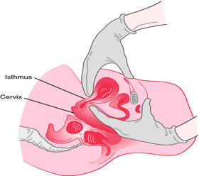
Hegar’s sign is the softening and compressibility of the lower uterine segment during pregnancy. To assess Hegar’s sign, the health care provider gently presses on the cervix with one hand while simultaneously pressing on the lower abdomen with the other hand. This maneuver allows the provider to feel for a softening and compressible area in the lower uterine segment, typically around six to eight weeks of gestation. However, connective tissue disorders can also cause the cervix and lower uterine segment to soften. See Figure 9.1[7] for an illustration of Hegar’s sign.

Enlargement of the uterus occurs during pregnancy due to fetal growth. However, uterine fibroids can also cause the uterus to enlarge and upon palpation can be mistaken as a fetus.
Skin hyperpigmentation can occur during pregnancy on the face, abdomen, axilla, areola, and nipples due to increased levels of estrogen, progesterone, and melanocyte-stimulating hormone. However, obesity and other medical conditions such as Addison’s disease can also cause hyperpigmentation.[8]
Urine or blood pregnancy testing is typically performed to confirm pregnancy. Urine tests are 95% accurate in detecting initially elevated levels of hCG if testing is completed on the first day of a missed period or after and using a first morning midstream urine collection. Blood tests can detect elevated levels of hCG as early as seven to eight days before a missed period. However, these tests can potentially be inaccurate due to a medical condition such as pituitary disease or ovarian cancer.[9]
Positive Signs of Pregnancy
Positive signs of pregnancy directly confirm pregnancy and include auscultation of the fetal heart rate, palpable fetal movement by the examiner, and visualization of the fetus via ultrasound. Fetal heart tones (FHT) can be heard by Doppler as early as ten weeks’ gestation. Fetal movement can be felt by the examiner when palpating the uterus.
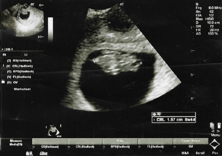
Visualization of the fetus is obtained by an ultrasound that confirms cardiac activity and the size and location of the fetus. An ultrasound is a safe and painless diagnostic procedure that uses high frequency sound waves and allows health care providers to visualize the inside of the uterus and examine the developing fetus. During pregnancy, two types of ultrasounds may be used called transvaginal and abdominal. During an abdominal ultrasound exam, gel is spread over the abdomen, and the ultrasound technician moves the transducer over the abdomen to produce the picture. The client can be positioned to see the images if desired. The client is often requested to come to the ultrasound exam with a full bladder to help displace the intestines and evaluate the uterus with better visibility. During a transvaginal ultrasound, a transvaginal probe encased in a disposable cover and coated with gel will be inserted into the vagina. The client may be asked to insert this probe themselves. A transvaginal ultrasound is often performed during the first trimester of pregnancy or later in pregnancy if complications arise, such as pain or bleeding. See Figure 9.2[10] for an ultrasound image obtained during the first trimester.

- Giles, A., Prusinski, R., & Wallace, L. (2024). Maternal newborn nursing. OpenStax. https://openstax.org/books/maternal-newborn-nursing/pages/1-introduction ↵
- Giles, A., Prusinski, R., & Wallace, L. (2024). Maternal newborn nursing. OpenStax. https://openstax.org/books/maternal-newborn-nursing/pages/1-introduction ↵
- Giles, A., Prusinski, R., & Wallace, L. (2024). Maternal newborn nursing. OpenStax. https://openstax.org/books/maternal-newborn-nursing/pages/1-introduction ↵
- Giles, A., Prusinski, R., & Wallace, L. (2024). Maternal newborn nursing. OpenStax. https://openstax.org/books/maternal-newborn-nursing/pages/1-introduction ↵
- Mater. (2022, May 18). Understanding your baby’s movements during pregnancy | Mater Mothers’ [Video]. YouTube. All rights reserved. https://www.youtube.com/watch?v=GKtUY36B80A ↵
- Giles, A., Prusinski, R., & Wallace, L. (2024). Maternal newborn nursing. OpenStax. https://openstax.org/books/maternal-newborn-nursing/pages/1-introduction ↵
- “Palpation-of-uterus.png” by unknown author is licensed under CC BY-NC-SA 4.0. Access for free at https://pressbooks.pub/nurs323 ↵
- Giles, A., Prusinski, R., & Wallace, L. (2024). Maternal newborn nursing. OpenStax. https://openstax.org/books/maternal-newborn-nursing/pages/1-introduction ↵
- Giles, A., Prusinski, R., & Wallace, L. (2024). Maternal newborn nursing. OpenStax. https://openstax.org/books/maternal-newborn-nursing/pages/1-introduction ↵
- “332276c58435fb3a362a0e94f749dea90f67e826” by OpenStax is licensed under CC BY 4.0. Access for free at https://openstax.org/books/maternal-newborn-nursing/pages/13-1-prenatal-testing-during-the-first-trimester ↵
Subjective cues of early pregnancy. Symptoms noticed by the client including quickening, amenorrhea, nausea and vomiting, fatigue, and breast enlargement and tenderness.
Objective cues discoverable by the healthcare provider. Signs that are objectively noticed by the health care provider and include Chadwick sign, Goodell sign, Hegar sign, enlargement of the uterus, skin hyperpigmentation, and palpation of the fetus.
Cues provided by the fetus. Signs that directly confirm pregnancy and include auscultation of the fetal heart rate, palpable fetal movement by the examiner, and visualization of the fetus via ultrasound.
The first movements of the fetus felt by the pregnant person, typically occurring around the 16th to 20th week of pregnancy.
The absence of menstruation. It can be primary (when menstruation never begins) or secondary (when menstruation stops after previously occurring).
The bluish discoloration of the vagina and cervix due to the vasocongestion needed to support the growing uterus during pregnancy, typically noticeable around six to eight weeks of gestation.
Softening of the cervix and vagina and the increase in vaginal mucus discharge during pregnancy, typically noticeable around six to eight weeks of gestation.
A chronic condition in which tissue similar to the lining of the uterus (endometrium) grows outside the uterus, often on the ovaries, fallopian tubes, or other pelvic organs. This can cause pain, inflammation, and infertility.
Occurs when endometrial tissue grows into the muscular wall of the uterus, causing the uterus to thicken.
The softening and compressibility of the lower uterine segment during pregnancy.
A measure of the rhythm and rate of a fetus’s heart.
A safe and painless diagnostic procedure that uses high frequency sound waves and allows health care providers to visualize the inside of the uterus and examine the developing fetus.

