8.4 Reproduction
Fertilization
There are two types of gametes involved in reproduction: the male gametes, called sperm, and female gametes, called ova. The male gametes are produced in the testes through a process called spermatogenesis, which begins at about 12 years of age. The female gametes are present at birth but are immature. They are stored in the ovaries until puberty, when one ovum ripens and is released about every 28 days in a process called oogenesis.[1]
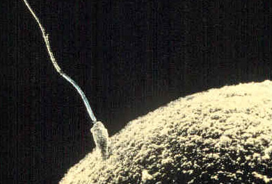
After the ovum is released from the ovary, it is drawn into the Fallopian tube. If sexual intercourse occurs, millions of sperm are ejaculated into the vagina, but only a few reach the ovum. Fertilization occurs when a sperm and an ovum combine, also referred to as conception. See Figure 8.14[2] for an image of conception.

After a single sperm has entered the ovum, the wall of the ovum becomes hard and prevents other sperm from entering. The head of the sperm, containing the genetic information from the father, unites with the nucleus of the ovum that contains genetic information from the mother, and a new cell is formed. Because each of these reproductive cells is a haploid cell containing half of the genetic material needed to form a human being, their combination forms a diploid cell called a zygote.[3]
During the first week after conception, the zygote divides and multiplies, going from a one-cell structure to two cells, then four cells, then eight cells, and so on. This process of cell division is called mitosis. The zygote continues to divide and travel through the Fallopian tube for about seven to ten days, at which point it arrives in the uterus. Once in the uterus, the zygote implants itself in the endometrium, the inner lining of the uterus, where the embryonic stage begins about three weeks after conception.[4]
Human chorionic gonadotropin (HCG) is a hormone produced by cells that will eventually make up the placenta in the early stages of pregnancy. HCG can be detected by a blood pregnancy test at around 7-12 days after conception and by a urine pregnancy test about two weeks after conception. HCG increases quickly (almost doubling every three days) for the first eight to ten weeks of pregnancy.[5]
View the following YouTube video[6] illustrating conception: Fertilization.
Embryonic Stage
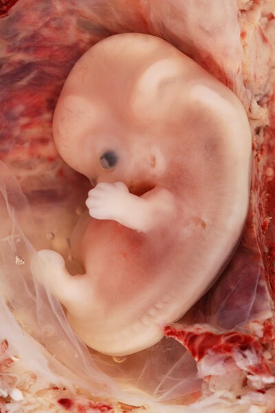
An embryo is an organism in its early stages of development, specifically from Day 16 through Week 8. See Figure 8.15[7] for an image of a human embryo.

During the embryonic stage, blood vessels grow, forming the placenta. The placenta is a structure that connects the fetus to the uterus, providing oxygen and nutrients from the mother to the developing embryo via the umbilical cord. Basic structures of the embryo start to develop into areas that will become the head, chest, and abdomen. During the embryonic stage, the heart begins to beat, and organs form and begin to function.[8]
The embryonic stage of fetal development begins with the completion of implantation and ends at the start of Week 9 of gestation. The weeks of gestation during pregnancy are calculated from the first day of the pregnant person’s last menstrual period rather than from the date of conception. In the final days of the pre-embryonic stage, the blastocyst develops into three germ layers called the ectoderm, mesoderm, and endoderm. The formation and development of the organs of the body, called organogenesis, starts at the beginning of the embryonic stage. The three germ layers develop into the organs, tissues, and structures of the body.[9]
Although the nervous system starts forming first, the embryo’s cardiovascular system begins functioning first. The hemoglobin formed by the embryo has a higher affinity for oxygen than adult hemoglobin because the embryo is dependent on the parent for oxygen. The embryo also requires more oxygen to support its rapid growth. The nervous system works within the first eight weeks of gestation but is not fully developed at birth. This is evident by a newborn’s lack of neuromuscular coordination, developed senses, or speech. The brain is not fully developed until five years of age. In most cases, the gonads differentiate into male or female by eight weeks. The testes stay in the inguinal canal, fully descending between 34 and 36 weeks of gestation.[10]
Exposure to hazardous agents called teratogens during organogenesis increases risk of malformations in all systems of the embryo’s body. For example, during organogenesis, the trachea and esophagus are one tube, and the separation into esophagus and trachea is not complete until five weeks of gestation. If the pregnant person is exposed to a teratogen at this critical point, then a congenital anomaly called a tracheal-esophageal (TE) fistula could result. In this anomaly, the esophagus is attached to the trachea rather than to the stomach.[11] Table 8.4 summarizes the weekly changes in embryo development.
Table 8.4. Embryonic Stage of Development[12]
| Week of Gestation | Structure and System Development |
|---|---|
| Week 3 |
|
| Week 4 |
|
| Week 5 |
|
| Week 6 |
|
| Week 7 |
|
| Week 8 |
|
Fetal Period
When the embryo reaches nine weeks’ gestation, it is called a fetus until birth. At the beginning of this stage, the fetus is about the size of a kidney bean and begins to take on the recognizable form of a human being. See Figure 8.16[13] for an image of an early fetus.

Mothers experience quickening, the first feeling of movement of the fetus in utero, around 16 to 24 weeks. By the time the fetus reaches the sixth month of development (23-24 weeks), it weighs up to 1.4 pounds. Hearing has developed, so the fetus can respond to sounds. The internal organs, such as the lungs, heart, stomach, and intestines, have formed enough that a fetus born prematurely at this point has a chance of survival with intensive medical care.
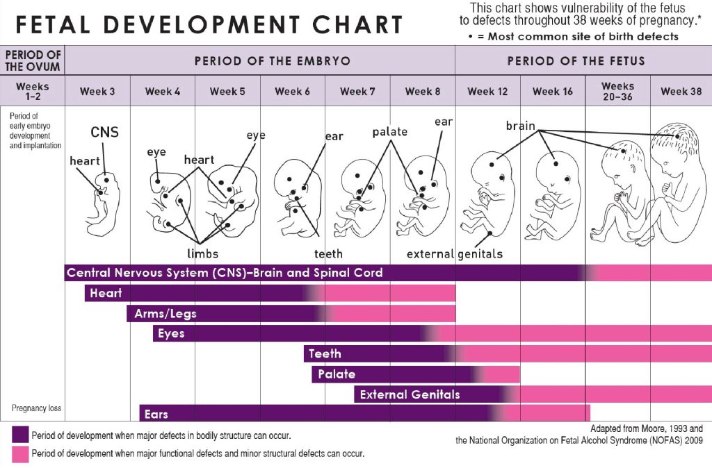
Between the seventh and ninth months, the fetus is primarily preparing for birth. It is exercising its muscles, and its lungs begin to expand and contract. Around 36 weeks, the fetus is almost ready for birth. It weighs about six pounds and is about 18.5 inches long. By Week 37 all organ systems are developed enough that the fetus could survive outside the mother’s uterus without many of the risks associated with premature birth. The fetus continues to gain weight and grow in length until approximately 40 weeks. See Figure 8.17[14] for an illustration of embryonic and fetal development. This illustration shows the development of the organs, structures, and tissues of the embryo and fetus, as well as when organs or structures are most vulnerable to teratogens.

View the following TED Talk video[15] showing human development from conception: Conception to Birth – Visualized.
Pregnancy
A full-term pregnancy lasts approximately 270 days (approximately 38.5 weeks) from conception to birth. Because it is easier to remember the first day of the last menstrual period (LMP) than to estimate the date of conception, obstetricians set the due date as 284 days (approximately 40.5 weeks) from the LMP. This assumes that conception occurred on Day 14 of the woman’s cycle. These 40 weeks of pregnancy are usually discussed in terms of three trimesters, each trimester lasting approximately 14 weeks. The first trimester is from the first day of the woman’s LMP to 13 weeks and 6 days. The second trimester is from Week 14 to 27 weeks and 6 days. The third trimester is from Week 28 to Week 40 and 6 days.[16] Providing nursing care for pregnant women is further discussed in the “Antepartum” chapter. See Figure 8.18[17] for an image of a pregnant woman.

- Lazzara, J. (2020). Lifespan development. Maricopa Community Colleges. https://open.maricopa.edu/devpsych/ ↵
- “Sperm-egg.jpg” by unknown author is licensed in the Public Domain. ↵
- Lazzara, J. (2020). Lifespan development. Maricopa Community Colleges. https://open.maricopa.edu/devpsych/ ↵
- Lazzara, J. (2020). Lifespan development. Maricopa Community Colleges. https://open.maricopa.edu/devpsych/ ↵
- Cleveland Clinic. (2024). Human chorionic gonadotropin. https://my.clevelandclinic.org/health/articles/22489-human-chorionic-gonadotropin ↵
- Nucleus Medical Media. (2013, January 31). Fertilization [Video]. YouTube. All rights reserved. https://youtu.be/_5OvgQW6FG4?si=siTDwkcpfKtYJ9LC ↵
- “9-Week_Human_Embryo_from_Ectopic_Pregnancy.jpg” by Ed Uthman from Houston, TX, USA is licensed under CC BY 2.0 ↵
- Lazzara, J. (2020). Lifespan development. Maricopa Community Colleges. https://open.maricopa.edu/devpsych/ ↵
- Giles, A., Prusinski, R., & Wallace, L. (2024). Maternal-newborn nursing. OpenStax. https://openstax.org/books/maternal-newborn-nursing/pages/1-introduction ↵
- Giles, A., Prusinski, R., & Wallace, L. (2024). Maternal newborn nursing. OpenStax. https://openstax.org/books/maternal-newborn-nursing/pages/1-introduction ↵
- Giles, A., Prusinski, R., & Wallace, L. (2024). Maternal newborn nursing. OpenStax. https://openstax.org/books/maternal-newborn-nursing/pages/1-introduction ↵
- Giles, A., Prusinski, R., & Wallace, L. (2024). Maternal newborn nursing. OpenStax. https://openstax.org/books/maternal-newborn-nursing/pages/1-introduction ↵
- “Human_fetus_10_weeks_with_amniotic_sac_-_therapeutic_abortion.jpg” by suparna sinha is licensed under CC BY-SA 2.0 ↵
- “10-4-fetal-growth-and-development” by Centers for Disease Control and Preventions (CDC) is in the Public Domain. Access for free at https://openstax.org/books/maternal-newborn-nursing/pages/1-introduction ↵
- Tsiaras, A. (2010, December). Conception to birth -- Visualized [Video]. Ted.com. All rights reserved. https://www.ted.com/talks/alexander_tsiaras_conception_to_birth_visualized ↵
- American College of Obstetricians and Gynecologists. (2024). How your fetus grows during pregnancy. https://www.acog.org/womens-health/faqs/how-your-fetus-grows-during-pregnancy ↵
- “PregnantWoman.jpg” by Ken Hammond is licensed in the Public Domain. ↵
The process of egg cell (ovum) development in the ovaries.
The process where a sperm cell penetrates and merges with an egg cell, resulting in the formation of a zygote.
The moment when a sperm fertilizes an egg, leading to the formation of an embryo.
Fertilized egg cell that results from the union of a female gamete (egg, or ovum) with a male gamete (sperm).
The process of cell division that results in two identical daughter cells, each with the same number of chromosomes as the parent cell.
The inner lining of the uterus that thickens during the menstrual cycle to prepare for a potential pregnancy. If fertilization does not occur, it is shed during menstruation.
A hormone produced during pregnancy that maintains the corpus luteum and is the basis for most pregnancy tests.
An early stage of development in multicellular organisms. In humans, it refers to the first eight weeks after conception.
An organ that develops in the uterus during pregnancy, providing oxygen and nutrients to the growing fetus while removing waste products from the fetus's blood.
The cord connecting the developing fetus to the placenta, allowing the exchange of oxygen, nutrients, and waste products between the mother and fetus.
The period in prenatal development that lasts from fertilization to the end of the eighth week, during which the major organs and structures begin to form.
The early phase of prenatal development, from fertilization through the second week, before the embryo is fully implanted in the uterus.
The phase of embryonic development where the organs of the body begin to form.
Substances or environmental factors that can cause birth defects or abnormal development in a fetus.
The term for a developing human from the ninth week of pregnancy until birth.
The first movements of the fetus felt by the pregnant person, typically occurring around the 16th to 20th week of pregnancy.
The first day of a woman's most recent menstrual cycle, often used to calculate gestational age in pregnancy.
0 to 13 weeks and six days of gestation.
14 to 27 weeks and six days of gestation.
28 weeks until delivery.

