7.4 Cardiac Muscle
Cardiac Muscle[1]
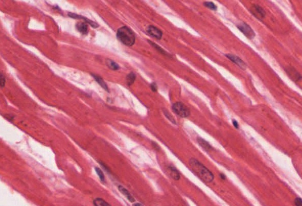
Cardiac muscle is an involuntary muscle that forms the walls of the heart. The cells of cardiac muscle, known as cardiomyocytes, are striated and they contain sarcomeres. The fibers are shorter than skeletal muscle fibers and typically have just one centrally located nucleus. Special cardiomyocytes called pacemaker cells stimulate the heart to contract on its own without any nerve impulse. Cardiomyocytes also possess many mitochondria to provide the energy needed for contraction. Cardiac muscle fibers are extensively branched and connect to one another at their ends by intercalated discs. An intercalated disc allows the cardiac muscle cells to coordinate contraction so that the heart can work as a pump. See Figure 7.32[2] for an image of cardiac muscle tissue.

View the University of Michigan WebScope to explore a slide of cardiac muscle in greater detail.
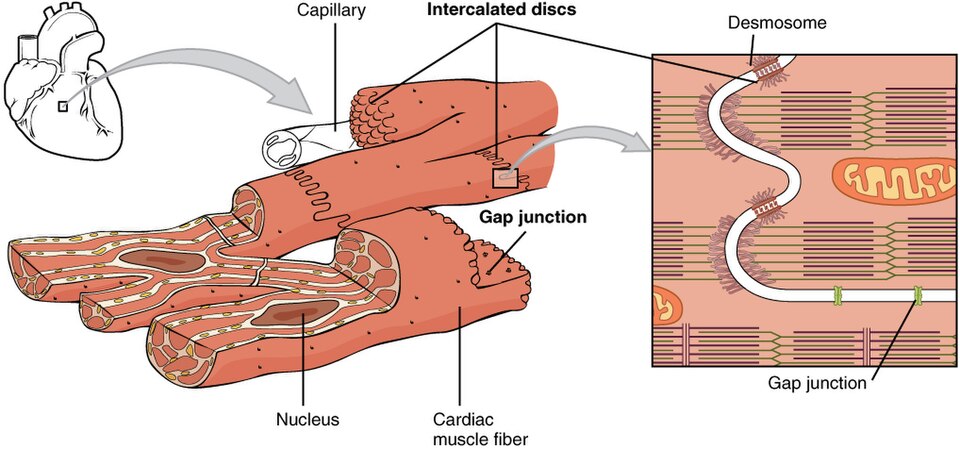
Intercalated discs are part of the sarcolemma and contain two structures important in cardiac muscle contraction: gap junctions and desmosomes. A gap junction forms channels between cardiac muscle fibers to allow the depolarizing current to flow from one cardiac muscle cell to the next. This joining is called electric coupling, and in cardiac muscle it allows the coordinated contraction of the entire heart. The remainder of the intercalated disc is composed of desmosomes. A desmosome is a cell structure that anchors the ends of cardiac muscle fibers together, so the cells do not pull apart during the stress of individual fibers contracting. See Figure 7.33[3] for an illustration of a desmosome in cardiac muscle.

Contractions of the heart (heartbeats) are controlled by specialized cardiac muscle cells called pacemaker cells that directly control heart rate. Although cardiac muscle is involuntary and cannot be consciously controlled, the pacemaker cells respond to signals from the autonomic nervous system (ANS) to speed up or slow down the heart rate. The pacemaker cells can also respond to various hormones that adjust heart rate to control blood pressure.
- Betts, J. G., Young, K. A., Wise, J. A., Johnson, E., Poe, B., Kruse, D. H., Korol, O., Johnson, J. E., Womble, M., & DeSaix, P. (2022). Anatomy and physiology 2e. OpenStax. https://openstax.org/books/anatomy-and-physiology-2e/pages/1-introduction ↵
- “414c_Cardiacmuscle” by OpenStax College is licensed under CC BY 3.0 ↵
- “1020_Cardiac_Muscle” by OpenStax is licensed under CC BY 4.0 ↵
Involuntary, striated muscle tissue found only in the heart.
Allows the cardiac muscle cells to coordinate contraction so that the heart can work as a pump.
Channels between cardiac muscle fibers to allow the depolarizing current to flow from one cardiac muscle cell to the next.
A cell structure that anchors the ends of cardiac muscle fibers together, so the cells do not pull apart during the stress of individual fibers contracting.

