3.4 Cytoplasm, Organelles, and Cytoskeleton
Cytoplasm, Organelles, and Cytoskeleton[1]
Now that we have discussed the cell membrane and substances crossing the cell membrane via diffusion, osmosis, and active transport, let’s examine the inside of cells. All living cells in the human body contain many important parts, each with a very specific job.
Cytoplasm
The inside of a cell is called cytoplasm that includes cytosol and organelles. Cytosol is a jelly-like substance that fills the cell and is made of water, salts, and proteins. All animal cells are eukaryotic cells, meaning they have a nucleus. Eukaryotic cells also contain several organelles. Organelles, which means “little organs,” work together to keep the cell healthy and perform important functions. The cytoskeleton, also considered part of the cytoplasm, is a group of fibrous proteins that provide structural support for cells and perform other functions.
Organelles of the Cell
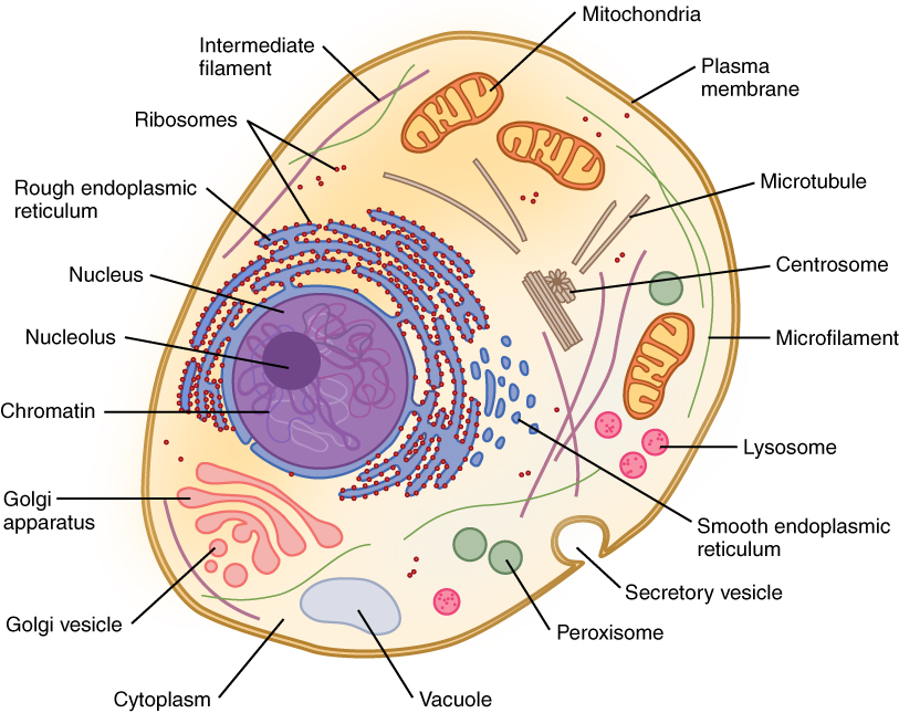
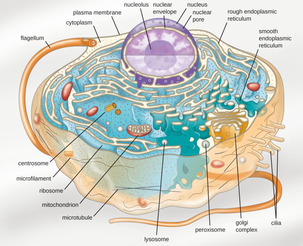
See Figure 3.11[2] for an illustration of primary organelles and structures in a human cell. This subsection will discuss the nucleus, endoplasmic reticulum, ribosomes, Golgi apparatus, lysosome, mitochondria, centrosomes, and peroxisomes.
Nucleus
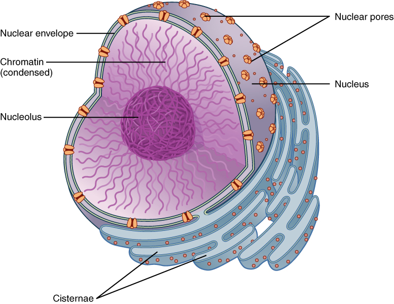
The nucleus is found in the center of the cell and contains the cell’s DNA. The DNA is the genetic material that determines the entire structure and function of that cell, so the nucleus is often referred to as the control center of the cell. Inside the nucleus, the DNA exists in a loosely tangled form called chromatin. The nuclear membrane (or nuclear envelope) is the membrane covering the nucleus. It contains holes called nuclear pores that allow substances to enter or leave the nucleus. The nucleolus is a structure found within the nucleus. Its function is to produce ribosomes (discussed later in this chapter) for the cell. See Figure 3.12[3] for an illustration of the structures inside the nucleus.
Endoplasmic Reticulum and Ribosomes
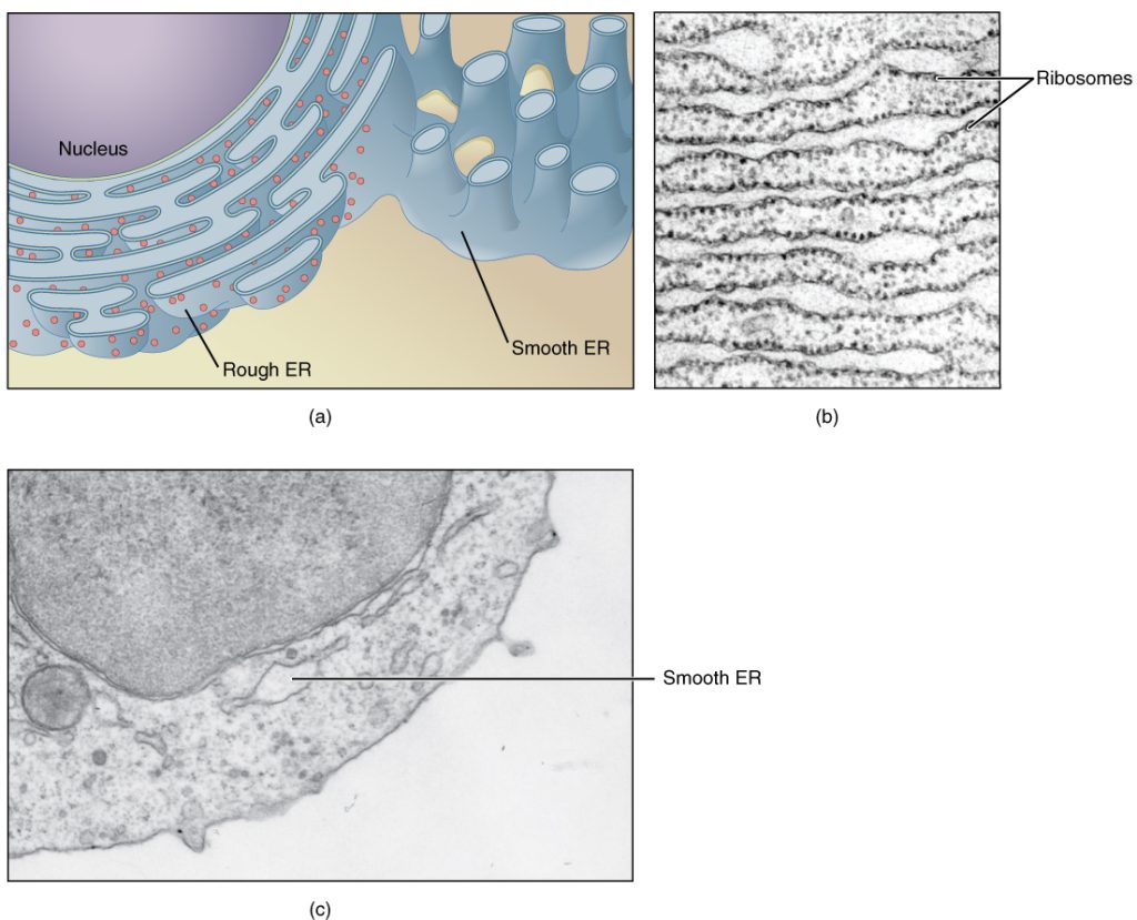
The endoplasmic reticulum (ER) is a system of channels that is continuous with the nuclear membrane covering the nucleus and is composed of the same lipid bilayer material. It provides passages throughout much of the cell that help with transporting, making, and storing materials. There are two parts to the endoplasmic reticulum, called the smooth and rough endoplasmic reticulum. The smooth endoplasmic reticulum makes lipids and phospholipids (the main component of the cell membrane). The rough endoplasmic reticulum is dotted with many ribosomes that attach to the endoplasmic reticulum, giving it a “rough” appearance. Ribosomes are organelles that make proteins. The rough ER will then modify the newly synthesized proteins produced by the ribosomes. See Figure 3.13[4] for an illustration of these parts of the endoplasmic reticulum.
Golgi Apparatus
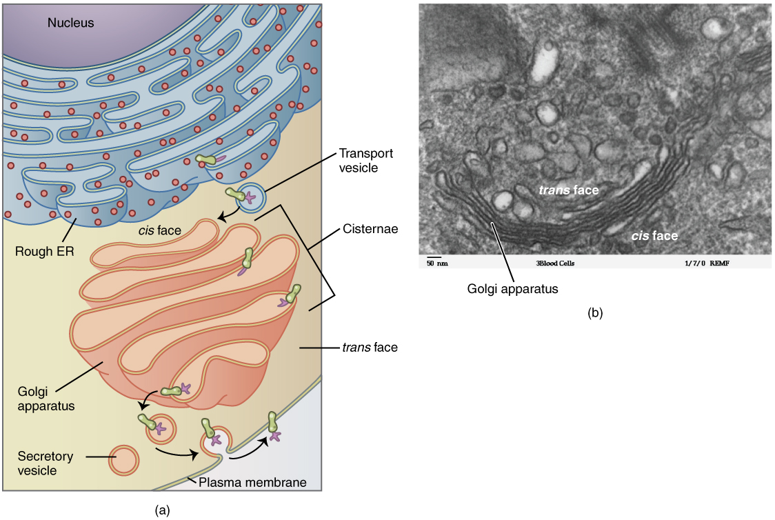
The Golgi apparatus is responsible for sorting, modifying, and shipping off the products that come from the rough ER, much like a post office. It also makes lysosomes. The Golgi apparatus looks like a stack of oddly shaped pancakes. See Figure 3.14[5] for an illustration of the Golgi apparatus.
Lysosomes
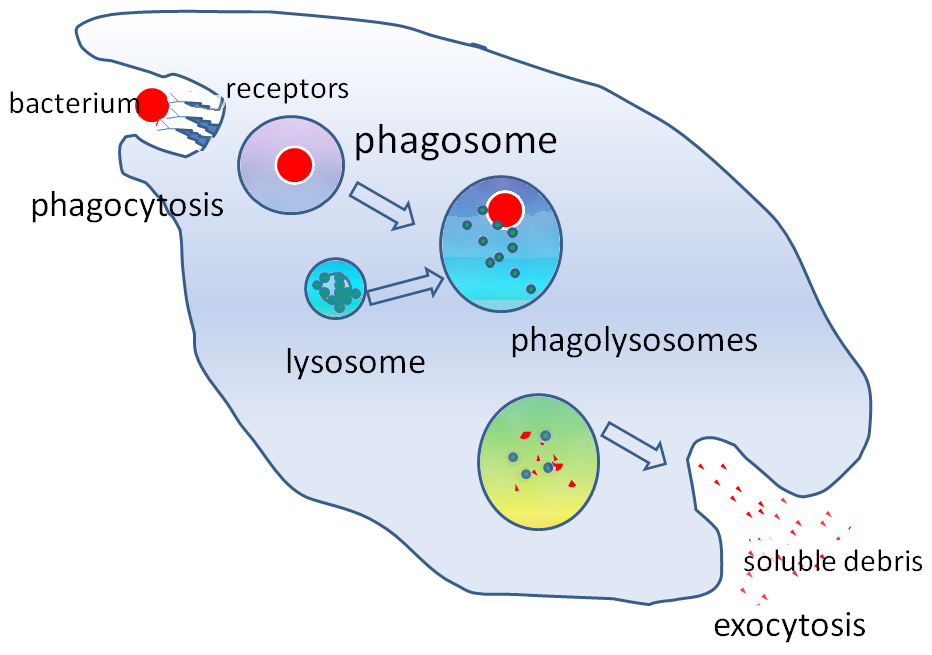
A lysosome is an organelle that contains enzymes that break down and digest unneeded things, such as foreign material or a damaged organelle. For example, when a white blood cell phagocytizes a pathogen like a virus, the virus is digested by the enzymes released by lysosomes. See Figure 3.15[6] for an illustration of a lysosome inside a white blood cell digesting a particle.
Mitochondria
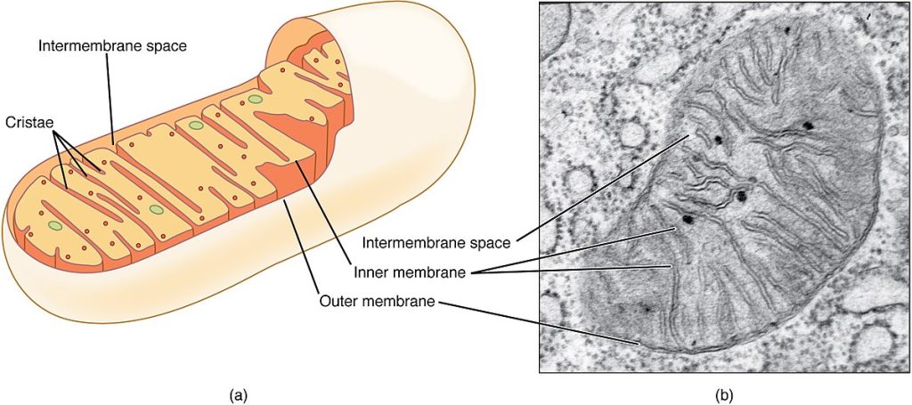
A mitochondrion (the singular form of mitochondria) is a bean-shaped organelle that is the “energy supplier” of the cell. Mitochondria convert energy stored in nutrients (such as glucose) into adenosine triphosphate (ATP), using a process called cellular respiration, which provides usable energy to the cell. In fact, cellular respiration is the reason why your body requires oxygen. Without enough oxygen, cells can’t generate enough ATP to maintain life for very long. Cells constantly use ATP to function, so mitochondria are constantly producing ATP. Different cells have different numbers of mitochondria depending on their energy needs. For example, muscle cells have more mitochondria than bone cells. See Figure 3.16[7] for an illustration of mitochondria.
Centrosomes
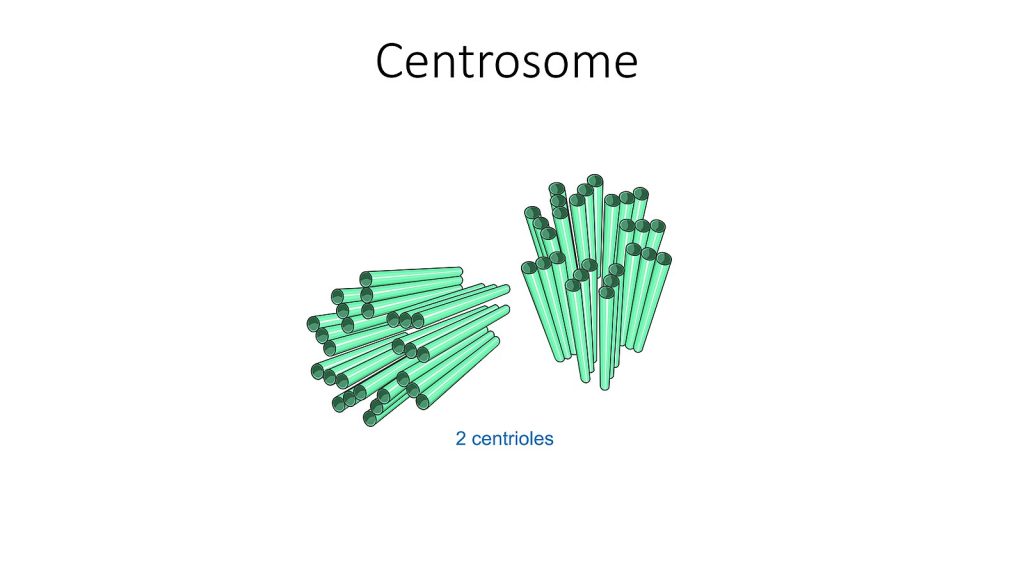
Centrosomes are a pair of cylindrical structures called centrioles that look like gears or pinwheels. They are located near the nucleus in the cell. Their function is to form a structure called the spindle during the process of cell division. See Figure 3.17[8] for an illustration of a centrosome.
Peroxisomes
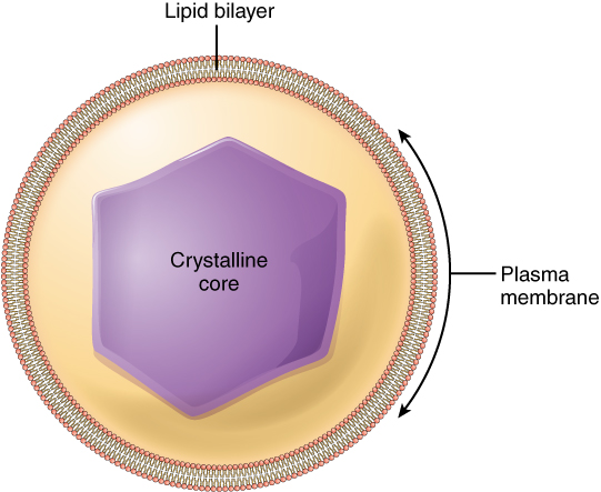
Like lysosomes, a peroxisome is an organelle that contains mostly enzymes. Peroxisomes perform different functions, including metabolizing lipids and detoxifying harmful substances. Peroxisomes are like tiny sewage treatment plants that neutralize harmful toxins so that they do not wreak havoc in the cells. For example, there are a high number of peroxisomes in the liver, an organ primarily responsible for detoxifying the blood before it travels throughout the body. See Figure 3.18[9] for an illustration of a peroxisome.
View supplementary YouTube videos[10],[11] that review cell organelles:
Introduction To The Cell | Organelles | Animated Music Video |
Cytoskeleton
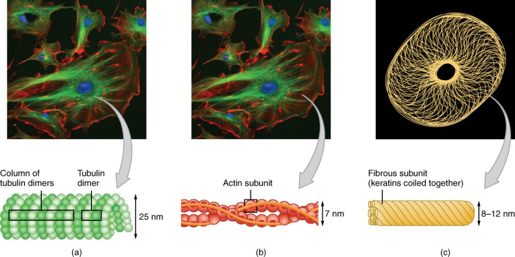
Similar to how bones support the human body, the cytoskeleton helps support the cell. The cytoskeleton is a group of proteins that provide structural support for cells. It also helps with cell movement, reproduction, and transportation of substances within the cell. The cytoskeleton consists of microtubules, microfilaments, and intermediate filaments.
Microtubules are the thickest of the three components and are made from a protein called tubulin. Microtubules perform several functions, including the following:
- Maintain the shape and structure of the cell
- Prevent the cell from being compressed or squashed
- Keep the organelles in the right places within the cell
- Make up two cellular appendages used for motion:
- Cilia are found on the surface of many cells of the body, including cells that line the airways of the respiratory system. Cilia move wastes such as dust, mucus, and bacteria up through the airways, away from the lungs and to the mouth. Cilia also help move egg cells from the ovary to the uterus in the female reproductive system.
- Flagella are a tail-like structure for movement. The only cells in humans that have flagella are sperm cells.
See Figure 3.19[12] for an illustration of the cytoskeleton.
Microfilaments are the thinner cytoskeletal filament, primarily composed of the protein actin. Muscle tissue is made up of actin fibers that, along with another protein called myosin, are responsible for muscle contraction.
Intermediate filaments are intermediate in thickness and made up of a protein called keratin. These filaments are wound together like a rope and help maintain cell shape and structure. See Figure 3.20[13] for an illustration of the components of the cytoskeleton.
Complete a supplementary Wisc-Online learning activity[14] on cell organelles: A Typical Animal Cell.
- Betts, J. G., Desaix, P., Johnson, E., Johnson, J. E., Korol, O., Kruse, D., Poe, B., Wise, J., Womble, M. D., & Young, K. A. (2022). Anatomy and physiology, 2e. OpenStax. https://openstax.org/books/anatomy-and-physiology-2e/pages/1-introduction ↵
- “0312_Animal_Cell_and_Components” by OpenStax is licensed under CC BY 4.0 ↵
- “0318_Nucleus” by OpenStax is licensed under CC BY 4.0 ↵
- “0313_Endoplasmic_Reticulum” by OpenStax is licensed under CC BY 4.0 ↵
- “0314_Golgi_Apparatus” by OpenStax is licensed under CC BY 4.0 ↵
- “Phagocytosis2” by en:User:Graham Beards is licensed under CC BY-SA 4.0 ↵
- “0315_Mitochondrion_new” by OpenStax is licensed under CC BY 4.0 ↵
- “Centrosome_-_Two_centrioles_--_Smart-Servier” by Laboratoires Servier is licensed under CC BY-SA 3.0 ↵
- “0316_Peroxisome” by OpenStax is licensed under CC BY 4.0 ↵
- Biology Music Videos. (2024, September 6). Introduction to the cell | Organelles | Animated music video | [Video]. YouTube. All rights reserved. https://www.youtube.com/watch?v=CNjNhNRAcIM ↵
- Forciea, B. (2015, March 20). Cell organelles for anatomy and physiology [Video]. YouTube. All rights reserved. https://www.youtube.com/watch?v=njiAmKZomIg ↵
- “OSC_Microbio_03_04_eukcell” by OpenStax is licensed under CC BY 4.0 ↵
- “0317_Cytoskeletal_Components” by OpenStax is licensed under CC BY 4.0 ↵
- Heins, G. (n.d.). A typical animal cell [Video]. Wisc-Online. All rights reserved. https://www.wisc-online.com/learn/natural-science/life-science/ap11403/a-typical-animal-cell ↵
The gel-like substance inside the cell membrane, excluding the nucleus, that contains organelles and the cytosol, where most cellular processes occur.
The gel-like fluid inside a cell where organelles, proteins, and molecules are suspended.
Cells that contain a nucleus and other membrane-bound organelles, such as mitochondria and the endoplasmic reticulum.
Specialized structures within a cell that perform specific functions, such as energy production (mitochondria), protein synthesis (ribosomes), and waste disposal (lysosomes).
A network of protein fibers that provides structural support, facilitates cell movement, and plays a role in intracellular transport and cell division.
The control center of a eukaryotic cell, which houses the genetic material (DNA) and is involved in regulating cell activities like growth and reproduction.
A complex of DNA and proteins (histones) found in the nucleus, which condenses into chromosomes during cell division.
The double-layered membrane that surrounds the nucleus, separating the nuclear contents from the cytoplasm.
Another term for the nuclear membrane, consisting of two lipid bilayers that protect the genetic material inside the nucleus.
Protein complexes embedded in the nuclear envelope that regulate the transport of molecules (such as RNA and proteins) between the nucleus and cytoplasm.
A dense region inside the nucleus where ribosomal RNA (rRNA) is synthesized and combined with proteins to form ribosomal subunits.
An extensive network of membranes involved in protein and lipid synthesis.
The part of the ER lacking ribosomes, involved in lipid synthesis, detoxification, and calcium storage.
The part of the ER that has ribosomes attached, primarily involved in protein synthesis and modification.
Small RNA and protein complexes responsible for protein synthesis. They can be found floating in the cytoplasm or attached to the rough ER.
A stack of membranous sacs involved in the modification, sorting, and packaging of proteins and lipids for secretion or delivery to other organelles.
A membrane-bound organelle containing digestive enzymes responsible for breaking down waste materials, cellular debris, and foreign substances.
The powerhouse of the cell, responsible for producing ATP through cellular respiration. It has its own DNA and double membrane.
Regions near the nucleus that contain centrioles, involved in the organization of the microtubules and spindle formation during cell division.
Membrane-bound organelles that contain enzymes for the breakdown of fatty acids and the detoxification of hydrogen peroxide.
Hollow tubes made of tubulin protein that provide structural support, facilitate intracellular transport, and are involved in cell division and cilia/flagella movement.
Short, hair-like projections from the cell surface that are involved in movement of the cell or the movement of substances along the cell surface.
Long, whip-like structures that enable the cell to move.
Thin protein fibers (primarily actin) that provide structural support, facilitate cell movement, and play a role in cytokinesis during cell division.
Protein fibers that provide structural support and mechanical strength to the cell, helping to maintain its shape and resist stress.

