3.2 Structure and Composition of the Cell Membrane
Structure and Composition of the Cell Membrane[1]
All living cells in the human body have a cell membrane surrounding them. Just as the outer layer of your skin separates your body from the environment, the cell membrane (also known as the plasma membrane) separates the inside of the cell from the outside of the cell, providing a protective barrier around the cell and regulating what can go into or out of the cell.
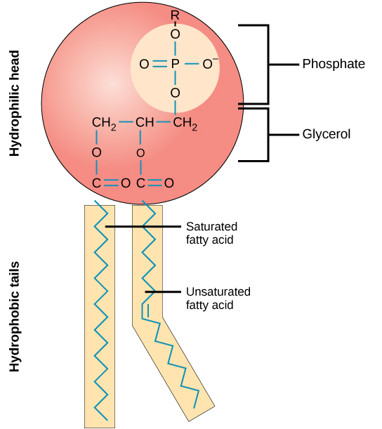
The cell membrane is a very flexible bilayer (meaning it has two layers) made up of molecules called phospholipids. A single phospholipid molecule has a phosphate group on one end, called the “head,” and two side-by-side chains of fatty acids that make up the “tails.” See Figure 3.1[2] for an illustration of a phospholipid molecule.

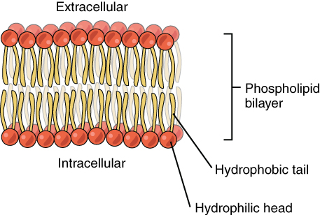
The phosphate “head” is negatively charged, making it polar and hydrophilic, meaning “water loving or attracted to water.” The phosphate heads are attracted to the watery environments outside of the cell (extracellular fluid) and inside of the cell (intracellular fluid or cytosol), so they naturally organize themselves to be in contact with these areas.
The lipid tails are uncharged, making them nonpolar and are hydrophobic, meaning “water fearing or repelling to water.” Because of this, the fatty acid tails naturally organize themselves away from the watery environments of the extracellular and intracellular fluids and point to the inside of the cell membrane. See Figure 3.2[3] for an illustration of the phospholipid bilayer.

Phospholipids are considered amphipathic molecules because they contain both a hydrophilic and a hydrophobic region. Another great example of an amphipathic substance is soap. Soap works to remove oil and grease stains because of its amphipathic properties. The hydrophilic portion can dissolve in water while the hydrophobic portion can trap grease that can then be washed away.
Membrane Proteins
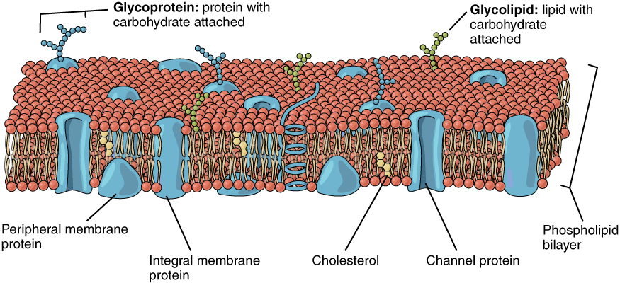
The phospholipid bilayer forms the basic structure of the cell membrane, but it also contains many types of proteins on and within the membrane. Two types of proteins that are commonly associated with the cell membrane are integral proteins and peripheral proteins as shown in Figure 3.3.[4]

Integral proteins are proteins that are embedded in the membrane. They include the following proteins:
- Carrier proteins: Proteins that change their shape to move substances across the cell membrane.
- Channel proteins: Proteins that selectively allow materials, such as certain ions, to pass into or out of the cell.
- Cell recognition proteins: Proteins that mark a cell’s identity so that it can be recognized by other cells.
- Dual receptor and ion channel proteins: Some integral proteins are receptors and channel proteins. For example, when a dopamine molecule binds to a dopamine receptor protein, a channel in the cell membrane opens to allow certain ions to flow into the cell.
- Glycoproteins: Proteins that have carbohydrate molecules attached that extend extracellularly (outside of the cell). The attached carbohydrate molecules on glycoproteins help with cell recognition.
Peripheral proteins are typically only found on the inner or outer surface of the cell membrane but can also be attached to the internal or external surface of an integral protein. These proteins usually perform a specific function for the cell. For example, some peripheral proteins on the surface of intestinal cells break down nutrients to sizes that can pass through the cells and into the bloodstream.
- Betts, J. G., Desaix, P., Johnson, E., Johnson, J. E., Korol, O., Kruse, D., Poe, B., Wise, J., Womble, M. D., & Young, K. A. (2022). Anatomy and physiology, 2e. OpenStax. https://openstax.org/books/anatomy-and-physiology-2e/pages/1-introduction ↵
- “Figure_05_01_02” by CNX OpenStax is licensed under CC BY 4.0 ↵
- “0302_Phospholipid_Bilayer” by CNX OpenStax is licensed under CC BY 4.0 ↵
- “0303_Lipid_Bilayer_With_Various_Components” by OpenStax is licensed under CC BY 4.0 ↵
Also called the plasma membrane, is a selectively permeable structure that surrounds the cell, providing protection and regulating the movement of substances in and out.
Also known as the cell membrane, the outer boundary of a cell that separates its internal environment from the external surroundings.
The main component of cell membranes, which protect cells from environmental damage and allow for cellular processes.
A substance or molecule that has an affinity for water.
Watery environments outside of the cell.
The fluid found inside cells, making up about two-thirds of the total body water.
The gel-like fluid inside a cell where organelles, proteins, and molecules are suspended.
A term used to describe substances that repel or do not dissolve in water.
Has both hydrophilic (water-attracting) and hydrophobic (water-repelling) regions.
Proteins that are embedded in the membrane.
Proteins that change their shape to move substances across the cell membrane.
Proteins that selectively allow materials, such as certain ions, to pass into or out of the cell.
Proteins that mark a cell’s identity so that it can be recognized by other cells.
Also known as a sensor, monitors a physiological value, such as temperature, respiratory rate, blood pressure, etc.
A molecule that binds to and activates a receptor.
A membrane receptor that can bind to two different ligands or perform two distinct functions within a signaling pathway.
Membrane proteins that form pores in the cell membrane, allowing specific ions (e.g., Na⁺, K⁺, Ca²⁺, Cl⁻) to pass through.
Proteins that have carbohydrate molecules attached that extend outside of the cell.
Outside the cell.
Typically only found on the inner or outer surface of the cell membrane but can also be attached to the internal or external surface of an integral protein.

