11.3 Conduction System of the Heart
Conduction System of the Heart[1]
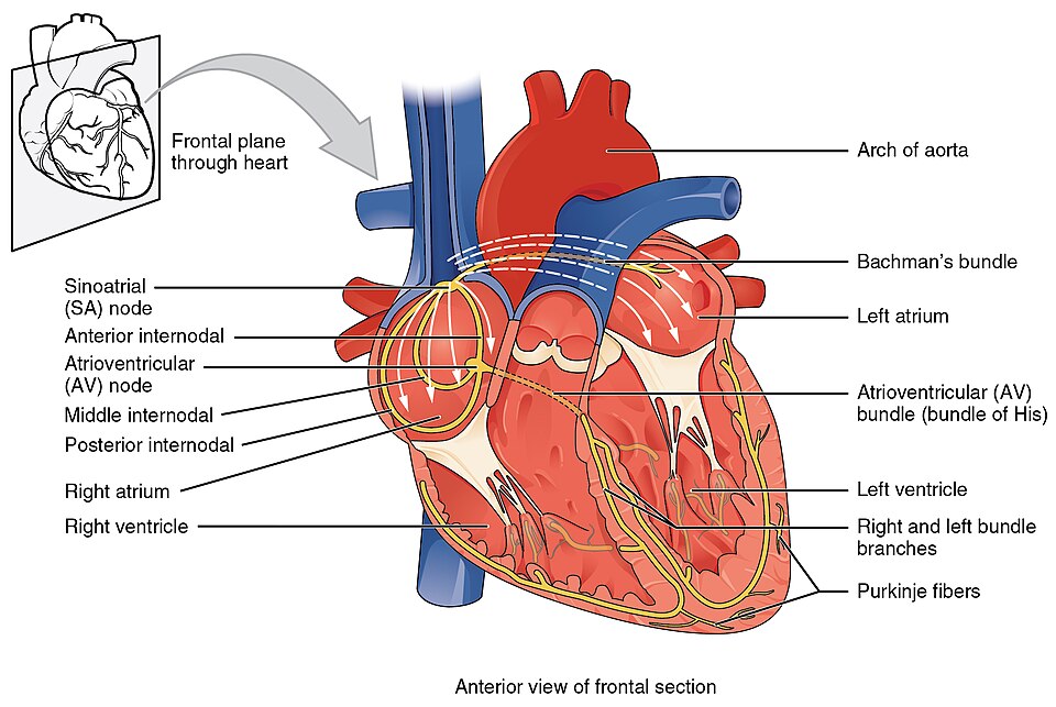
The heart generates its own electrical impulse as part of the cardiac conduction system. The components of the cardiac conduction system include the sinoatrial node, the atrioventricular node, the atrioventricular bundle, the bundle branches, and the Purkinje fibers. See Figure 11.14[2] for an illustration of the cardiac conduction system.

Sinoatrial (SA) Node
Normal cardiac rhythm is established by the sinoatrial (SA) node, a specialized clump of conducting cells located in the wall of the upper right atrium close to the opening of the superior vena cava. The SA node is known as the pacemaker of the heart. It is self-depolarizing, which means it generates its own electrical signal, stimulating the heart to beat. Unlike skeletal muscle, cardiac muscle doesn’t need a motor neuron to stimulate contraction.
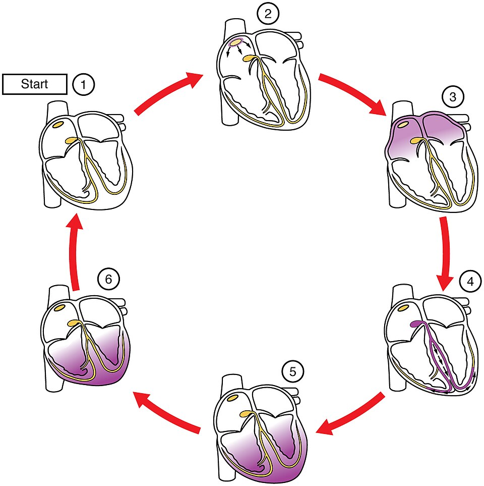
Once the SA node fires, the impulse spreads throughout the atria and causes them to contract. The signal also spreads to the atrioventricular node. The fibrous cardiac skeleton prevents the impulse from spreading into the myocardial cells in the ventricles except at the atrioventricular node.
Atrioventricular (AV) Node
The atrioventricular (AV) node is a second clump of specialized conductive cells located in the inferior portion of the right atrium within the atrioventricular septum. The septum prevents the impulse from spreading directly to the ventricles without passing through the AV node first.
Atrioventricular Bundle (Bundle of His), Bundle Branches, and Purkinje Fibers
Arising from the AV node, the atrioventricular (AV) bundle, or bundle of His, proceeds through the interventricular septum before dividing into the right and left bundle branches. The left bundle branch supplies the left ventricle, and the right bundle branch the right ventricle. Because the left ventricle is much larger than the right, the left bundle branch is also considerably larger than the right. Both bundle branches descend and reach the apex of the heart where they connect with the Purkinje fibers.
The Purkinje fibers are additional conductive fibers that spread the impulse to the contractile cells in the ventricles. They extend throughout the myocardium from the apex of the heart up the ventricles. Because the electrical stimulus begins at the apex, contraction of the ventricles also begins at the apex and travels up toward the base of the heart, similar to squeezing a tube of toothpaste from the bottom. This allows the blood to be pumped out of the ventricles and into the aorta and pulmonary trunk. See Figure 11.15[3] for an illustration of cardiac conduction.

View a supplementary video[4] on the conduction of the electrical impulse through the heart at Cardiac Conduction System.
Electrocardiogram
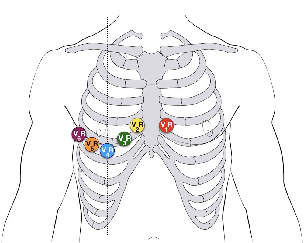
By placing electrodes on the skin in specific locations, we can record the heart’s electrical activity using an electrocardiogram (ECG), also commonly abbreviated EKG (“K” for kardiology, the German term for cardiology). Careful analysis of the ECG reveals a detailed picture of both normal and abnormal heart function and is an important clinical diagnostic tool. A 12-lead electrocardiograph uses electrodes placed in standard locations on the patient’s skin, as illustrated in Figure 11.16.[5]

Sometimes it is useful to monitor a patient’s cardiac activity over a longer period of time. A Holter monitor is a small, portable, battery-operated device that continuously monitors a patient’s cardiac electrical activity over a longer period of time during their normal daily routine. It is used to detect irregular heart rhythms.
- Betts, J. G., Young, K. A., Wise, J. A., Johnson, E., Poe, B., Kruse, D. H., Korol, O., Johnson, J. E., Womble, M., & DeSaix, P. (2022). Anatomy and physiology 2e. OpenStax. https://openstax.org/books/anatomy-and-physiology-2e/pages/1-introduction ↵
- “2018_Conduction_System_of_Heart” by OpenStax College is licensed under CC BY 3.0 ↵
- “2019 Cardiac ConductionN” by OpenStax College is licensed under CC BY 3.0 ↵
- A.D.A.M. (2024, May 8). Cardiac conduction system [Video]. MedlinePlus. All rights reserved. https://medlineplus.gov/ency/anatomyvideos/000021.htm ↵
- “Right-sided-12-lead-ECG-lead-placement” by unknown author, LITFL is licensed under CC BY-NC-SA 4.0. Access for free at https://litfl.com/ecg-lead-positioning/ ↵
A network of specialized heart cells that generate and transmit electrical impulses, controlling the heart’s rhythmic contractions.
A specialized clump of conducting cells located in the wall of the upper right atrium close to the opening of the superior vena cava.
Relating to the connection between the atria and ventricles of the heart.
A group of specialized heart muscle fibers that transmit electrical impulses from the AV node down to the ventricles.
A group of specialized heart muscle fibers that transmit electrical impulses from the AV node down to the ventricles.
Pathways that split from the AV bundle and carry impulses down the left and right sides of the interventricular septum to the ventricles.
Specialized fibers that spread the electrical impulse throughout the ventricles, causing them to contract.
A test that records the electrical activity of the heart over time using electrodes placed on the skin, producing a waveform that helps diagnose heart conditions.

