1.7 Major Body Regions and Their Subdivisions
Major Body Regions and Their Subdivisions[1],[2]
Body Cavities
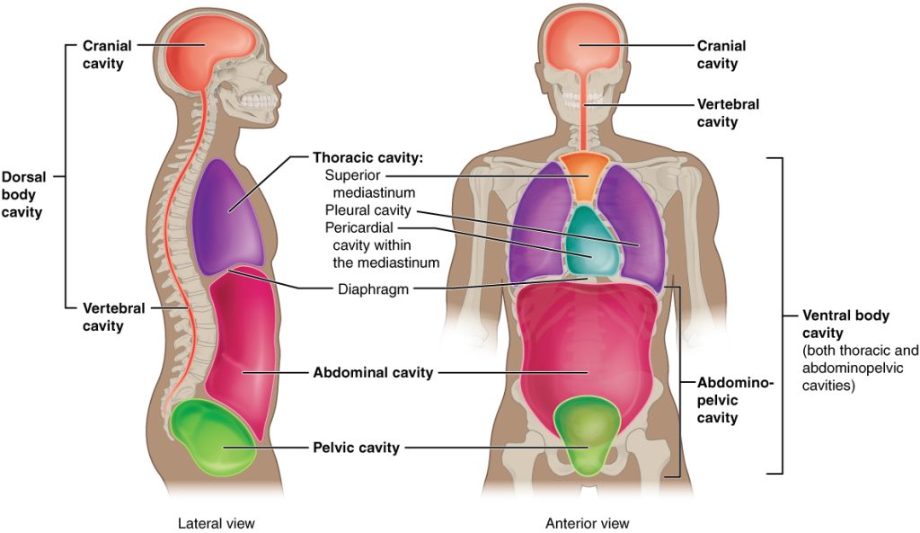
The body uses membranes, sheaths, and other structures to create cavities or compartments. The ventral (anterior) cavity and dorsal (posterior) cavity are the largest body compartments that contain and protect internal organs. See Figure 1.8[3] for an illustration of the ventral (anterior) and dorsal (posterior) cavities.

The ventral (anterior) cavity contains the lungs, heart, stomach, and intestines and allows for significant changes in the size and shape of these organs as they do their work. The anterior (ventral) cavity has two main subdivisions:
- Thoracic cavity: The thoracic cavity is the chest cavity and it contains the heart and lungs. It’s surrounded and protected by the rib cage. The heart is located in the central portion of the thoracic cavity, which is called the mediastinum. The diaphragm forms the floor of the thoracic cavity and separates it from the abdominopelvic cavity.
- The abdominopelvic cavity is the largest cavity in the body. Although no membrane divides the abdominopelvic cavity, it can also be subdivided into the abdominal cavity, which contains the digestive organs, and the pelvic cavity, which contains the urinary bladder and reproductive organs.
The posterior (dorsal) cavity is divided into two smaller cavities:
- The cranial cavity, which contains the brain.
- The spinal cavity (or vertebral cavity), which contains the spinal cord.
Just as the brain and spinal cord make up a continuous, uninterrupted structure, the cranial and spinal cavities are also continuous. The brain and spinal cord are protected by the bones of the skull and the spine and by cerebrospinal fluid, a colorless fluid that provides a cushion.
Complete a supplementary Wisc-Online learning activity on body sections and divisions of the abdominal pelvic cavity: Ventral (Anterior) and Dorsal (Posterior) Cavities. Click on “Next” at the bottom of the black box in this activity to proceed through this activity.
Serous Membranes
A serous membrane (also referred to as serosa) is a thin membrane that covers the walls and organs of the thoracic and abdominopelvic cavities. Serous membranes have two layers:
- The parietal layer lines the walls of the body cavity.
- The visceral layer covers the organs; it is also called the viscera (viscer means “organ”).
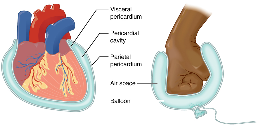
Between the parietal and visceral layers is a very thin, fluid-filled space, or cavity. Both the parietal and visceral layers secrete slippery, watery fluid into this serous cavity, filling it with fluid. This serous cavity cushions and reduces friction on organs when they move, such as when the lungs inflate or the heart beats. See Figure 1.9[4] for an illustration of a serous membrane surrounding the heart, called the pericardium. The pericardium is a double-layered membrane that surrounds the heart, similar to a fluid-filled balloon surrounding a fist.

There are three serous cavities and associated membranes:
- Pleura: The pleura is the serous membrane surrounding the lungs, reducing friction between the lungs and the chest wall.
- Pericardium: The pericardium is the serous membrane that surrounds the heart, reducing friction caused by the beating of the heart.
- Peritoneum: The peritoneum is the serous membrane that surrounds several organs in the abdominopelvic cavity, reducing friction between the organs and the abdominal wall.
Regions and Quadrants of the Abdominopelvic Cavity
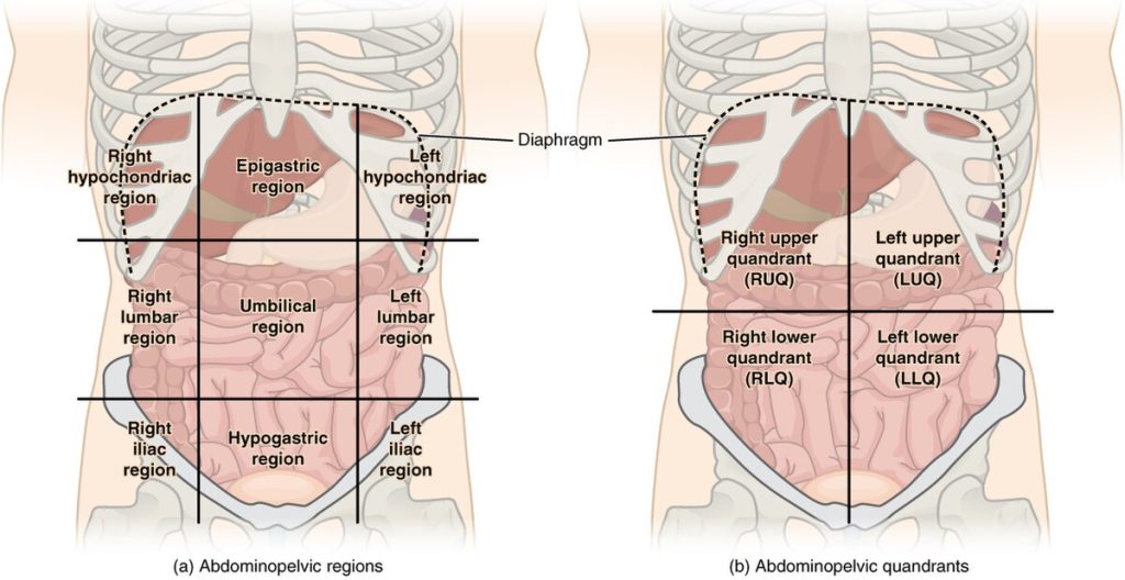
To maintain clear communication among health care professionals, the abdominopelvic cavity is divided into either nine regions or four quadrants. See Figure 1.10[7] for an illustration of the abdominopelvic cavity subdivisions.

The more detailed regional approach divides the cavity into nine regions. One horizontal line lies just below the ribs, one line lies just above the pelvis, and two vertical lines lie as if they were dropped from the middle of each clavicle (collarbone).
A simpler quadrants approach is commonly used in health care and divides the cavity into four regions. One horizontal line and one vertical line intersect at the patient’s umbilicus (also called naval or “belly button”).
Axial and Appendicular Regions of the Body
Regional terms describe anatomy by dividing the body into regions that contain structures that do similar things. There are two main regions of the body:
- Axial Region: The axial region makes up the main axis of the body, including the head, neck, chest, and abdomen.
- Appendicular Region: The appendicular region includes the arms and legs (called appendages) that connect to the axial region.
- Betts, J. G., Young, K. A., Wise, J. A., Johnson, E., Poe, B., Kruse, D. H., Korol, O., Johnson, J. E., Womble, M., & DeSaix, P. (2022). Anatomy and physiology, 2e. OpenStax. https://openstax.org/books/anatomy-and-physiology-2e/pages/1-introduction ↵
- Boundless. (n.d.). Boundless anatomy and physiology. Lumen Learning. https://university.pressbooks.pub/test456/ ↵
- “Dorsal_Ventral_Body_Cavities” by Connexions is licensed under CC BY 3.0 ↵
- “Serous_Membrane” by Connexions is licensed under CC BY 3.0 ↵
- Forciea, B. (2015, May 12). Anatomy and physiology: Body cavities (v2.0) [Video]. YouTube. Used with permission. All rights reserved. https://www.youtube.com/watch?v=tq22iGDHTTI ↵
- RegisteredNurseRN. (2019, July 12). Body cavities and membranes (Dorsal, Ventral) - Anatomy and physiology [Video]. YouTube. Used with permission. All rights reserved. https://www.youtube.com/watch?v=4GLMjSj4k9U ↵
- “Abdominal_Quadrant_Regions” by OpenStax is licensed under CC BY 4.0 ↵
Ventral (anterior) cavity contains the lungs, heart, stomach, and intestines and allows for significant changes in the size and shape of these organs as they do their work.
The chest cavity containing the heart and lungs.
Central portion of the thoracic cavity.
The largest cavity in the body containing the abdominal and pelvic areas
Cavity which contains the abdominal organs.
Cavity which contains the urinary bladder and reproductive organs.
A body cavity located on the back (dorsal) side of the body.
Cavity which contains the brain.
Cavity which contains the spinal cord; also known as the vertebral cavity.
A thin membrane that covers the walls and organs of the thoracic and abdominopelvic cavities; also known as serosa.
Serious membrane layer which lines the walls of the body cavity.
Serous mebrane which covers the organs; it is also called the viscera.
The serous membrane that surrounds the heart, reducing friction caused by the beating of the heart.
The serous membrane surrounding the lungs, reducing friction between the lungs and the chest wall.
The serous membrane that surrounds several organs in the abdominopelvic cavity, reducing friction between the organs and the abdominal wall.
Collarbone.
Naval or “belly button”.
The axial region makes up the main axis of the body, including the head, neck, chest, and abdomen.
Region which includes the arms and legs (called appendages) that connect to the axial region.

