4.2 Categories of Tissues
Categories of Tissues[1]
The term tissue is used to describe a group of cells that work together in the human body. Although there are many types of cells in the body, they are organized into four main categories of tissues:
- Epithelial
- Connective
- Muscle
- Nervous
See Figure 4.1[2] for an illustration of these four categories of tissues.

Each category of tissue has specific functions that contribute to the overall health and maintenance of the body. Histology is the study of tissues, including their appearance, organization, and function. The remaining subsections of this section will discuss these four categories of tissue.
View a supplementary YouTube video[3] overview about of types of tissues:
Epithelial Tissue
Epithelial tissue, also referred to as epithelium, is a large sheet of cells that cover surfaces of the body that are exposed to the surrounding environment (such as the skin), line the insides of organs and blood vessels, internal body cavities and passageways, and form certain glands. For this reason, epithelium can be remembered as a “covering.”
Epithelial tissue is primarily composed of cells, with little to no material between the cells. The apical surface is the top layer of the epithelial tissue that is exposed to the environment. The basal surface is the bottom layer that is attached to a basement membrane, which connects the epithelial tissue to structures deep in the tissue.
Epithelial tissue is mostly avascular, meaning it doesn’t have a blood supply. Therefore, nutrients must diffuse or be absorbed from the underlying tissue or from the surface of the epithelial tissue.
Epithelial tissue goes through rapid mitosis (cell division) to replace damaged and/or dead cells. The sloughing off of dead cells is a characteristic of surface epithelium and allows our skin, airways, and digestive tracts to rapidly replace damaged cells with new cells.
Functions of Epithelial Tissue
There are three main functions of epithelial tissue, including protection, secretion, and movement:
- Protection: Epithelial tissue’s main job is to protect the body from physical, chemical, and biological wear and tear. Epithelial cells act as a physical barrier because all substances that enter the body must cross an epithelium.
- For example, the skin on your hands protects the tissues underneath from damage when you handle or touch something.
- Secretion: Many epithelial cells are capable of secretion and release mucus and chemicals onto their apical surfaces.
- For example, the epithelium of the small intestine releases digestive enzymes to help digest food, and cells lining the respiratory tract secrete mucus that traps dust and bacteria.
- Movement: Some epithelial cells help move particles and secretions in the body. Cilia are tiny hair-like extensions of the apical cell membrane of these types of epithelial tissue. Cilia beat in unison and move fluids, as well as trapped particles.
- For example, respiratory airways have a ciliated epithelium that sweeps particles of dust and pathogens trapped in the mucus up toward the throat, where they can be swallowed and broken down in the acidic environment of the stomach. In the Fallopian tubes, the cilia help move the oocyte (egg cell) from the ovary to the uterus.
Classifications of Epithelial Tissues
Epithelial tissues are classified based on two characteristics: the number of cell layers and the shape of the cells in these layers.
Layers of epithelial tissue can be classified as simple, stratified, or pseudostratified:
- Simple epithelium: Simple epithelium has one layer of cells.
- Stratified epithelium: Stratified epithelium has more than one layer of cells.
- Transitional epithelium: Transitional epithelium is a specialized type of stratified epithelium in which the shape of the cells can change.
- Pseudostratified epithelium: Pseudostratified epithelium (where the term pseudo means “false”) contains a single layer of cells that appears like more than one layer due to the various locations of cell nuclei in different areas of the layer. These “staggered” nuclei make this tissue look as though it has more than one layer of cells.
Simple and stratified epithelium contain the following types of cells:
- Squamous epithelium: Squamous cells are irregular-shaped, flat, thin cells. They look “squashed.”
- Cuboidal epithelium: Cuboidal cells are box- or cube-shaped cells.
- Columnar epithelium: Columnar cells are rectangular-shaped cells, meaning they are taller than they are wide. They look like little columns or rectangles standing on end.
Pseudostratified epithelium only contains columnar cells.
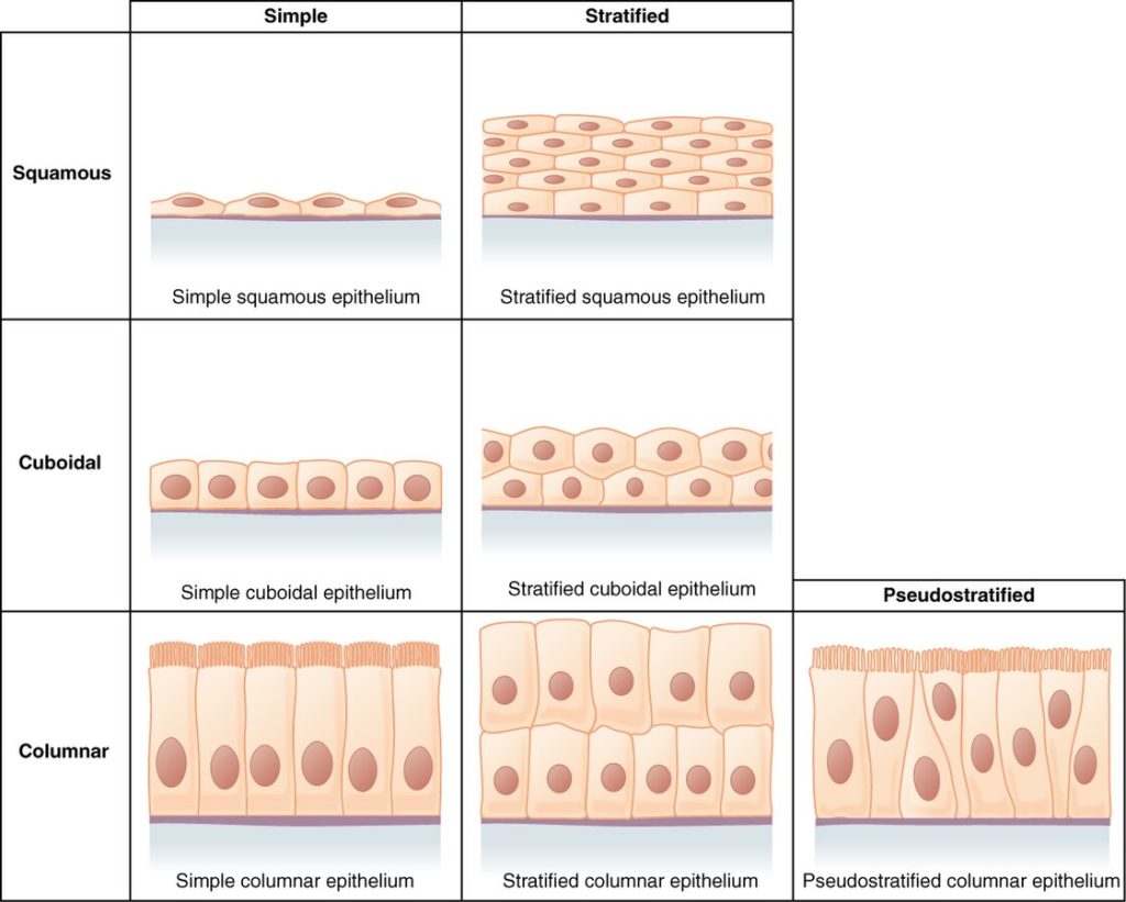
See Figure 4.2[4] for an illustration of different types of cell layers and cell types.
Simple Epithelium
Simple epithelium has one layer of cells with different or unique shapes that reflect tissue function. Simple epithelium can be further divided into squamous, cuboidal, or columnar types of cells:
- Simple squamous epithelium: A single layer of irregular-shaped cells that are thin and flat, like the scales of a fish. The nuclei are similar to the shape of the cell and are flat, horizontal, and oval.
- For example, endothelium is composed of simple squamous epithelium tissue that lines blood vessels. Air sacs in the lungs and blood capillary walls also contain simple squamous epithelium. This very thin layer of squamous cells allows oxygen and carbon dioxide to easily diffuse across during gas exchange.
- Simple cuboidal epithelium: A single layer of cells that are cube or box shaped. Their nuclei are round and located near the center of the cell.
- For example, kidney tubules and sweat glands are made of simple cuboidal epithelium tissue. Their cuboidal shape is important for secretion and absorption.
- Simple columnar epithelium: A single layer of cells that are rectangular shaped. The nuclei are elongated and located near the bottom (basal surface) of the cells.
- For example, some parts of the digestive system are composed of non-ciliated simple columnar epithelium. Like the cuboidal epithelium, these cells are active in absorption and secretion.
- Another example is ciliated columnar epithelium. These simple columnar epithelial cells have cilia on their apical surfaces and are found in parts of the respiratory system and the lining of the Fallopian tubes in the female reproductive system.
- Pseudostratified columnar epithelium: A single layer of columnar cells that appear to be stratified (i.e., have multiple layers) but consist of a single layer of irregularly shaped and different sized cells. The nuclei appear in different areas of the cells, giving the tissue the appearance of multiple layers, but all the cells are in contact with the basal surface. However, some columnar cells do not reach the apical surface.
- For example, pseudostratified columnar epithelium is found in the respiratory tract, where some of these cells also have cilia.
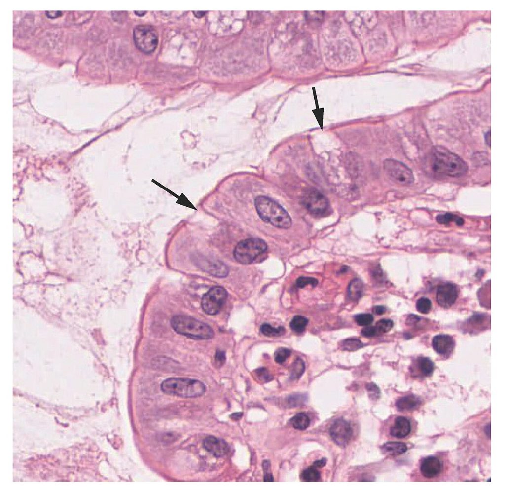
Both simple and pseudostratified columnar epithelia may also contain additional types of cells. For example, the goblet cell, a mucous-secreting cell (also called a “gland”), is found in the columnar epithelial cells of mucous membranes. See Figure 4.3[5] for an illustration of a goblet cell.
Stratified Epithelium
Stratified epithelium consists of two or more layers of cells. This epithelium protects the body against physical and chemical wear and tear. Stratified epithelium is named by the shape of its apical layer of cells (i.e., those closest to the surface) and includes the following types of cells:
-
- Stratified squamous epithelium: The apical cells of stratified squamous epithelium are squamous, whereas the basal layer contains either columnar or cuboidal cells. This is the most common type of stratified epithelium in the human body and is often found in areas that encounter friction. The top layer may or may not be covered with dead cells filled with keratin (a protein that makes skin strong).
- For example, skin is composed of keratinized stratified squamous epithelium, but the lining of the mouth is composed of nonkeratinized stratified squamous epithelium.
- Stratified cuboidal epithelium and stratified columnar epithelium: Stratified cuboidal epithelium consists of two to three layers of cube-shaped cells. Stratified columnar epithelium is composed of two layers of column-shaped cells. These two tissue types are rare in the human body and found only in certain glands and ducts.
- For example, stratified columnar epithelium is found in the urethra.
- Stratified squamous epithelium: The apical cells of stratified squamous epithelium are squamous, whereas the basal layer contains either columnar or cuboidal cells. This is the most common type of stratified epithelium in the human body and is often found in areas that encounter friction. The top layer may or may not be covered with dead cells filled with keratin (a protein that makes skin strong).
-
- Transitional stratified epithelium: Transitional stratified epithelium consists of apical cells that change shape in response to certain conditions.
-
-
- For example, the urinary bladder is lined with transitional stratified epithelium. When the bladder is empty, the cells in the apical layer are cuboidal. As the bladder fills with urine, the apical cells transition from cuboidal to squamous. The transitional stratified epithelium appears thicker and multi-layered when the bladder is empty and more stretched out and less stratified when the bladder is full.
-
Figure 4.4[6] summarizes the locations and functions of simple, stratified, and pseudostratified epithelium.

Glandular Epithelium
A gland is a structure made up of one or more cells that can synthesize (make) and secrete substances. Most glands are made up of groups of epithelial cells. Glands are classified as endocrine or exocrine:
- Endocrine glands: Endocrine glands are ductless glands that release secretions called hormones directly into the blood (with endo meaning “inside”). Hormones are released into the bloodstream and transported to cells with receptors for that particular hormone.
- Examples of endocrine glands include the pituitary gland, thyroid gland, adrenal gland, and the pancreas.
- Examples of hormones include the human growth hormone (hGH), insulin, adrenaline, melatonin, estrogen, and testosterone.
- Exocrine glands: Exocrine glands release secretions through a duct (or tube) that opens to the surface (with exo meaning “outside”).
- Examples of secretions from exocrine glands are mucous, sweat, saliva, digestive enzymes, and breast milk.
View a supplementary YouTube video[7] on epithelial tissue:
Connective Tissue
Connective tissue is the second category of tissue. Connective tissue, as its name implies, connects cells and organs to help protect, support, and bind together all parts of the body. Most of the body is composed of connective tissue. The basic structure of connective tissues is cells embedded in a matrix, similar to how fruit pieces appear in gelatin salad. Matrix is made up of a ground substance (or “filler”) and almost always contains protein fibers. Matrix can be a fluid, gel, or solid.
Classification of Connective Tissue
Connective tissue comes in many forms that are classified based on the characteristics of their cells, matrix, and fibers.
Cells
Different types of cells found in connective tissue include the following:
- Fibroblasts: Fibroblasts secrete substances that combine with fluids to form the matrix. They are the most abundant cell in connective tissue and are present in all types.
- Adipocytes: Adipocytes store fat. The number and type of adipocytes depend on their location and vary among individuals.
- Mesenchymal cells: Mesenchymal cells are adult stem cells. These cells can differentiate into any type of connective tissue cell that is needed for repair and healing of damaged tissue.
- Macrophages: Macrophages are large cells that develop from white blood cells called monocytes. Macrophages are an important part of the immune system that defends the body against pathogens, disease-causing organisms, such as bacteria and fungi.
- Mast cells: When irritated or damaged, mast cells release histamine, which causes vasodilation and increased blood flow at the site of injury or infection, along with itching, swelling, and redness.
See Figure 4.5[8] for an illustration of different types of cells in connective tissue.

Matrix
Matrix is a second component used to classify connective tissue that is secreted by fibroblasts. It is composed of a gel-like material called ground substance that is clear and viscous.
Protein Fibers
Protein fibers are a third component used to classify connective tissue. Three main types of protein fibers include collagen, elastic, and reticular fibers:
- Collagen fibers are made from fibrous protein subunits linked together to form a long and straight fiber. Collagen fibers, while flexible, have great strength, resist stretching, and give ligaments and tendons their characteristic resilience and strength.
- Elastic fibers contain the protein elastin, along with smaller amounts of other proteins and glycoproteins. The main property of elastin is that it returns to its original shape after being stretched or compressed. Elastic fibers are found in elastic tissues in skin and the elastic ligaments of the vertebral column.
- Reticular fibers are formed from the same protein subunits as collagen fibers, but these fibers remain narrow and are arranged in a branching pattern. They are found throughout the body but are most abundant in the reticular tissue of soft organs, such as the liver and spleen.
Functions of Connective Tissues
Connective tissues perform many functions in the body. Major functions include the following:
- Support and connect tissues: For example, tendons attach muscles to bones, and bones support the soft tissues of the body.
- Protect: For example, fibrous capsules and bones protect delicate organs.
- Transport: For example, fluids, nutrients, wastes, and chemical messengers are moved throughout the body by blood and lymph.
- Storage: For example, adipose cells store surplus energy in the form of fat and help insulate the body.
Categories of Connective Tissue
There are three categories of connective tissue based on the types of their cells, the characteristics of their matrix, and the types of protein fibers found within the matrix. These categories include connective tissue proper, supportive, and fluid.
Connective tissue proper includes loose connective tissue and dense connective tissue. Both subtypes of tissue have a variety of cell types and protein fibers suspended in matrix. Dense connective tissue is reinforced by bundles of fibers that provide tensile strength, elasticity, and protection. In loose connective tissue, the fibers are loosely organized, leaving large spaces in between. Supportive connective tissue includes bone and cartilage that provides structure and strength to the body and protects soft tissues. In bone connective tissue, the matrix is rigid and described as calcified because of the deposited calcium salts. Fluid connective tissue includes blood and lymph that contain specialized cells that circulate throughout the body in a watery fluid containing salts, nutrients, and dissolved proteins. See Table 4.1 for a table summarizing the major types of connective tissue. Each of these categories of connective tissue is described in further detail in the following subsections.
Table 4.1. Types of Connective Tissue
| Connective Tissue Proper | Supportive Connective Tissue | Fluid Connective Tissue |
|---|---|---|
Loose Connective Tissue
Dense Connective Tissue
|
Cartilage
Bone
|
Blood
Lymph |
Loose Connective Tissue
Loose connective tissue is found between many organs to absorb shock and connect tissues together. In loose connective tissue, the protein fibers are loosely organized, leaving large spaces between them. There are three types of loose connective tissue called adipose, areolar, and reticular.
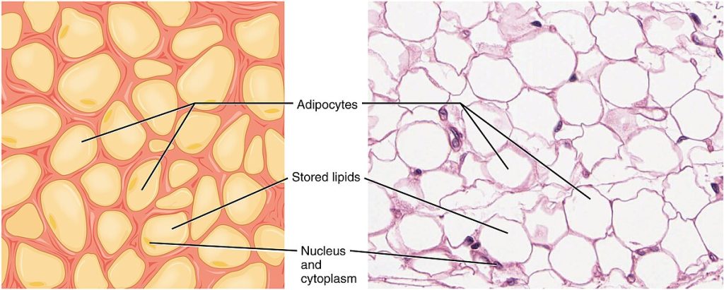
Adipose tissue contains mostly fat storage cells with only a small amount of matrix. In addition to fat storage, adipose tissue also insulates the body and protects organs from injury. See Figure 4.6[9] for an image of adipose tissue.
Areolar tissue fills the spaces between muscle fibers, surrounds blood and lymph vessels, and supports organs in the abdominal cavity. Areolar tissue is found underneath most epithelial tissues and is the connective tissue component of epithelial membranes. See Figure 4.7[10] for an image of areolar tissue.
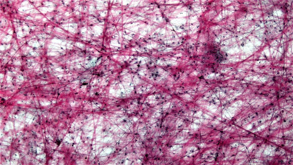
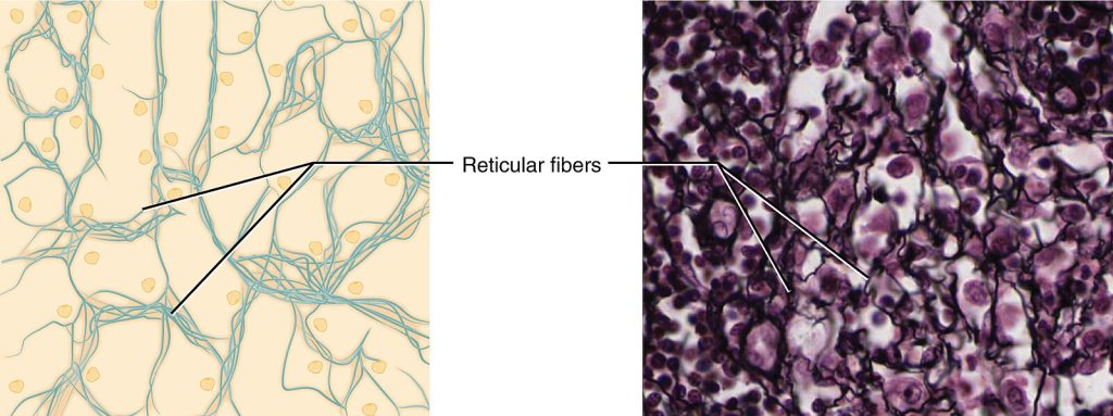
Reticular tissue is a mesh-like, supportive framework for soft organs such as the spleen and the liver. Reticular cells produce the reticular fibers that form a network for other cells to attach. It derives its name from the Latin word reticulus, which means “little net.” See Figure 4.8[11] for an image of reticular tissue.
Dense Connective Tissue
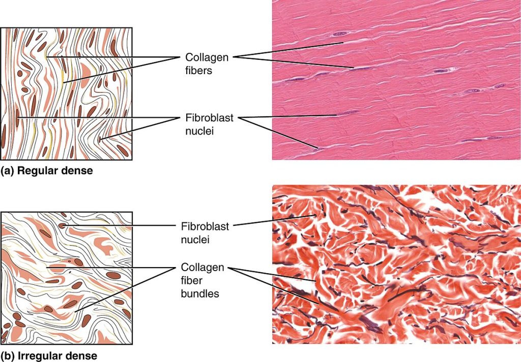
Dense connective tissue is reinforced by bundles of fibers that provide strength, elasticity, and protection. Dense connective tissue contains more collagen fibers than loose connective tissue, so it has a greater resistance to stretching. There are two major categories of dense connective tissue called dense regular connective tissue and dense irregular connective tissue.
- Dense regular connective tissue organizes fibers parallel to each other, which makes it very strong and resistant to stretching. However, it is poorly vascularized, which lengthens healing time for injuries. Ligaments and tendons are made of dense regular connective tissue.
- Dense regular elastic tissue contains elastin fibers, in addition to collagen fibers, which allows the ligament to return to its original length after stretching. The ligaments in the vocal cords and between the vertebrae are elastic.
- Dense irregular connective tissue fibers are randomly organized. This arrangement gives the tissue greater strength in all directions. In some tissues, fibers crisscross and form a mesh. The dermis of the skin is an example of dense irregular connective tissue rich in collagen fibers.
- Dense irregular elastic tissues give arterial walls the strength and the ability to regain their original shape after stretching.
See Figure 4.9[12] for images comparing regular dense connective tissue to irregular dense connective tissue.
Supportive Connective Tissue
Supportive connective tissue provides structure and strength to the body and protects soft tissues. It is characterized by a few distinct cell types and densely packed protein fibers in a matrix. Two major forms of supportive connective tissue are bone and cartilage.
Bone
Bone is the hardest connective tissue. It provides protection to internal organs and supports the body. Bone’s hard extracellular matrix contains mostly collagen fibers in a mineralized ground substance. Without collagen, bones would be brittle and shatter easily. Without minerals, bones would flex and provide little support. Unlike cartilage, bone is highly vascularized and can recover from injuries in a relatively short time. Bone is divided into two main types:
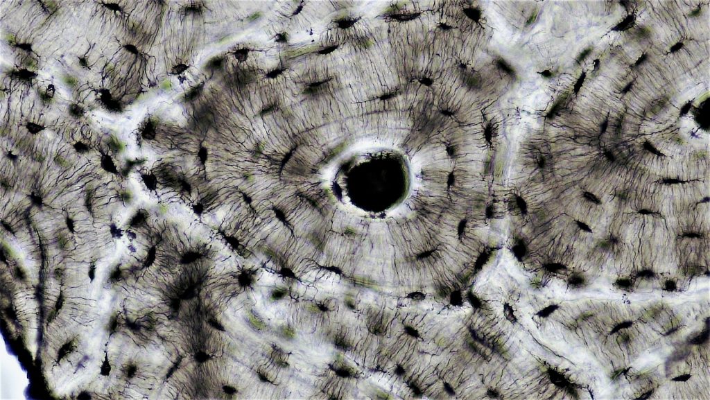
- Compact bone (also known as cortical bone) is solid and has a lot of structural strength. It is arranged into basic units called osteons. To learn more about compact bone osteons, refer to the “Skeletal System” chapter.
- Spongy bone (also known as cancellous bone) looks like a sponge under the microscope. It contains empty spaces and is lighter than compact bone. It is found in the interior of some bones and at the ends of long bones.
See Figure 4.10[13] for an image of compact bone tissue.
Cartilage
Embedded within the cartilage matrix are chondrocytes, or cartilage cells. Cartilage tissue is avascular (without a blood supply), so all nutrients must diffuse through the matrix to reach the chondrocytes. This characteristic results in the very slow healing of injured cartilage tissue.
Three main types of cartilage tissue are hyaline cartilage, fibrocartilage, and elastic cartilage:
- Hyaline cartilage is the most common type of cartilage in the body. The surface of hyaline cartilage is smooth. It is strong and flexible and is found in the rib cage, the nose, and the ends of long bones.
- Fibrocartilage is the strongest of the three cartilage types. It is tough because it has thick bundles of collagen fibers in its matrix. Examples of fibrocartilage are menisci in the knee joint and the intervertebral discs between the vertebrae.
- Elastic cartilage contains elastic fibers, as well as collagen. This tissue gives support, as well as elasticity. For example, the external ear contains elastic cartilage. Tug gently at your ear lobe and notice how it returns to its original shape.
See Figure 4.11[14] for illustration of these types of cartilage.

Fluid Connective Tissue
Fluid connective tissue has various specialized cells that circulate in a watery fluid containing salts, nutrients, and dissolved proteins. The two types of fluid connective tissues are blood and lymph.
Blood
In blood, cells circulate in a liquid matrix. The cells circulating in blood are all derived from stem cells located in bone marrow.
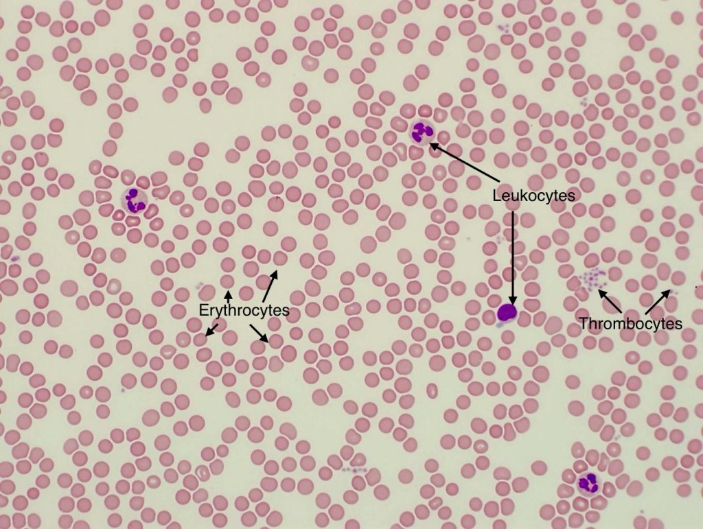
- Erythrocytes: Erythrocytes, also called red blood cells (RBCs), transport oxygen and some carbon dioxide.
- Leukocytes: Leukocytes, also called white blood cells (WBCs), are responsible for defending against potentially harmful microorganisms.
- Thrombocytes: Thrombocytes, commonly called platelets, are involved in blood clotting.
Nutrients, salts, and wastes are also dissolved in this liquid matrix and transported through the body.
See Figure 4.12[15] for an image of blood cells.
Lymph
Lymph is a clear, colorless fluid that plays a vital role in the body’s immune system. It is composed of a liquid matrix that contains leukocytes (white blood cells). Excess fluid is collected from the body tissues by lymphatic capillaries and travels through lymph nodes, where it is filtered and cleaned before returning to the bloodstream. This process helps to protect the body from infections.
View a supplementary YouTube video[16] on connective tissue:
Tissue Membranes
A tissue membrane is a thin layer or sheet of cells that covers the outside of the body (for example, skin), the organs (for example, pericardium), internal passageways that lead to the exterior of the body (for example, mucosa of stomach), and the lining of the moveable joint cavities. There are two basic types of tissue membranes: connective tissue and epithelial membranes. See Figure 4.13 [17] for an image of tissue membranes.

Connective Tissue Membranes
The connective tissue membranes are formed solely from connective tissue. These membranes encapsulate organs, such as the kidneys, and line our movable joints. A synovial membrane is a type of connective tissue membrane that lines the cavity of a freely movable joint. For example, synovial membranes surround the joints of the shoulder, elbow, and knee. Synovial membranes secrete synovial fluid, a natural lubricant that enables the bones of a joint to move freely against one another without much friction.
Epithelial Membranes
The epithelial membrane is composed of epithelium attached to a layer of connective tissue, for example, your skin. The mucous membrane is also a combination of connective and epithelial tissues. Sometimes called mucosae, these epithelial membranes line the body cavities and hollow passageways that open to the external environment and include the digestive, respiratory, urinary, and reproductive tracts. Mucus covers the epithelial layer. The underlying connective tissue, called the lamina propria (literally meaning “own layer”), helps support the fragile epithelial layer.
A serous membrane is an epithelial membrane that lines cavities that do not open to the outside, and they cover the organs located within those cavities. Serous fluid is a watery, slippery fluid that lubricates the membrane and reduces abrasion and friction between organs. Serous membranes are identified according to locations. Three serous membranes line the thoracic cavity: the two pleura that cover the lungs and the pericardium that covers the heart. A fourth, the peritoneum, is the serous membrane that lines the abdominopelvic cavity and covers the organs found there.
The skin is an epithelial membrane also called the cutaneous membrane. It is a stratified squamous epithelial membrane resting on top of connective tissue. The apical surface of this membrane is exposed to the external environment and is covered with dead, keratinized cells that help protect the body.
View a supplementary YouTube video[18] on connective tissue:
Muscle Tissue
Muscle tissue is the third category of tissue.
Functions of Muscle Tissue
Muscle tissue is characterized as an excitable tissue that responds to electrical stimulation and contracts to produce movement.
Types of Muscle Tissue
There are three major types of muscle tissue:
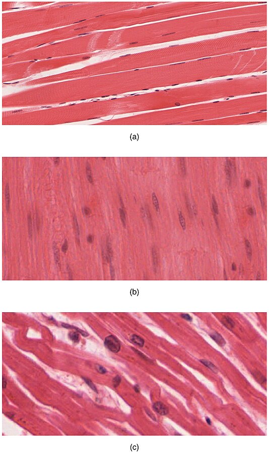
- Skeletal Muscle: Skeletal muscle is voluntary, meaning you can consciously control it. For example, muscles in the arms and legs are skeletal muscles. Skeletal muscle tissue is arranged in bundles surrounded by connective tissue. Under the light microscope, muscle cells appear striated with many nuclei squeezed along the sarcolemmas (cell membranes). The striations are due to the regular arrangement of the contractile proteins actin and myosin, along with structural proteins.
- Smooth Muscle: Smooth muscle is involuntary, meaning you cannot choose to control it. For example, the muscles in the intestines and blood vessels are smooth muscle. Each smooth muscle cell is spindle shaped with a single nucleus and no visible striations
- Cardiac muscle: Cardiac muscle is also involuntary and makes up the contractile walls of the heart. The cells of cardiac muscle are called cardiomyocytes. They are single cells with a single centrally located nucleus that also appear striated under the microscope
See Figure 4.14[19] for an image of muscle tissue.
Read more details about muscle tissue in the “Muscular System” chapter.
Nervous Tissue
Nervous tissue is the fourth category of tissue.
Functions of Nervous Tissue
Nervous tissue is characterized as being excitable and capable of sending and receiving electrical signals throughout the body.
Types of Nervous Tissue
There are two main classes of nervous tissue called neurons and neuroglia:
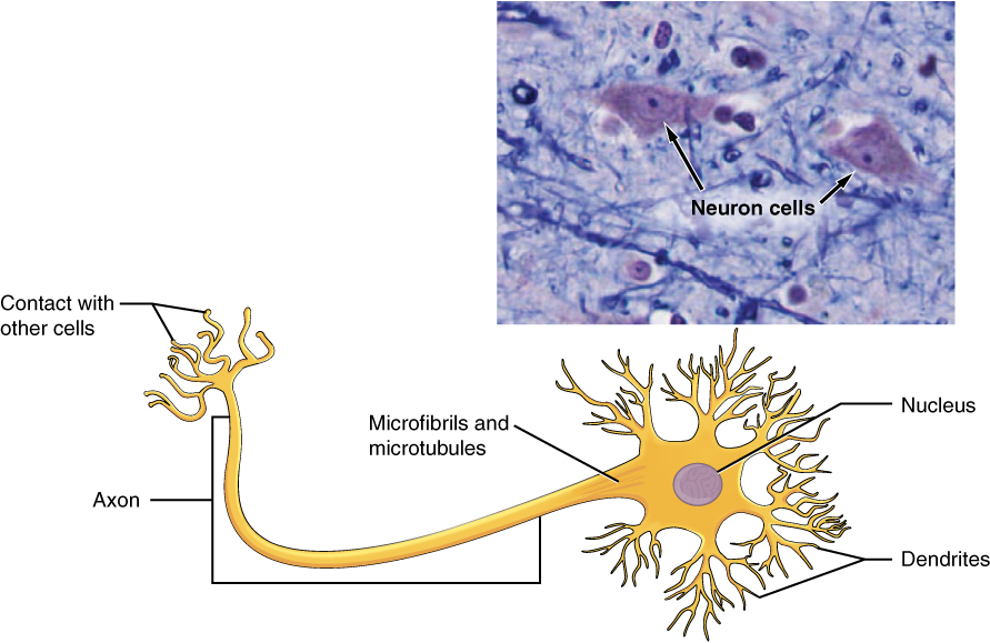
- Neurons: Neurons collect sensory information from the environment, send motor commands to muscles, and transmit electrical signals called nerve impulses throughout the body.
- Neuroglia (also called glial cells) include several different types of cells that each play an essential role in supporting neurons.
See Figure 4.15[20] for an image of neurons.
Read more details about nervous tissue in the “Nervous System” chapter.
- Betts, J. G., Desaix, P., Johnson, E., Johnson, J. E., Korol, O., Kruse, D., Poe, B., Wise, J., Womble, M. D., & Young, K. A. (2022). Anatomy and physiology 2e. OpenStax. https://openstax.org/books/anatomy-and-physiology-2e/pages/1-introduction ↵
- “401_Types_of_Tissue” by OpenStax College is licensed under CC BY 3.0 ↵
- Forciea, B. (2022, December 24). Short topic: Tissues [Video]. YouTube. Used with permission. All rights reserved. https://www.youtube.com/watch?v=LYslovpayXk ↵
- “403_Epithelial_Tissue” by OpenStax College is licensed under CC BY 3.0 ↵
- “404b_Goblet_Cell_new” by OpenStax Anatomy and Physiology is licensed under CC BY 4.0 ↵
- “423_Table_04_02_Summary_of_Epithelial_Tissue_CellsN” by OpenStax College is licensed under CC BY 3.0 ↵
- Forciea, B. (2015, May 12). Anatomy and physiology: Epithelial tissue (v2.0) [Video]. YouTube. Used with permission. All rights reserved. https://www.youtube.com/watch?v=gaYms0ZjPRc ↵
- “408_Connective_Tissue” by OpenStax College is licensed under CC BY 3.0 ↵
- “409_Adipose_Tissue” by OpenStax College is licensed under CC BY 3.0 ↵
- “Connective_Tissue_Loose_Aerolar_(39977986150)” by Fayette A Reynolds M.S, Berkshire Community College Bioscience Image Library is licensed under CC0, Public Domain ↵
- “410_Reticular_Tissue” by OpenStax College is licensed under CC BY 3.0 ↵
- “411_Reg_Dense-Irregular_Dense” by OpenStax College is licensed under CC BY 3.0 ↵
- “Connective_Tissue_Compact_Bone_(41068142774)” by Fayette A Reynolds M.S, Berkshire Community College Bioscience Image Library is licensed under CC0, Public Domain ↵
- “412_Types_of_Cartilage-new” by OpenStax College is licensed under CC BY 3.0 ↵
- “Labelled Blood Slide” by Mary Bauer, Chippewa Valley Technical College is licensed under CC BY 4.0 ↵
- Forciea, B. (2015, May 12). Anatomy and physiology: Connective tissue (v2.0) [Video]. YouTube. Used with permission. All rights reserved. https://www.youtube.com/watch?v=yi9kG_MhJrc ↵
- “413_Types_of_Membranes” by OpenStax College is licensed under CC BY 3.0 ↵
- Forciea, B. (2015, May 12). Anatomy and physiology: Connective tissue (v2.0) [Video]. YouTube. Used with permission. All rights reserved. https://www.youtube.com/watch?v=yi9kG_MhJrc ↵
- "414_Skeletal_Smooth_Cardiac" by OpenStax College is licensed under CC BY 3.0 ↵
- “415_Neuron” by OpenStax College is licensed under CC BY 3.0 ↵
A group of many similar cells that work together to perform a specific function.
The study of tissues.
A tissue type that covers body surfaces and lines cavities.
A layer of epithelial cells that covers internal and external surfaces of the body.
The bottom layer of epithelial cells that attaches to the basement membrane.
A thin, fibrous layer that anchors epithelial tissue to underlying connective tissue.
Lacking blood vessels.
A process of nuclear division that results in two identical daughter cells with the same number of chromosomes as the parent cell.
Short, hair-like projections from the cell surface that are involved in movement of the cell or the movement of substances along the cell surface.
A single layer of epithelial cells.
Multiple layers of epithelial cells.
A type of epithelium that can stretch and change shape.
Epithelial tissue that looks stratified but is actually a single layer.
Flat, scale-like epithelial cells.
A type of epithelial tissue composed of cube-shaped cells.
A type of epithelial tissue composed of column-shaped cells.
A single layer of flat epithelial cells.
Simple squamous epithelium tissue that lines blood vessels.
A single layer of cube-shaped epithelial cells.
A single layer of column-shaped epithelial cells.
A type of epithelial tissue that appears layered but consists of a single layer of cells with varying heights.
A mucus-secreting cell found in epithelial tissue.
A single layer of flat epithelial cells.
A structural protein found in skin, hair, and nails.
Multiple layers of cube-shaped epithelial cells.
Multiple layers of columnar epithelial cells.
Stratified epithelium that allows for stretching and shape changes.
A structure that secretes substances for various functions.
Glands that secrete hormones directly into the bloodstream.
Chemical messengers secreted by endocrine glands.
Glands that release their secretions through ducts to the surface of an organ or tissue.
Tissue that connects, supports, and binds other tissues and organs.
The extracellular material in connective tissues composed of fibers and ground substance.
Cells that produce fibers and matrix in connective tissue.
Fat cells that store energy in the form of lipids.
Stem cells that can differentiate into various connective tissue types.
Immune cells that engulf and digest pathogens and debris.
Disease-causing microorganisms.
Cells that release histamine and other chemicals during inflammation.
Structural fibers found in connective tissues, including collagen, elastic, and reticular fibers.
Strong, fibrous proteins that provide structural support in connective tissues.
Fibers composed of elastin that provide elasticity to tissues.
A protein that gives connective tissues their elasticity.
Thin collagen fibers that form a supportive network in tissues.
A classification of connective tissue that includes loose and dense connective tissues.
Connective tissue that provides structural support, including bone and cartilage.
A type of connective tissue, including blood and lymph, that transports substances.
A connective tissue with loosely arranged fibers that provide support and flexibility.
A type of connective tissue that stores energy in the form of fat.
A loose connective tissue that provides support and flexibility to surrounding structures.
A type of connective tissue with a network of reticular fibers that support cells.
A type of connective tissue with tightly packed, parallel collagen fibers that provide strength in one direction.
A specialized connective tissue that contains elastic fibers, allowing for stretch and recoil.
Connective tissue with randomly arranged collagen fibers that provide strength in multiple directions.
A type of connective tissue with irregularly arranged elastic fibers that allow for flexibility.
Dense, hard bone tissue that provides strength and support, also known as cortical bone.
Structural units of compact bone.
Porous bone tissue that provides structural support and houses bone marrow, also known as cancellous bone.
Cartilage cells.
The most common type of cartilage in the body. It has a smooth surface, providing strength and flexibility. This cartilage is found in the rib cage, nose, and at the ends of long bones, where it helps cushion joints and support structural integrity.
The strongest type of cartilage, known for its toughness due to thick bundles of collagen fibers in its matrix. It provides strength and shock absorption in high-stress areas, such as the menisci of the knee joint and the intervertebral discs between vertebrae.
A flexible type of cartilage that contains both elastic fibers and collagen. It provides support while maintaining elasticity, allowing structures to return to their original shape after bending. An example is found in the external ear.
Red blood cells responsible for oxygen transport.
White blood cells involved in immune response.
Platelets involved in blood clotting.
A fluid that circulates in the lymphatic system and aids immune function.
Thin layer or sheet of cells that covers the outside of the body (for example, skin), the organs (for example, pericardium), internal passageways that lead to the exterior of the body (for example, mucosa of stomach), and the lining of the moveable joint cavities.
A type of body membrane that is made entirely of connective tissue, unlike epithelial membranes (like mucous or serous membranes) which include both epithelial and connective tissue layers.
A type of connective tissue membrane that lines the cavity of a freely movable joint.
Membrane composed of epithelium attached to a layer of connective tissue.
Membrane layer which is composed of a combination of connective and epithelial tissues.
Underlying layer of connective tissue which helps to support the epithelial layer.
A thin membrane that covers the walls and organs of the thoracic and abdominopelvic cavities; also known as serosa.
A stratified squamous epithelial membrane resting on top of connective tissue.
Tissue that contracts to produce movement.
Striated muscle tissue attached to bones that enables movement, facial expressions, posture, and other voluntary actions.
Involuntary, non-striated muscle tissue found in the walls of organs.
Involuntary, striated muscle tissue found only in the heart.
Nerve cells that transmit electrical signals.
Supportive cells in the nervous system.

