3.5 Cell Cycle
Cell Cycle[1]
Beginning at conception, your cells have reproduced themselves many times during growth and development. Even in adulthood, most cells throughout the human body go through regular cell division to replace cells that are old or damaged. (There are a few cells in the body that do not go through routine cell division such as red blood cells, nerve cells, and some muscle cells).
A somatic cell is a general term for all human cells, except for egg and sperm, which are referred to as germ (or sex) cells. Somatic cells contain 46 individual chromosomes that pair together to make 23 pairs of homologous chromosomes (with homo meaning “same”). One member of each chromosome pair comes from each parent, so half of your chromosomes come from the egg and the other half from the sperm. The human is a diploid organism, having 23 homologous pairs of chromosomes in each of the somatic cells, or 46 total chromosomes.
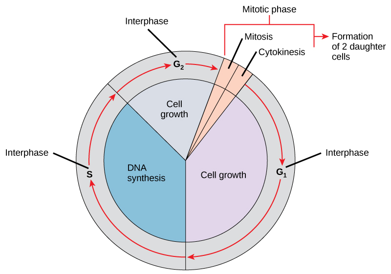
Billions of cells in the human body divide every day. The cell cycle is an ordered series of events involving cell growth and cell division that produces two new daughter cells. Cells on the path to cell division proceed through a series of precisely timed and carefully regulated stages of growth, DNA replication, and division that produce two genetically identical cells. The cell cycle consists of three major parts: interphase, mitosis, and cytokinesis. During interphase, the cell grows and DNA is replicated. During the mitotic phase, the replicated DNA is separated, and in cytokinesis, the cytoplasm is divided up as the cell splits into two new cells.
- Interphase is the time during the cell cycle when the cell is not dividing. Cells spend most of their time in interphase. Cells grow, carry out their normal activities, and prepare for cell division in this phase.
- Mitosis is the time during the cell cycle where the genetic material (DNA) is divided or “divvied up” in an organized series of steps or phases between the two newly forming daughter cells. It occurs relatively quickly, over one to two hours.
- Cytokinesis divides the cytoplasm into two new cells.
Interphase: Synthesis Phase
While in interphase, a cell grows and carries out its normal functions, in addition to getting ready for division. Chromosomes are not visible during interphase because DNA is uncoiled into chromatin, a material that resembles coffee grounds.
Interphase is divided into three parts:
- Growth 1 phase (G1 phase): The cell is growing and performing its functions.
- Synthesis phase (S phase): The cell enters the S phase when it is no longer functioning well and needs to divide. During this period, the cell replicates its DNA.
- Growth 2 phase (G2 phase): A phase where the cell continues to make preparations for mitosis.
During the synthesis phase (S phase) of interphase, the cell replicates its DNA in its nucleus (i.e., makes an identical copy) because one copy will be needed for each new cell that will be created during mitosis and cytokinesis. It also duplicates a microtubule-organizing structure called the centrosome. The centrosomes help separate DNA during the mitosis phase.
See Figure 3.21[2] for an illustration of the cell cycle.

Mitosis and Cytokinesis Phases
The mitosis and cytokinesis phases represent cell division. During the mitosis phase, replicated chromosomes are separated and organized into two new nuclei. After mitosis, the cytoplasm divides into two new cells during cytokinesis.
Mitosis consists of four major phases called prophase, metaphase, anaphase, and telophase that are further described in the following subsections.
Prophase
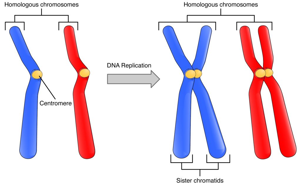
Prophase is the longest phase of mitosis. The nucleus and nucleolus disappear, and the chromatin (the previously replicated DNA) condenses into visible X-shaped chromosomes with sister chromatids. Each copy of the chromosome is referred to as a chromatid that is physically attached to the other copy by a centromere at its center, resulting in its familiar “X” shape.
Recall that a typical human cell contains 46 chromosomes that are organized into 23 pairs of homologous chromosomes. See Figure 3.22[3] for an illustration of a homologous pair of chromosomes before and after DNA replication.

In addition to the appearance of sister chromatids in the nucleus, changes also occur in the cytoplasm of the cell during prophase. The centrosomes begin to move to opposite poles of the cell and form spindle fibers that will elongate and fan out during the mitosis phase. Spindle fibers are used to support the division and movement of the sister chromatids during mitosis.
Metaphase
During metaphase the centrosomes complete their movement to the opposite poles of the cell, and the chromatids line up in the middle of the cell, often called its equator. The lining up of chromatids at the equator results in one sister chromatid being on each side of the equator.
Anaphase
During anaphase the spindle fibers separate the pairs of sister chromatids at their centromere and pull them to opposite ends of the cell into two separate groups. Once separated from each other, each chromatid is now called a chromosome.
Telophase
During telophase a nuclear membrane forms around each set of chromosomes. The chromosomes then spread out into loose chromatin, and the nucleus and nucleolus become visible again. The cell begins to develop a cleavage furrow, a band that squeezes the two cells apart until they separate.
Cytokinesis
Mitosis is followed by cytokinesis, the separation of the cytoplasm into two distinct daughter cells that are identical to each other, as well as to the original cell that underwent mitosis. The cleavage furrow that formed near the end of mitosis will continue pinching inwards until it splits the original cell into two new cells.
The phases of mitosis and cytokinesis are illustrated in Figure 3.23.[4]

View supplementary YouTube videos[5],[6],[7],[8]:
- The cell cycle: The Cell Cycle
- The mitosis phase: M Phase of the Cell Cycle
- Chromatin, Chromosomes, and Chromatids: Chromosomes vs Chromatids vs Chromatin (Different Forms of DNA)
- Mitosis: The Amazing Cell Process that Uses Division to Multiply! (Updated)
Cell Cycle Control
The cell cycle, from interphase through mitosis, is controlled by molecules within the cell, as well as by external triggers providing “stop” and “go” signals for the cell. Precise regulation of the cell cycle is important for the health of the body, and loss of cell cycle control can lead to uncontrolled cell division or cancer.
Mechanisms of Cell Cycle Control
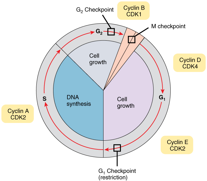
As a cell moves through its cycle, each phase has certain processes that must be completed before the cell advances to the next phase. A checkpoint is a point in the cell cycle where the cell can either be allowed to continue to the next phase or stopped. See Figure 3.24[9] for an illustration of checkpoints during the cell cycle. At the G1 checkpoint, the cell must be ready for DNA synthesis to occur. At the G2 checkpoint, the cell must be fully prepared for mitosis. Even during mitosis, a crucial checkpoint in metaphase ensures that the cell is fully prepared to complete cell division. Cells proceed through the cell cycle under the control of a variety of regulatory proteins called cyclins. Each cyclin determines whether or not the cell is prepared to move to the next stage.

Implications of a Cell Cycle Out of Control
Tumors and cancer are caused by abnormal cells that continuously multiply. If they continue to divide in an uncontrolled manner, these abnormal cells can damage the tissues around them, spread to other parts of the body, and eventually cause death to the individual.
In healthy cells, the tight control of the cell cycle prevents this overgrowth of abnormal cells from occurring. Failure to control the cell cycle can cause unwanted and excessive cell division resulting in cancer. These control failures can be caused by inherited genetic abnormalities that disrupt the function of certain “stop” and “go” signals. Environmental factors, such as UV rays, can also damage DNA and cause malfunctioning of the signals. Often, a combination of both genetic and environmental factors leads to cancer. For example, someone who smokes and is also genetically predisposed to cancer is at higher risk for lung cancer.
Cell cycle control is an example of a homeostatic mechanism that maintains correct cell function and health. While going through the phases of the cell cycle, molecules provide “stop” and “go” signals to regulate movement to the next phase. This homeostatic control of the cell cycle can be thought of like a car’s cruise control. Cruise control will continually apply just the right amount of acceleration to maintain a desired speed, unless the driver hits the brakes, in which case the car will slow down. Similarly, the cell uses specific molecules that push the cell forward in the cell cycle. Normally, a cell that does not meet its checkpoints is programmed to self-destruct in a process called apoptosis. A delicate homeostatic balance controls the cell cycle and ensures that only healthy cells replicate.
If cells escape the normal control system, they can divide and grow excessively. Normally, the immune system will find and destroy these rogue cells. However, if these cells remain undetected, they can become tumors or cancer. If an abnormal growth of cells does not pose a threat to surrounding tissues, it is called benign and can often be surgically removed. However, if abnormal cells divide uncontrollably and invade nearby tissues, they are called malignant. If these abnormal cells spread to other places in the body, the cancer has metastasized.[11],[12]
View a supplementary YouTube video[13] about cancer: 3D Medical Animation – What is Cancer?
- Betts, J. G., Desaix, P., Johnson, E., Johnson, J. E., Korol, O., Kruse, D., Poe, B., Wise, J., Womble, M. D., & Young, K. A. (2022). Anatomy and physiology, 2e. OpenStax. https://openstax.org/books/anatomy-and-physiology-2e/pages/1-introduction ↵
- “Figure_10_02_01” by OpenStax is licensed under CC BY 4.0 ↵
- “591e6fe431b1c89f4d2c37465291b0363b63ebce” by OpenStax is licensed under CC BY 4.0. Access for free at https://openstax.org/books/anatomy-and-physiology-2e/pages/3-5-cell-growth-and-division ↵
- “0331_Stages_of_Mitosis_and_Cytokinesis” by OpenStax is licensed under CC BY 4.0 ↵
- Nucleus Biology. (2021, November 4). The cell cycle [Video]. YouTube. All rights reserved. https://www.youtube.com/watch?v=e6N9_RhD10Q ↵
- Nucleus Biology. (2021, November 4). M phase of the cell cycle [Video]. YouTube. All rights reserved. https://www.youtube.com/watch?v=5bq1To_RKEo ↵
- Beverly Biology. (2024, November 14). Chromosomes vs chromatids vs chromatin (different forms of DNA) [Video]. YouTube. All rights reserved. https://www.youtube.com/watch?v=ud9mavLOgXE ↵
- Amoeba Sisters. (2016, April 14). Mitosis: The amazing cell process that uses division to multiply! (updated) [Video]. YouTube. All rights reserved. https://www.youtube.com/watch?v=f-ldPgEfAHI ↵
- “0332_Cell_Cycle_With_Cyclins_and_Checkpoints” by OpenStax is licensed under CC BY 4.0 ↵
- Beverly Biology. (2014, May 4). Mitosis vs meiosis [Video]. YouTube. All rights reserved. https://www.youtube.com/watch?v=C6hn3sA0ip0 ↵
- National Cancer Institute. (n.d.). NCI Dictionary of cancer terms: Malignancy. https://www.cancer.gov/publications/dictionaries/cancer-terms/def/malignancy ↵
- National Cancer Institute. (n.d.). NCI Dictionary of cancer terms: Metastatic. https://www.cancer.gov/publications/dictionaries/cancer-terms/def/metastatic ↵
- BioDigital, Inc. (2008, October 14). 3D medical animation - What is cancer? [Video]. YouTube. All rights reserved. https://www.youtube.com/watch?v=LEpTTolebqo ↵
Any body cell that is not involved in reproduction, containing a full set of chromosomes (diploid, 2n). Examples include skin cells, muscle cells, and liver cells.
Reproductive cells (e.g., sperm and eggs) that carry half the genetic information (haploid) and combine during fertilization to form a zygote.
Refers to chromosomes that are similar in structure, size, and genetic content, one inherited from each parent.
A cell that contains two full sets of chromosomes (2n), one set from each parent. Somatic cells are diploid.
The series of stages through which a cell goes to prepare for division, involving growth, DNA replication, and cell division. It includes interphase, mitosis, and cytokinesis.
The phase of the cell cycle where the cell grows, replicates DNA, and prepares for mitosis.
A process of nuclear division that results in two identical daughter cells with the same number of chromosomes as the parent cell.
The final step of cell division, where the cytoplasm divides and two distinct daughter cells are formed.
A complex of DNA and proteins (histones) found in the nucleus, which condenses into chromosomes during cell division.
The first phase of interphase, where the cell grows, synthesizes proteins, and prepares for DNA replication.
The phase of interphase where the cell replicates its DNA, ensuring each daughter cell receives an identical set of chromosomes.
The final phase of interphase, where the cell continues to grow and prepares for mitosis by synthesizing proteins necessary for cell division.
The process of duplicating a molecule, cell, or organelle to create an exact replica.
The phase of the cell cycle in which the nucleus divides into two identical nuclei, each containing the same number of chromosomes.
The first stage of mitosis where the chromatin condenses into visible chromosomes, the nuclear membrane breaks down, and spindle fibers begin to form.
One of the two identical halves of a chromosome after DNA replication.
The region where two sister chromatids are joined together.
Protein structures that form during mitosis to separate chromatids by attaching to the centromeres and pulling the chromatids to opposite poles of the cell.
The stage of mitosis where chromosomes align at the equator of the cell, preparing for separation.
The stage of mitosis where sister chromatids are pulled apart to opposite sides of the cell, ensuring each daughter cell gets a full set of chromosomes.
The final stage of mitosis where chromatids reach the poles, the nuclear envelope reforms, and the cell starts preparing to split into two.
A groove that forms in the cell membrane during cytokinesis in animal cells, helping to divide the cell into two daughter cells.
Control points in the cell cycle where the cell ensures that it is ready to proceed with DNA replication or cell division. If errors are detected, the cell cycle may be halted.
Programmed cell death, a controlled process that allows the body to eliminate damaged or unnecessary cells, often during development or in response to DNA damage.
Refers to a tumor or growth that is non-cancerous, non-invasive, and does not spread to other parts of the body. Benign tumors typically have a favorable prognosis.
Refers to a tumor or growth that is cancerous, capable of invading surrounding tissues and spreading to other parts of the body (a process known as metastasis)
Refers to the process by which cancer cells spread from their original (primary) site to other parts of the body, forming secondary tumors.

