8.3 Anatomy of the Nervous System
Anatomy of the Nervous System[1]
Nervous System Divisions
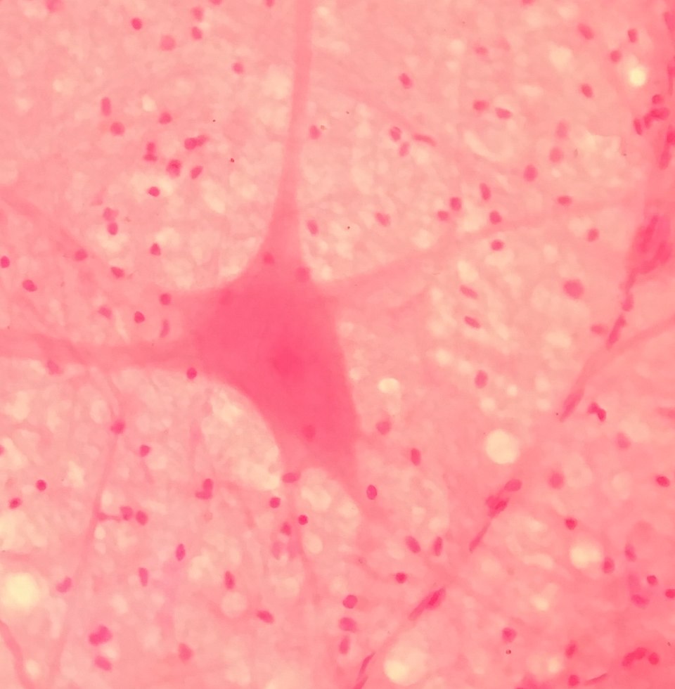
The nervous system can be divided into two major regions: the central nervous system and the peripheral nervous system. The central nervous system (CNS) includes the brain and spinal cord, and the peripheral nervous system (PNS) includes all of the parts of the nervous system found outside of the brain and spinal cord. The peripheral nervous system is so named because it is on the periphery—meaning beyond the brain and spinal cord. See Figure 8.1[2] for an illustration of the parts of a neuron and Figure 8.3[3] for a microscopic image of a neuron.
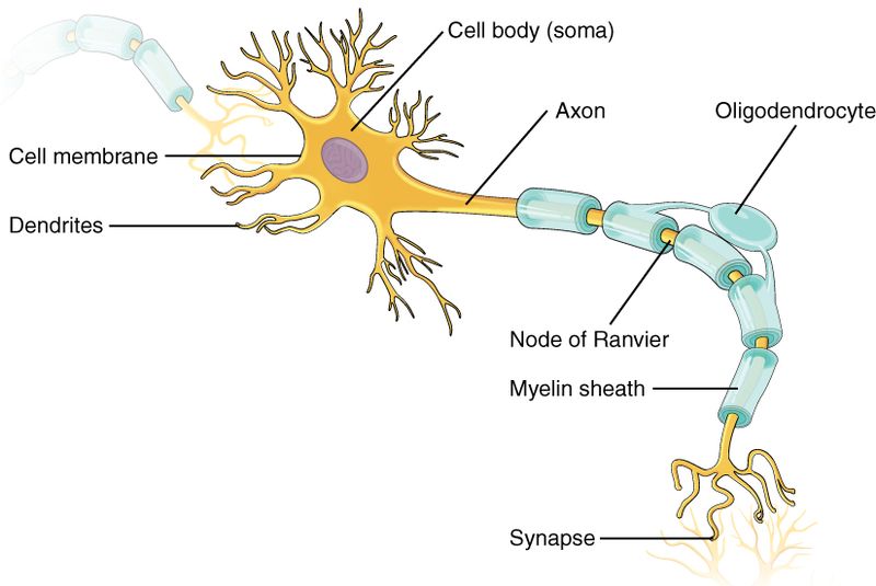
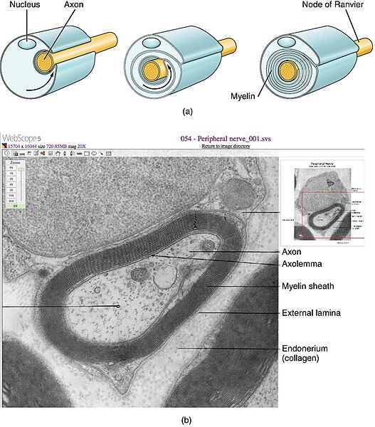
Some axons have a lipid-rich, insulating substance called myelin made from neuroglia. Myelin acts as insulation much like the plastic or rubber that is used to insulate electrical wires. A key difference between myelin and the insulation on a wire is that there are gaps in the myelin covering an axon. Each gap is called a node of Ranvier and is important to the way that electrical signals travel down the axon. At the end of the axon are the axon terminals that are several branches extending toward the target cell. Each axon terminal has an enlarged tip called a synaptic end bulb. These bulbs are what make the connection with the target cell at the synapse.
Gray Matter and White Matter
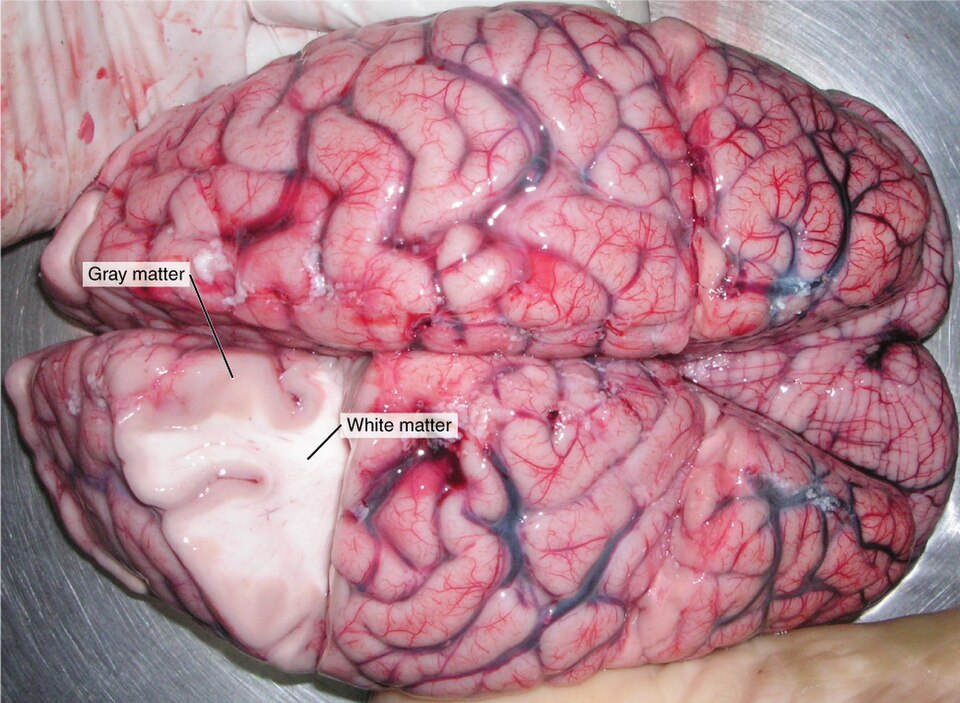
There are regions of nervous tissue that contain mostly cell bodies and regions that are largely composed of just axons. These two regions within nervous system structures are often referred to as gray matter (the regions with many cell bodies and dendrites) or white matter (the regions with many myelinated axons).
The colors used to describe these regions are the colors in unstained nervous tissue. Gray matter is not necessarily gray. It can be pinkish because of blood content, or slightly tan, depending on how long the tissue has been preserved. White matter is white because axons are insulated by myelin and lipids appear as white (“fatty”) material, similar to fat on a raw piece of beef. See Figure 8.4[4] for an image of the appearance of white and gray matter in a brain removed during autopsy.
Neuron Terminology in the CNS and PNS
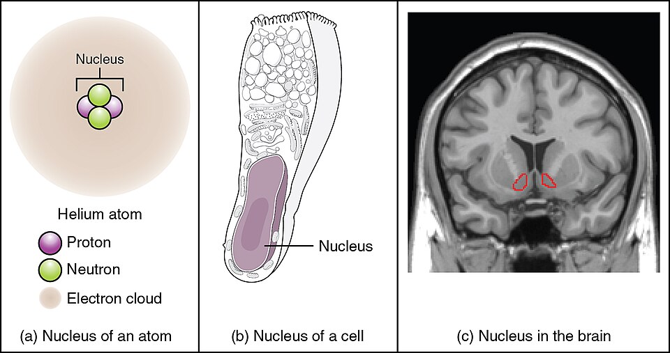
The cell bodies and axons of neurons can be located in the central nervous system (CNS) or the peripheral nervous system (PNS); therefore, separate names are given to identify their location. A collection of neuron cell bodies in the CNS is referred to as a nucleus. In the PNS, a collection of neuron cell bodies is referred to as a ganglion (plural = ganglia).
Note that the word nucleus has several different meanings in anatomy and physiology. It is the center of an atom, where protons and neutrons are found; it is the control center of a cell, where the DNA is found; and it is a collection of nerve cell bodies in the CNS. Figure 8.5[5] illustrates these three different meanings of the word nucleus within anatomy and physiology.
Terminology applied to bundles of axons also differs depending on location. A bundle of axons, or fibers, found in the CNS is called a tract. Ascending tracts carry sensory information up the spinal cord, and descending tracts carry motor information down the spinal cord. A bundle of axons in the PNS is called a nerve. An important point to make about these two terms is that they can both be used to refer to the same bundle of axons. When those axons are in the PNS, the term is nerve, but if they are in the CNS, the term is tract. For example, axons that project from the retina to the brain are called the optic nerve as they leave the eye, but when they enter the brain, they are referred to as the optic tract. See Figure 8.6[6] for an illustration of the optic nerve and optic tract.
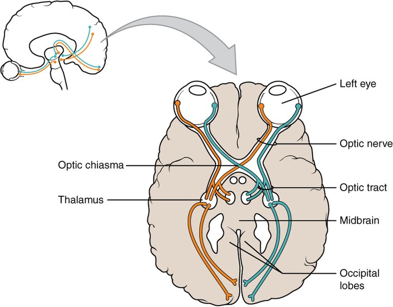
Table 8.3a helps to clarify which of these terms apply to the central or peripheral nervous systems. Read more about nerves in the “Nerves” subsection of the “Peripheral Nervous System” subsection.
Table 8.3a. Structures of the CNS and PNS[7]
Structure |
CNS |
PNS |
|---|---|---|
| Group of Neuron Cell Bodies (i.e., gray matter) | Nucleus | Ganglion |
| Bundle of Axons (i.e., white matter) | Tract | Nerve |
Types of Neurons
There are trillions of neurons in the nervous system that can be classified by many different criteria. The first way is to classify them by function.
Classification by Function
Sensory neurons are also known as afferent neurons (the prefix af- means “toward”). These neurons carry sensory information to the spinal cord or brain. Motor neurons are known as efferent neurons (the prefix ef- means “away from”). These neurons carry information to other motor neurons, muscles, or glands. Both sensory and motor neurons are found in the PNS. Interneurons are also referred to as association neurons. They connect the sensory neurons to the motor neurons and are responsible for interpreting sensory information, storing information, and for “decision-making” or determining what response, if any, is needed. Interneurons are found within the CNS. Most neurons in the nervous system are interneurons.
Classification by Structure
Another way to classify neurons is by the number of processes attached to the cell body. Using the standard model of neurons, one of these processes is the axon, and the rest are dendrites. Because information flows through the neuron from dendrites and cell bodies toward the axon, these names are based on the neuron's polarity.
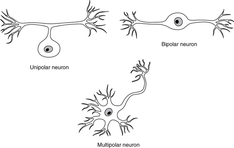
Unipolar cells have only one process emerging from the cell (uni- = “one”). True unipolar cells are only found in invertebrate animals, so the unipolar cells in humans are more appropriately called “pseudo-unipolar” cells. Bipolar cells have two processes (bi- = “two”), which extend from each end of the cell body, opposite to each other. One is the axon, and one is the dendrite. Bipolar cells are not very common. They are found mainly in the olfactory epithelium (where smell stimuli are sensed) and as part of the retina. Multipolar neurons are any neurons that are not unipolar or bipolar. They have one axon and two or more dendrites (usually many more) (multi- = “many"). Most of the neurons in the human body are multipolar. See Figure 8.7[8] for an illustration of unipolar, bipolar, and multipolar neurons.
Other Ways to Classify Neurons
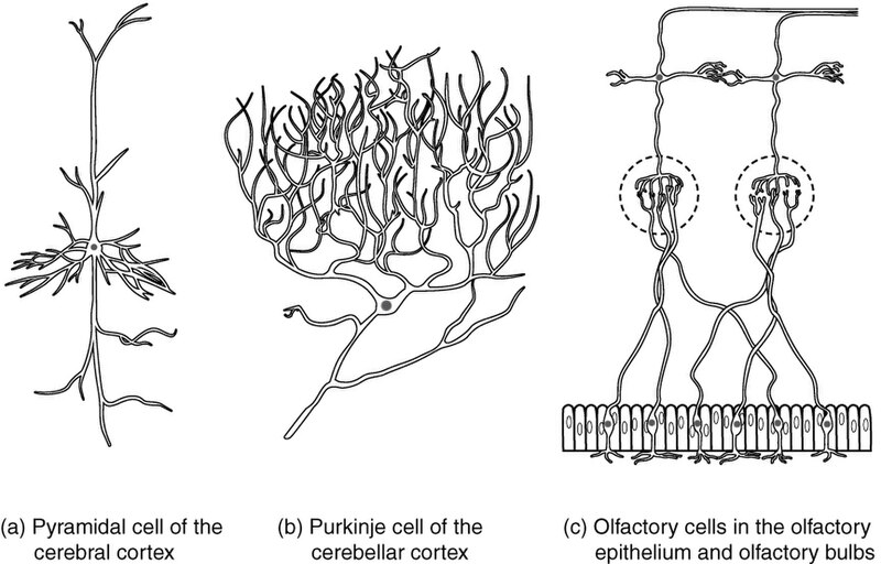
Neurons can also be classified by where they are found, who found them, what they do, or even what chemicals they use to communicate with each other. For example, a multipolar neuron that has a very important role in the cerebellum is known as a Purkinje cell, named after the anatomist who discovered it. See Figure 8.8[9] for an illustration of other ways to classify neurons.
Neuroglia
In addition to neurons, neuroglia, also referred to as glial cells or simply glia, are the other type of nervous tissue. They are support cells that help neurons communicate. There are six types of neuroglial cells, each with specific jobs relative to their location. Unlike neurons, neuroglia are able to go through mitosis. Because of this, most brain tumors (uncontrolled cell growth) are glial cells not neuronal cells.
Neuroglia of the CNS
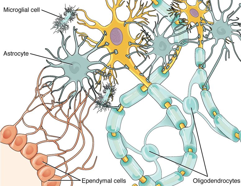
The CNS has four different neuroglia called astrocytes, oligodendrocytes, microglia, and ependymal cells. The astrocyte is named because it appears to be star-shaped under the microscope (astro- = “star”). It provides support to neurons of the CNS. Astrocytes have many processes extending from their main cell body. These processes extend to interact with neurons, blood vessels, or the connective tissue covering the CNS that is called the pia mater. Astrocytes support neurons in the central nervous system by maintaining the concentration of chemicals in the extracellular space, removing excess signaling molecules, reacting to tissue damage, and contributing to the blood-brain barrier (BBB). The blood-brain barrier is a physical barrier that restricts what can cross from the blood into the brain. Nutrients, such as glucose or amino acids, can pass through the BBB, but other molecules cannot. While this barrier protects the CNS from exposure to toxins and pathogens, it also keeps out cells that could protect the brain and spinal cord from disease and damage. The BBB also makes it challenging for medications to be developed that can treat brain diseases.
Also found in the CNS is the oligodendrocyte, which is the neuroglia that insulates axons with myelin. The name means “cell of a few branches” (oligo- = “few”; dendro- = “branches”; -cyte = “cell”). There are only a few processes that extend from the cell body. Each one reaches out and surrounds an axon to insulate it with a myelin sheath. The function of myelin will be discussed below.
Microglia, as the name implies (micro- = “small”), are smaller than most of the other neuroglia. Microglia are a type of macrophage. When macrophages encounter diseased or damaged cells, they ingest and digest those cells or the pathogens that cause disease. Microglia are also referred to as CNS-resident macrophages.
The ependymal cell is a neuroglial cell that filters blood to make cerebrospinal fluid (CSF), the fluid that circulates through and around the brain and spinal cord. Because of the blood-brain-barrier (BBB), the extracellular space in nervous tissue does not easily exchange components with the blood. Ependymal cells line the four spaces in the brain called ventricles. The choroid plexus is a specialized structure in the ventricles where ependymal cells come in contact with blood vessels and filter and absorb components of the blood to produce cerebrospinal fluid. The neuroglia appears similar to epithelial cells with only a single layer. They also have cilia on their apical surface to help move the CSF through the ventricular space. See Figure 8.9[10] for an illustration of astrocyte, oligodendrocytes, microglia, and ependymal cells.
Neuroglia of the PNS
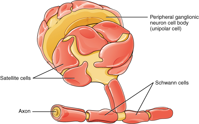
The peripheral nervous system has two types of neuroglia: satellite cells and Schwann cells. Satellite cells provide support to neurons, performing similar functions in the PNS as astrocytes do in the CNS—except for establishing the BBB. The second type of neuroglia in the PNS is the Schwann cell, which insulates axons with myelin. See Figure 8.10[11] for an illustration of satellite cells and Schwann cells.
Myelin Sheath
The insulation for axons in the nervous system is provided by two different neuroglia or glial cells, oligodendrocytes in the CNS and Schwann cells in the PNS. Myelin is a lipid-rich covering that surrounds the axon creating a myelin sheath that helps increase the speed of electrical signals along the axon. The appearance of the myelin sheath can be thought of like pastry wrapped around a hot dog for creating “pigs in a blanket.” See Figure 8.11[12] for an illustration of the myelin sheath.
Central Nervous System
The brain and the spinal cord are the two main components of the central nervous system. The spinal cord is a single structure, whereas the brain has four major regions: cerebrum, diencephalon, brain stem, and cerebellum.
Functions of the Central Nervous System
The central nervous system is responsible for integration. This integration process requires the analysis of sensory information and development of thoughts, emotions, and conscious experiences in the brain. It is the location where memories are stored. The regulation of homeostasis throughout the body is also governed by the CNS. Decisions are made whether and how to respond. Finally, the coordination of reflexes depends on the integration of sensory and motor pathways in the spinal cord.
Anatomy of the Central Nervous System
The adult brain is separated into four major regions: the cerebrum, the diencephalon, the brain stem, and the cerebellum.
Cerebrum
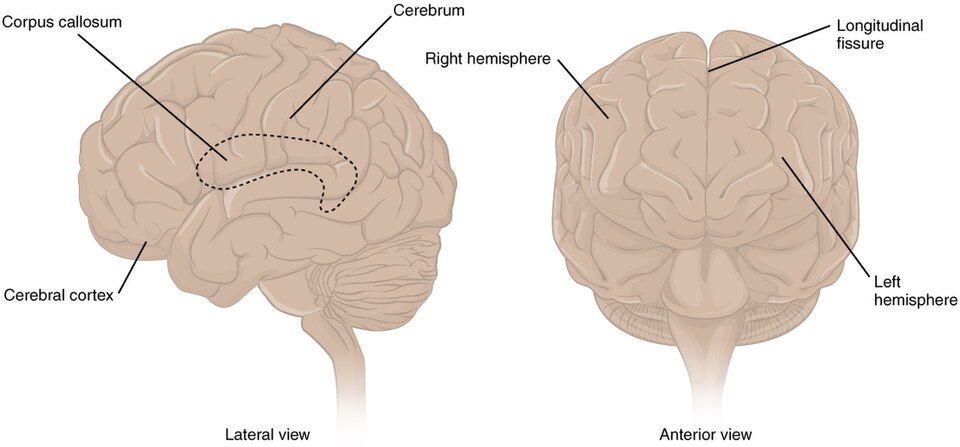
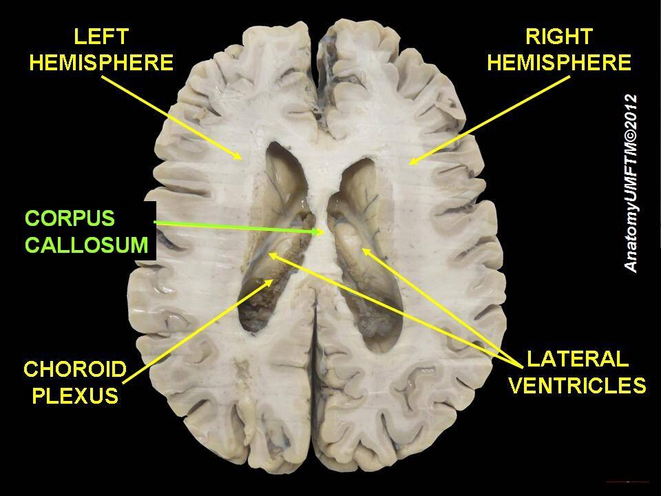
The iconic gray structure of the human brain, which appears to make up most of the mass of the brain, is the cerebrum. It is responsible for higher neurological functions such as memory, emotion, and consciousness. The wrinkled outer portion is the cerebral cortex, and the rest of the cerebrum is deep to the cortex. The large groove between the two sides of the cerebrum is called the longitudinal fissure. It separates the cerebrum into two distinct halves, a right and left cerebral hemisphere. Deep within the cerebrum, the white matter of the corpus callosum provides the major pathway for communication between the two hemispheres of the cerebrum. See Figure 8.12[13] for an illustration of the cerebrum and its associated structures and Figure 8.13[14] for an image of the corpus callosum.
The Myth of Left Brain/Right Brain
There is a persistent myth that people are “right-brained” or “left-brained,” which is an oversimplification of an important concept about the cerebral hemispheres. There is some lateralization of function, in which the left side of the brain is devoted to language and math function and the right side is devoted to spatial and nonverbal reasoning. While these functions are predominantly associated with those sides of the brain, there is no monopoly by either side on these functions. For example, many functions, such as language, are distributed throughout the cerebrum. Damage to the left side of the brain may result in aphasia, a loss of speech function. However, there are also well-documented cases of language functions lost from damage to the right side of the brain. Right-side damage can result in a loss of ability to understand figurative aspects of speech, such as jokes, irony, or metaphors. Nonverbal aspects of speech can be affected by damage to the right side, such as facial expression or body language, and right-side damage can lead to a “flat affect” in speech, or a loss of emotional expression in speech—sounding like a robot when talking.
Cerebral Cortex
As mentioned earlier, the cerebrum is covered by a continuous wrinkled layer of gray matter called the cerebral cortex. Extensive folding in the cerebral cortex enables more gray matter to fit into the limited space of the cranium. If the gray matter of the cortex were peeled off the cerebrum and laid out flat, its surface area would be roughly equal to one square meter. This thin, extensive region is responsible for the higher functions of the nervous system.
Gyri and Sulci
A gyrus (plural = gyri) is the ridge of one of those wrinkles, and a sulcus (plural = sulci) is the groove between two gyri. The pattern of these folds of tissue indicates specific regions of the cerebral cortex.
Lobes of the Cerebral Cortex
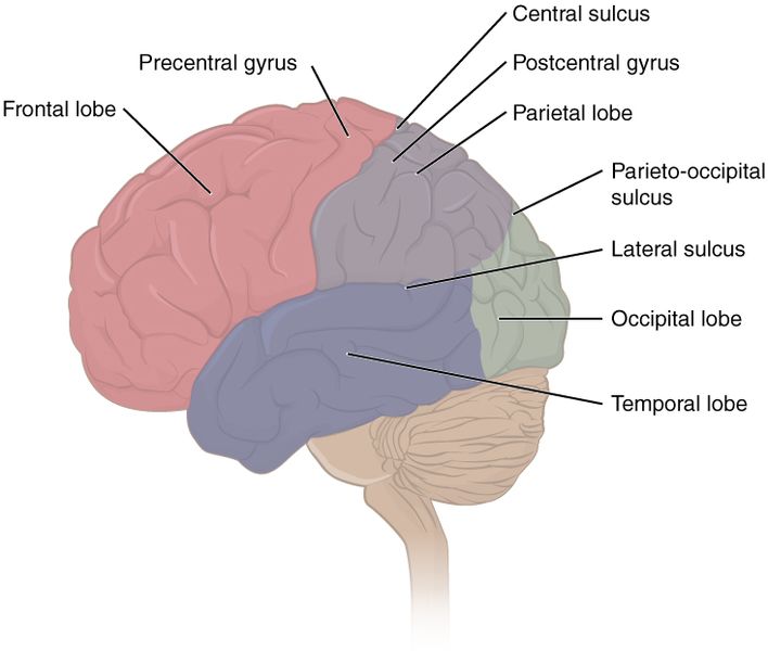
The surface of the brain can be mapped on the basis of the locations of large gyri and sulci. Using these landmarks, the cortex can be separated into four major regions or lobes. Notice that these lobes have the same names as the bones that cover them:
- Temporal lobes: The temporal lobes, one on each side of the brain, are the regions of the cerebral cortex deep to the temporal bones. The lateral sulcus is the landmark that separates the temporal lobe from the other regions.
- Parietal lobes: The parietal lobes are regions of the cerebral cortex deep to the parietal bones. They are superior to the lateral sulcus and directly behind the frontal lobe.
- Frontal lobes: The frontal lobes are regions of the cerebral cortex deep to the frontal bone. The frontal lobe is also superior to the lateral sulcus. It is separated from the parietal lobe by the central sulcus.
- Occipital lobes: The occipital lobes are regions of the cerebral cortex deep to the occipital bone. It is in the posterior region of the cortex and has no obvious anatomical border between it and the parietal or temporal lobes on the lateral surface of the brain. From the medial surface, an obvious landmark separating the parietal and occipital lobes is called the parieto-occipital sulcus. The fact that there is no obvious anatomical border between these lobes is consistent with the functions of these regions being interrelated.
See Figure 8.14[15] for an illustration of the lobes of the cerebral cortex.
Higher Mental Functions of the Cerebral Cortex
The cerebral cortex performs higher mental functions referred to as cognitive abilities. The responsibilities for these cognitive abilities are distributed across the regions of the cortex, and specific locations are responsible for particular functions. For this reason, if a particular area of the brain becomes injured or diseased, specific cognitive abilities are affected. Cognitive abilities can be classified by four areas: orientation and memory, language and speech, sensorium, and judgment and abstract reasoning:
- Orientation and Memory: Orientation is the patient’s awareness of their immediate circumstances. An adult with normal cognitive functioning is aware of their name, where they are and why, and the date. This function occurs in the prefrontal cortex (the anterior portion of the frontal lobe), and it can be temporarily or permanently affected by many medical conditions. Memory refers to how the brain stores and remembers information. It is a function of the temporal lobe.
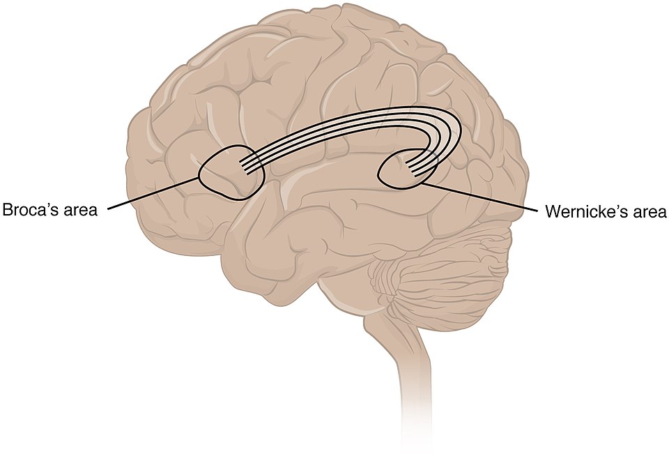
- Language and Speech: Wernicke’s area and Broca’s area are two separate areas involved in speech and language. Both areas are found in a person's dominant hemisphere, which for most people is the left side of the brain. See Figure 8.15[16] for an illustration of the location of Wernicke's area and Broca's area in the brain. Broca’s area is responsible for controlling movements of the structures of speech production. It is named after a French surgeon and anatomist who studied patients who could not produce speech. Patients could understand speech, but had difficulty producing speech sounds, suggesting a damaged or underdeveloped Broca’s area. This is known as an expressive aphasia, which means that speech production is compromised. This type of aphasia is often described as non-fluency because the ability to say some words leads to broken or halting speech. Grammar can also appear to be lost. Wernicke’s area is responsible for speech comprehension. The aphasia associated with Wernicke’s area is known as receptive aphasia, which is not a loss of speech production, but a loss of understanding. Patients, after recovering from acute forms of this aphasia, report not being able to understand what is said to them or what they are saying themselves, but they often cannot keep from talking.
- Sensorium: Sensorium refers to interpretation of sensory stimuli. The cerebral cortex has several regions responsible for sensory perception. From the primary cortical areas of the somatosensory (touch), visual, auditory, and gustatory senses to the association areas that process information, the cerebral cortex is the source of conscious sensory perception. In contrast, sensory information can also be processed by deeper brain regions, which we may vaguely describe as subconscious. For instance, we are not constantly aware of the proprioceptive stimuli (information concerning the position and movement of the body) that the cerebellum uses to maintain balance.
- Judgment and abstract reasoning: Judgment and abstract reasoning refer to making sense of concepts and making appropriate decisions. For example, when your alarm goes off in the morning, you must decide whether to hit the snooze button or jump out of bed. You use judgment and abstract reasoning to decide if the ten extra minutes of sleep is worth rushing through your morning routine or risk being late. The prefrontal cortex is responsible for these functions of planning and making decisions.
Brodmann’s Areas
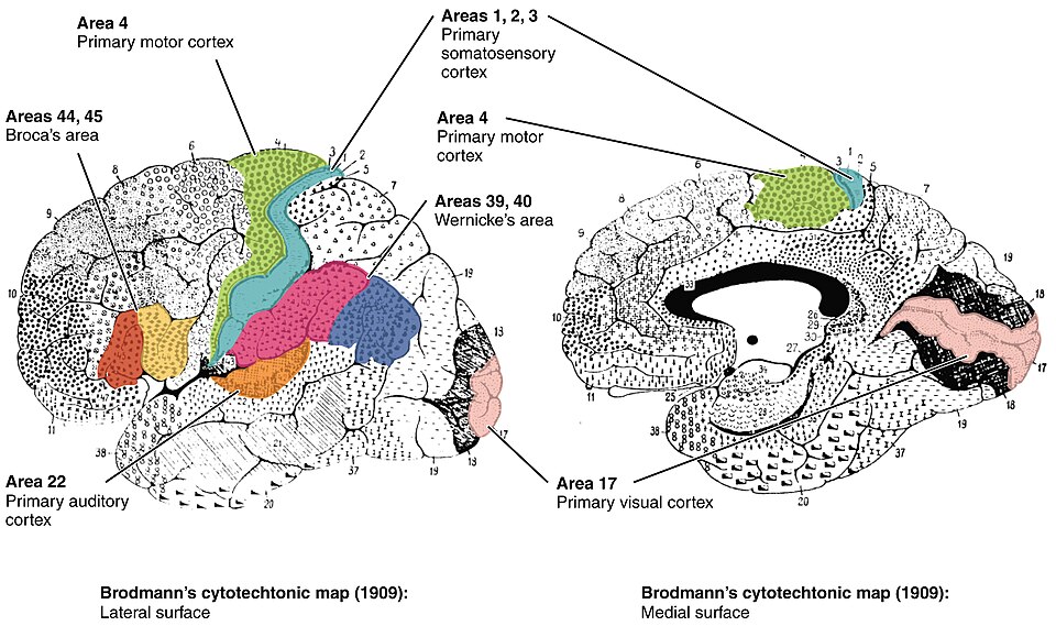
Different regions of the cerebral cortex can be associated with particular functions, a concept known as localization of function. In the early 1900s, a German neuroscientist named Korbinian Brodmann performed an extensive study of the cerebral cortex. His work resulted in a system of classification known as Brodmann’s areas, which is still used today to describe the anatomical distinctions within the cortex. See Figure 8.16[17] for an illustration of Brodmann's areas of the cerebral cortex.
Sensory Areas in the Cortex
The main sensation associated with the parietal lobe is somatosensation or touch, meaning the general sensations associated with the body. Posterior to the central sulcus is the postcentral gyrus, the primary somatosensory area, which is identified as Brodmann’s areas 1, 2, and 3. All the tactile senses are processed in this area, including touch, pressure, tickle, pain, itch, and vibration, as well as more general senses of the body such as proprioception (body position) and kinesthesia (movement).
The primary visual area is found in the occipital lobe, specifically areas 17 and 18. Because visual information is complex, it is processed in the temporal and parietal lobes as well.
The temporal lobe contains the primary auditory (hearing) area and the primary olfactory (smell) area. Because regions of the temporal lobe are also part of the limbic system (the system related to emotional processing), memory is another important function associated with that lobe. Memory is essentially a sensory function; memories are recalled sensations, such as the smell of Grandma’s apple pie or the sound of a barking dog. Even memories of movement are really the memory of sensory feedback from those movements, such as stretching muscles or the movement of the skin around a joint. Structures in the temporal lobe are responsible for establishing long-term memory, but the ultimate location of those memories is usually in the region in which the sensory perception was processed.
The primary gustatory area (sense of taste) is located in the insula. The insula is a fifth lobe of the cerebrum that is located deep to the temporal lobes.
Motor Areas of the Cerebral Cortex
Anterior to the central sulcus is the precentral gyrus of the frontal lobe, which contains the primary motor cortex. The primary motor cortex has voluntary control over skeletal muscles. Cells from this region of the cerebral cortex are the upper motor neurons that tell cells in the spinal cord to move skeletal muscles.
Broca’s area is responsible for the production of language or controlling movements of muscles responsible for speech. In most people, it is located in the left frontal lobe.
Association Areas of the Cerebral Cortex
A number of other regions that extend beyond the primary sensory and motor areas are association areas of the cortex that are also referred to as integrative areas. These areas are found in the spaces between the primary areas for sensory and motor functions, and they integrate multisensory information, or process sensory or motor information in more complex ways. For example, these association areas can integrate visual and motor functions to be able to reach to pick up a glass. The somatosensory function is the proprioceptive feedback from moving the arm and hand. The weight of the glass, based on what it contains, will influence how those movements are made.
Subcortical Structures
The basal nuclei (also called the basal ganglia) are responsible for planning and coordinating movements. They also can prevent or suppress unwanted movements and regulate muscle tone. For example, while a student is sitting in a classroom listening to a lecture, the basal nuclei will keep the urge to jump up and scream from actually happening. Damage to the basal nuclei is seen in the movement disorder called Parkinson’s disease. The basal forebrain contains nuclei that are important in learning and memory. Alzheimer’s disease is associated with a loss of neurons in the basal forebrain. The limbic cortex is the region of the cerebral cortex that is part of the limbic system, a collection of structures involved in emotion, memory, and behavior. Read more about Parkinson’s disease and Alzheimer’s disease in the “Disorders of the Nervous System and Sense Organs” section.
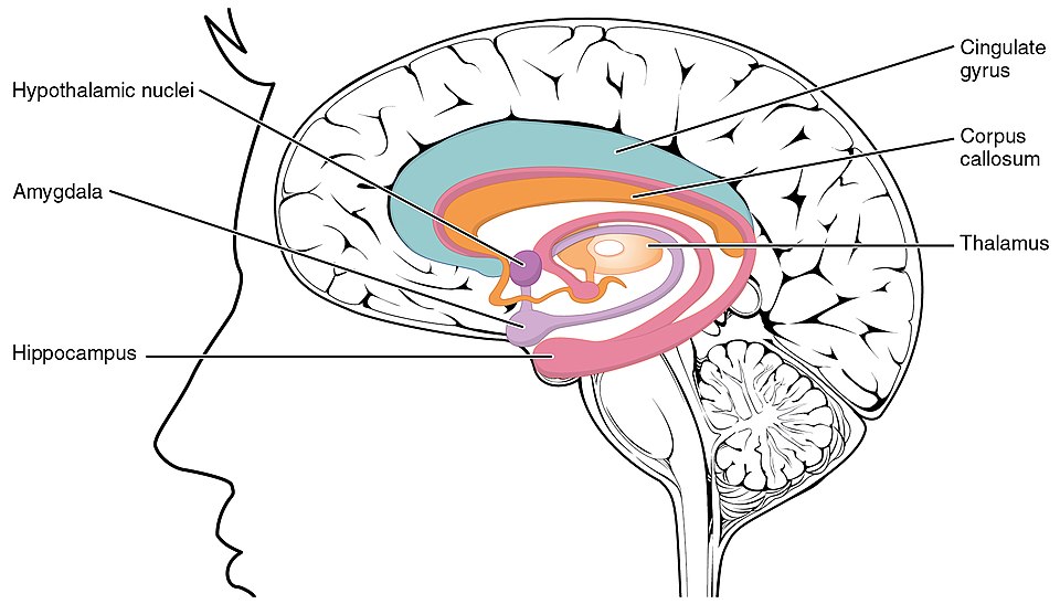
The hippocampus and amygdala are involved in long-term memory formation and emotional responses.
The amygdala is located in the temporal lobe that is part of the limbic lobe. See Figure 8.17[18] for an illustration of the limbic lobe. The limbic lobe includes structures that are involved in emotional responses, as well as structures that contribute to memory. The limbic lobe has strong connections with the hypothalamus and influences the state of its activity on the basis of emotional state. For example, when you are anxious or scared, the amygdala will send signals to the hypothalamus to stimulate the sympathetic fight-or-flight response. The hypothalamus will also stimulate the release of stress hormones through its control of the endocrine system in response to amygdala input.
The Diencephalon
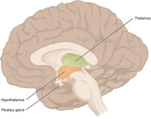
The diencephalon is located inferior to the cerebrum. The diencephalon can be described as any region of the brain with “thalamus” in its name. The two major regions of the diencephalon are the thalamus and the hypothalamus.
Thalamus
The thalamus relays information between the cerebral cortex and the brain stem, spinal cord, and nerves. All sensory information, except for the sense of smell, passes through the thalamus to the cortex. However, the thalamus does not just pass the information on; it also processes that information. For example, the portion of the thalamus that receives visual information will influence what visual stimuli are important, or what receives attention. The cerebrum also sends information down to the thalamus, which usually communicates motor commands.
Hypothalamus
Inferior and slightly anterior to the thalamus is the hypothalamus, the other major region of the diencephalon. The hypothalamus is the major regulator of homeostasis with its control of the autonomic nervous system and the endocrine system. The hypothalamus also controls body temperature, appetite, thirst, and sleep. Other parts of the hypothalamus are involved in memory and emotion as part of the limbic system. See Figure 8.18[19] for an illustration of the diencephalon, thalamus, and hypothalamus.
Brain Stem
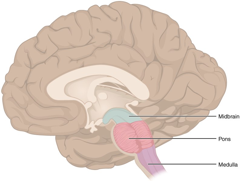
The midbrain and hindbrain (composed of the pons and the medulla oblongata) are collectively referred to as the brain stem. The brain stem connects the brain to the spinal cord.
The cranial nerves connect through the brain stem and provide the brain with the sensory input and motor output associated with the head and neck, including most of the special senses. The major ascending and descending tracts between the spinal cord and brain, specifically the cerebrum, pass through the brain stem.
An area of gray matter spread throughout the brain stem, known as the reticular formation (also called the reticular activating system), is related to sleep and wakefulness and general brain activity and attention. It also has a role in maintaining muscle tone. Damage to this structure in a person may result in a coma.
Midbrain
The midbrain is a small region between the thalamus and pons. The midbrain coordinates sensory representations of the visual, auditory, and somatosensory perceptual spaces.
The tectum is a region of the midbrain composed of four nuclei or “bumps” known as the colliculi (singular = colliculus), which means “little hill” in Latin. These colliculi are subdivided into inferior and superior colliculi.
The inferior colliculus is the inferior pair of these enlargements and is part of the auditory brain stem pathway. Neurons of the inferior colliculus project to the thalamus, which then sends auditory information to the cerebrum for the conscious perception of sound.
The superior colliculus is the superior pair and combines sensory information about visual space, auditory space, and somatosensory space. Activity in the superior colliculus is related to orienting the eyes to a sound or touch stimulus. For example, if you are walking along the sidewalk on campus and you hear chirping, the superior colliculus coordinates that information with your awareness of the visual location of the tree right above you.
Another structure found in the midbrain is the substantia nigra. This structure is responsible for regulating automatic skeletal muscle movements, such as swinging your arms when you walk. Cells in the substantia nigra degenerate in Parkinson’s disease.
Pons
The pons is the main connection between the cerebellum and the brain stem. The word pons comes from the Latin word for “bridge.” It is visible on the anterior surface of the brain stem as the thick bundle of white matter attached to the cerebellum. The pons, along with the medulla oblongata, regulates respiration.
Medulla Oblongata
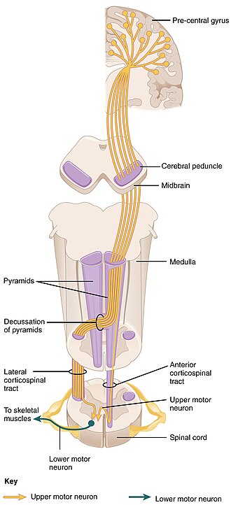
The medulla oblongata, sometimes just referred to as medulla, is the most inferior of the brain stem before it transitions into the spinal cord. It contains both the ascending sensory tracts and descending motor tracts. Upon entering the medulla, the descending motor tracts make up the two large white matter lateral bulges referred to as the pyramids. See Figure 8.19[20] for an illustration of the midbrain, pons, and medulla.
The defining landmark of the medullary-spinal border is the pyramidal decussation, which is where the motor tracts cross over to the opposite side of the brain. This is why the right side of the brain controls the left side of the body, and the left side of the brain controls the right side of the body. See Figure 8.20[21] for an illustration of the pyramids in the medulla oblongata.
The medulla oblongata is the cardiovascular control center, controlling heart rate, force of the heart’s contraction, and blood pressure.
Respiration rate is also controlled by the respiratory center in the medulla oblongata, which responds primarily to changes in hydrogen, carbon dioxide, and pH levels in the body.
Cerebellum
Attached to the brain stem, but considered a separate region of the brain, is the cerebellum. The cerebellum, as the name suggests, is the “little brain” as it accounts for approximately 10 percent of the brain’s mass. It is covered in folds and grooves like the cerebrum and looks like a miniature version of that part of the brain. See Figure 8.21[22] for an illustration of the cerebellum.
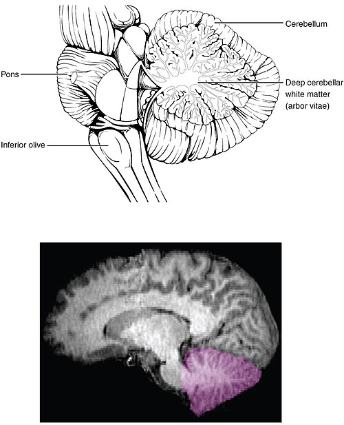
The cerebellum is largely responsible for balance, coordination, and complex movements. When the cerebellum does not work properly, coordination and balance are severely affected. The most dramatic example of this is during the overconsumption of alcohol. Alcohol inhibits the ability of the cerebellum to interpret proprioceptive feedback, making it more difficult to coordinate body movements, such as walking a straight line or guiding the movement of the hand to touch the tip of the nose.
The Spinal Cord
The spinal cord is another major organ of the central nervous system. The anterior midline of the spinal cord is marked by the anterior median fissure, and the posterior midline is marked by the posterior median sulcus.
The posterior or dorsal regions of the spinal cord are responsible for sensory functions, and the anterior or ventral regions are associated with motor functions. Axons enter the posterior side through the dorsal (posterior) nerve root, and the axons emerging from the anterior side do so through the ventral (anterior) nerve root. Note that it is common to see the terms dorsal (dorsal = “back”) and ventral (ventral = “belly”) used interchangeably with posterior and anterior in humans, particularly in reference to nerves and the structures of the spinal cord.
The length of the spinal cord is divided into regions that correspond to the regions of the vertebral column. Immediately below the brain stem is the cervical region, followed by the thoracic, then the lumbar, and finally the sacral region. The spinal cord is not the full length of the vertebral column because the spinal cord stops growing after the first or second year, but the skeleton continues to grow.
The long bundle of nerves that emerges from the end of the spinal cord resemble a horse’s tail and is named the cauda equina.
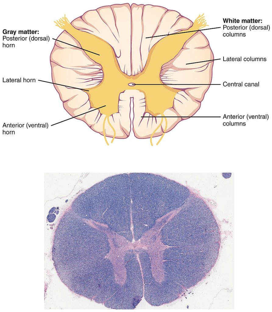
In a cross section, the gray matter of the spinal cord has the appearance of a butterfly and is divided into regions referred to as horns. The posterior horn is responsible for sensory processing, while the anterior horn sends out motor signals to the skeletal muscles. The lateral horn, which is only found in the thoracic, upper lumbar, and sacral regions, contains cell bodies of motor neurons of the autonomic nervous system.
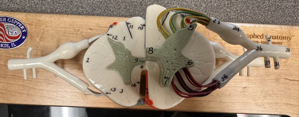
See Figure 8.22[23] for an illustration of the cross section of a spinal cord and Figure 8.23[24] for an image of a model of the spinal cord.
Gray Horns
White Columns (Tracts)
Just as the gray matter is separated into horns, the white matter of the spinal cord is separated into columns or tracts. Looking at the spinal cord longitudinally, the columns extend along its length as continuous bands of white matter. Between the two posterior horns of gray matter are the posterior columns. Between the two anterior horns are the anterior columns. The white matter on either side of the spinal cord, between the posterior horn and the anterior horn, are the lateral columns. Ascending tracts of nervous system fibers in these columns carry sensory information up to the brain, and descending tracts carry motor commands down from the brain.
View a supplementary YouTube video[25] about the gray matter of the spinal cord that receives input from fibers of the dorsal (posterior) root and sends information out through the fibers of the ventral (anterior) root: Spinal Cord Anatomy and Innervation.
As discussed in this video, these connections represent the interactions of the CNS with peripheral structures for both sensory and motor functions.
CNS Blood Supply
The central nervous system is crucial to survival, and any compromise in the brain and/or spinal cord can lead to severe difficulties. To protect this region from the toxins and pathogens that may be traveling through the bloodstream, there is strict control over what can pass into the brain and spinal cord. This begins with a unique arrangement of blood vessels carrying fresh blood into the CNS. The CNS filters that blood into cerebrospinal fluid (CSF), which then circulates both through and around the brain and spinal cord.
A lack of oxygen to the CNS can be devastating, and the cardiovascular system has specific regulatory mechanisms to ensure that the blood supply is not interrupted. There are multiple routes for blood to get into the CNS, with specializations to protect that blood supply and to maximize the ability of the brain to get an uninterrupted blood supply.
Neurons are very sensitive to oxygen deprivation and will start to deteriorate within one or two minutes, and permanent damage (cell death) could result within a few hours. The loss of blood flow to part of the brain is known as a stroke, or a cerebrovascular accident (CVA). Read more about CVAs in the “Disorders of the Nervous System and Sense Organs” section.
Meninges
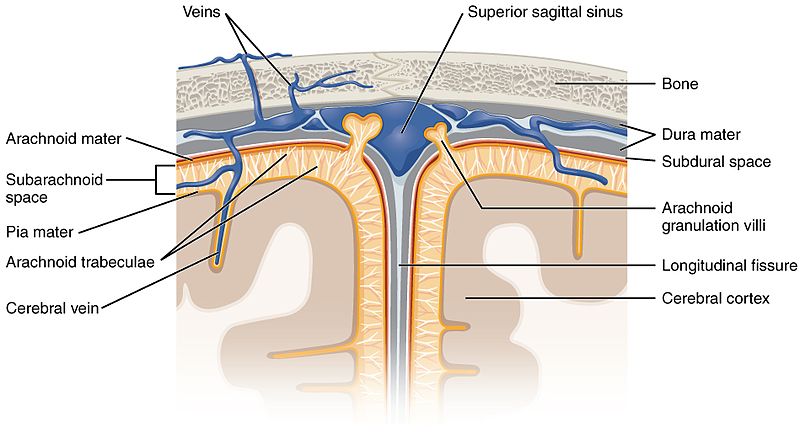
The outer surface of the CNS is covered and protected by three membranes composed of connective tissue called the meninges. The layers of the meninges are the dura mater, arachnoid mater, and pia mater.
The outermost layer, the dura mater, is a thick fibrous layer creating a strong protective sheath over the entire brain and spinal cord. The name comes from Latin for “tough mother” to represent its physically protective role. It encloses the entire CNS and the major blood vessels that enter the cranium and vertebral cavity. It is directly attached to the inner surface of the bones of the cranium and to the very end of the vertebral cavity. The epidural space (epi- = “on or upon” the dura) is the space between the dura and the vertebrae. The subdural space (sub- = “below” the dura) is the space between the dura and the middle layer or arachnoid mater.
The middle layer of the meninges is the arachnoid mater, a membrane of thin fibrous tissue that forms a loose sac around the CNS. Deep to the arachnoid is a thin, filamentous mesh called the arachnoid trabeculae, which looks like a spider web, giving this layer its name. The trabeculae are found in the subarachnoid space, which is filled with circulating CSF. CSF provides a liquid cushion for the brain and spinal cord.
Directly on the surface of the CNS is the pia mater, a thin fibrous membrane that follows the bumps and grooves of the gyri and sulci. The name pia mater comes from the Latin for “tender mother,” suggesting the thin membrane is a gentle covering for the brain. See Figure 8.24[26] for an illustration of the meningeal layers.
Lumbar Puncture
Because the spinal cord does not extend through the lower lumbar region of the vertebral column, a needle can be inserted through the dura and arachnoid layers to withdraw cerebrospinal fluid (CSF). This procedure is called a lumbar puncture and avoids the risk of damaging the spinal cord.
View a supplementary YouTube video[27] about lumbar puncture, a medical procedure used to sample the CSF and diagnose many neurological disorders: Lumbar Puncture: Everything You Need to Know.
Ventricles and Cerebrospinal Fluid (CSF)
Cerebrospinal fluid (CSF) is a colorless fluid that circulates throughout the brain and spinal cord. It is constantly being produced and reabsorbed into the blood. The fluid is essentially water, small molecules, and electrolytes. Oxygen and carbon dioxide are dissolved into the CSF, as they are in blood, and can diffuse between the fluid and the nervous tissue. In addition to supplying essential nutrients, CSF is also a cushion for the brain, protecting it from injury.
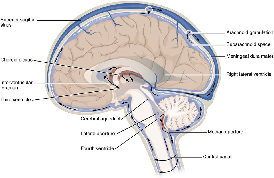
The ventricles are the open spaces within the brain where CSF circulates. In the ventricles, CSF is produced by filtering the blood with a choroid plexus, a specialized structure in the ventricles where ependymal cells come in contact with blood vessels and filter and absorb components of the blood to produce cerebrospinal fluid. The CSF circulates through all the ventricles and into the subarachnoid space where excess CSF is reabsorbed into the blood.
There are four ventricles within the brain. The first two are named the lateral ventricles and are located within each cerebral hemisphere. These ventricles are connected to the third ventricle, which is the space between the left and right sides of the diencephalon. The third ventricle opens into the cerebral aqueduct that passes through the midbrain and opens into the fourth ventricle, which is the space between the cerebellum and the pons and upper medulla oblongata. This fourth ventricle narrows into the central canal of the spinal cord. See 8.25[28] for an illustration of the ventricles.
Peripheral Nervous System
The peripheral nervous system is the division of the nervous system containing everything outside of the brain and spinal cord. The PNS is composed of groups of neuron cell bodies (ganglia) and bundles of axons (nerves), each discussed below.
Ganglia
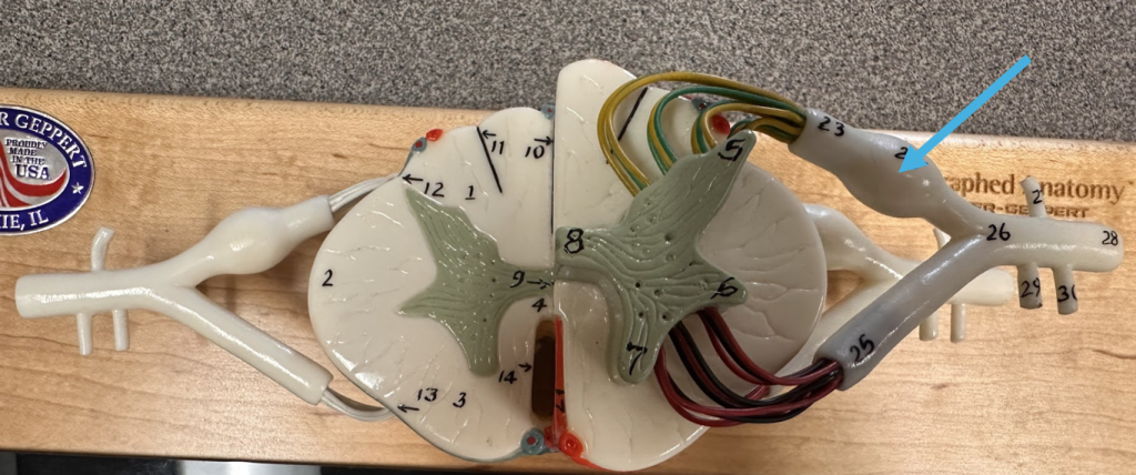
A ganglion (pleural = ganglia) is a group of neuron cell bodies located outside of the CNS or in the periphery. Ganglia can be categorized as either sensory ganglia or autonomic ganglia, referring to their primary functions. The most common type of sensory ganglion is a dorsal (posterior) root ganglion. These ganglia contain the cell bodies of neurons with axons that have sensory endings in the periphery, such as in the skin, and extend into the CNS through the dorsal nerve root. The ganglion is an enlargement of the nerve root. See Figure 8.26[29] for an image of a dorsal root ganglion.
Nerves
Bundles of axons in the PNS are referred to as nerves. Nerves are composed of more than just nervous tissue. They have connective tissue in their structure, as well as blood vessels that supply the tissues with nourishment.
The outer surface of a nerve is surrounded by a layer of fibrous connective tissue called the epineurium. Within the nerve, groups of axons are bundled into fascicles, which are surrounded by their own layer of fibrous connective tissue called the perineurium. Finally, individual axons are surrounded by loose connective tissue called the endoneurium. See Figure 8.27[30] for an illustration of these connective tissue layers. These three layers are similar to the connective tissue coverings for skeletal muscles.

Nerves are associated with the region of the CNS to which they are connected, either as cranial nerves connected to the brain or spinal nerves connected to the spinal cord.
Cranial Nerves[31]
The twelve pairs of nerves attached to the brain are the cranial nerves, which are primarily responsible for the sensory and motor functions of the head and neck, although one of these nerves targets organs in the thoracic and abdominal cavities as part of the parasympathetic nervous system. There are twelve cranial nerves, which are designated CN I through CN XII with CN abbreviated for “Cranial Nerve” and Roman numerals for nerves 1 through 12. Cranial nerves can be classified as sensory nerves, motor nerves, or a combination of both. Three of the nerves are solely composed of sensory fibers; five are strictly motor; and the remaining four are mixed nerves.
An overview of the cranial nerves; their functions; and whether they are a sensory, motor, or mixed nerve is provided in Table 8.3b.
Table 8.3b. Cranial Nerves
| Cranial Nerve | Function | Type of Nerve |
|---|---|---|
| CN I: Olfactory Nerve | Smell (olfaction) | Sensory nerve |
| CN II: Optic Nerve | Vision | Sensory nerve |
| CN III: Oculomotor Nerve | Eye movements, including lifting the upper eyelid when the eyes point up, and pupillary constriction | Motor nerve |
| CN IV: Trochlear Nerve | Up and down eye movements | Motor nerve |
| CN V: Trigeminal Nerve | Sensations of the face and control of chewing muscles | Both sensory and motor nerve |
| CN VI: Abducens Nerve | Abduction (movement to the outside) of eyes | Motor nerve |
| CN VII: Facial Nerve | Facial expressions, as well as part of the sense of taste and the production of saliva | Both sensory and motor nerve |
| CN VIII: Vestibulocochlear Nerve | Hearing and balance | Sensory nerve |
| CN IX: Glossopharyngeal Nerve | Contraction of muscles in the tongue and upper throat, as well as part of the sense of taste and the production of saliva | Both sensory and motor nerve |
| CN X: Vagus Nerve | Parasympathetic control of the organs of the thoracic and upper abdominal cavities | Both sensory and motor nerve |
| CN XI: Spinal Accessory (or Accessory) Nerve | Contraction of neck muscles | Motor nerve |
| CN XII: Hypoglossal Nerve | Responsible for controlling the muscles of the lower throat and tongue | Motor nerve |
See Figure 8.28[32] for an illustration of the cranial nerves.
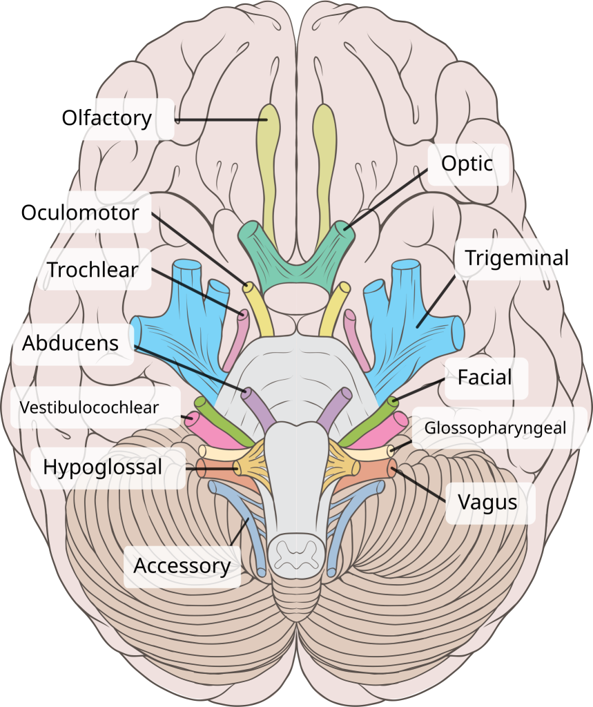
The first, second, and eighth nerves are purely sensory: the olfactory (CN I), optic (CN II), and vestibulocochlear (CN VIII) nerves. The three eye-movement nerves are only motor: the oculomotor (CN III), trochlear (CN IV), and abducens (CN VI). The spinal accessory (CN XI) and hypoglossal (CN XII) nerves are also strictly motor. The remainder of the nerves contain both sensory and motor fibers and are mixed nerves. They are the trigeminal (CN V), facial (CN VII), glossopharyngeal (CN IX), and vagus (CN X) nerves. The nerves that are mixed are often related to each other. For example, the trigeminal and facial nerves both concern the face; one concerns the sensations and the other concerns the muscle movements. The facial and glossopharyngeal nerves are both responsible for conveying gustatory or taste sensations, as well as controlling salivary glands. The vagus nerve is involved in visceral responses to taste, namely the gag reflex.
Spinal Nerves
The nerves connected to the spinal cord are the spinal nerves. The arrangement of these nerves is much more regular than that of the cranial nerves. All the spinal nerves are mixed nerves with combined sensory and motor axons that separate into two nerve roots. The sensory axons enter the spinal cord as the dorsal nerve root. The motor axons emerge as the ventral nerve root.
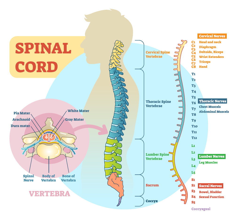
There are 31 pairs of spinal nerves, named for the level of the spinal cord at which each one emerges. There are eight pairs of cervical nerves designated C1 to C8, twelve thoracic nerves designated T1 to T12, five pairs of lumbar nerves designated L1 to L5, five pairs of sacral nerves designated S1 to S5, and one pair of coccygeal nerves. See Figure 8.29[33] for an illustration of the spinal nerves.
Nerve Plexuses
Spinal nerves extend outward from the vertebral column into the periphery. Axons from different spinal nerves come together and form networks of nerve fibers. This occurs at four places along the length of the vertebral column, each identified as a nerve plexus. Of the four nerve plexuses, two are found at the cervical level, one at the lumbar level, and one at the sacral level. See Figure 8.30[34] for an illustration of the nerve plexus locations.
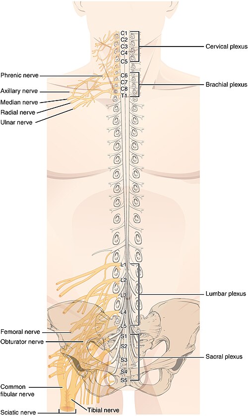
The cervical plexus is composed of axons from spinal nerves C1 through C5 and branches into nerves in the posterior neck and head, as well as the phrenic nerve, which connects to the diaphragm at the base of the thoracic cavity. The other plexus from the cervical level is the brachial plexus. Spinal nerves C4 through T1 reorganize through this plexus to give rise to the nerves of the arms, as the name brachial suggests.
The lumbar plexus arises from all the lumbar spinal nerves and gives rise to nerves innervating the pelvic region and the anterior leg. The femoral nerve is one of the major nerves from this plexus. The sacral plexus comes from the lower lumbar nerves L4 and L5 and the sacral nerves S1 to S4 to supply nerves to the posterior leg. The most significant nerve to come from this plexus is the sciatic nerve. The sciatic nerve extends across the hip joint and is most commonly associated with the condition sciatica, which is the result of compression or irritation of the nerve or any of the spinal nerves giving rise to it.
Divisions of the PNS
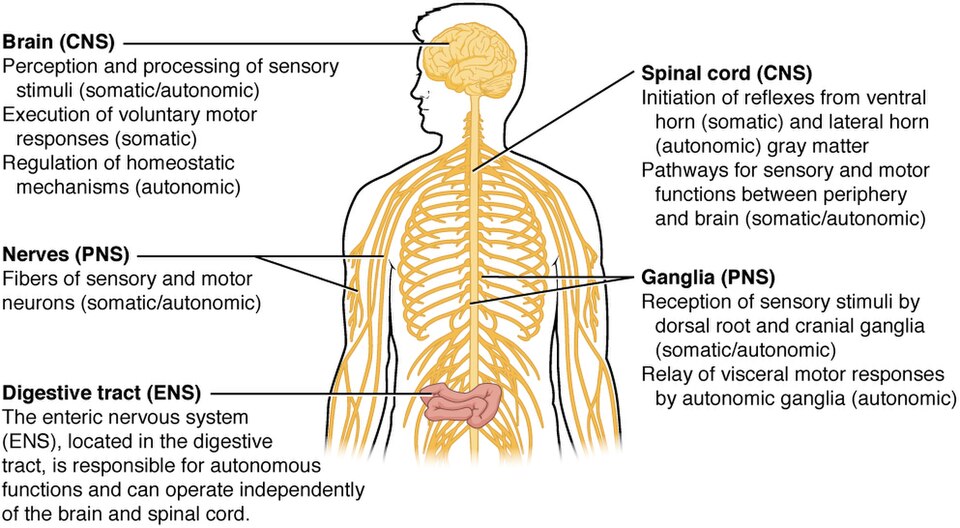
The PNS (peripheral nervous system) can be divided into three main divisions - the somatic nervous system (SNS) responsible for voluntary skeletal movements and senses, the autonomic nervous system (ANS) responsible for sympathetic (flight, fight, or fright) and parasympathetic (rest and digest) functions, and the enteric nervous system (ENS) responsible for digestive functions. See Figure 8.31[35] for examples of where these divisions of the nervous system are found.
Somatic Nervous System
The somatic nervous system (SNS) is responsible for our conscious perception of the environment (senses) and voluntary motor responses to that perception by the contraction of skeletal muscles, such as moving your leg or raising your hand. It also includes reflexes and automatic muscle control.
Senses
Sensory receptors help us to take in and understand both the external environment and the internal environment inside the body. Different types and functions of the senses are discussed in more detail in the “Senses” section.
Skeletal Muscle Movement
Somatic function includes conscious or voluntary (skeletal) muscle movement. For example, reading of this text starts with visual sensory input to the retina, which then projects to the thalamus and on to the cerebral cortex. A sequence of regions of the cerebral cortex processes the visual information, starting in the primary visual cortex of the occipital lobe, and resulting in the conscious perception of these letters. Subsequent cognitive processing results in understanding of the content. As you continue reading, regions of the cerebral cortex in the frontal lobe plan how to move the eyes to follow the lines of text. The output from the cortex causes activity in motor neurons in the brain stem that cause movement of the extraocular muscles through the third, fourth, and sixth cranial nerves. This example also includes sensory input (the retinal projection to the thalamus), central processing (the thalamus and subsequent cortical activity), and motor output (activation of neurons in the brain stem that lead to coordinated contraction of extraocular muscles).
Reflexes
Somatic motor responses also include reflexes. A reflex is a quick predictable automatic response to a stimulus and occurs without a conscious decision by the brain to perform it. For example, if something startles you, you might scream or jump backwards. Even though you may not consciously decide to perform those actions, they are the result of reflexes that involve skeletal muscle contractions.
Another example of a reflex is the withdrawal reflex. When you touch a hot stove, you pull your hand away. Sensory receptors in the skin sense extreme temperature. This triggers a nerve impulse, which travels along the sensory nerve from the skin through the dorsal spinal root to the processing center of the spinal cord, which then activates a motor neuron in the ventral horn of the spinal cord. That motor neuron sends a signal along its axon to stimulate the biceps brachii, causing contraction of the muscle and flexion of the forearm at the elbow to withdraw the hand from the hot stove.
The basic withdrawal reflex explained above includes sensory input (the painful stimulus of the hot stove), central processing (the interneuron in the spinal cord deciding what to do), and motor output (activation of a ventral motor neuron that causes the contraction of the biceps brachii to remove the hand from the stove).
Automatic (Unconscious) Movement
Other motor responses can become automatic or unconscious as a person learns motor skills (referred to as “habit learning” or “procedural memory”), for example, driving a car or riding a bike. Some other aspects of the somatic system also use voluntary muscles without conscious control. One example is the ability of our breathing to switch to involuntary control while we are focused on another task. The muscles that are responsible for the basic process of breathing are also utilized for speech, which is entirely voluntary.
Autonomic Nervous System
The autonomic nervous system (ANS) is responsible for involuntary control of the body to maintain homeostasis. Sensory input for autonomic functions can be from sensory structures tuned to external or internal environmental stimuli. The motor output extends to smooth and cardiac muscle, as well as glandular tissue. The role of the autonomic nervous system is to regulate the organ systems of the body. Sweat glands, for example, are controlled by the ANS. When you are hot, sweating helps cool your body down as a homeostatic mechanism. However, you may also start sweating when you are nervous, which is not a homeostatic mechanism but a physiological response to an emotional state.
The autonomic nervous system is further divided into the sympathetic nervous system and the parasympathetic nervous system. The autonomic nervous system is discussed in more detail in the “Autonomic Nervous System” section.
Enteric Nervous System
Another division of the nervous system, called the enteric nervous system (ENS), controls the smooth muscle and glandular tissue in the digestive system. It is not dependent on the CNS. The enteric system is also considered part of the autonomic system because the neural structures that make up the enteric system are a component of the autonomic output that regulates digestion.
- Betts, J. G., Young, K. A., Wise, J. A., Johnson, E., Poe, B., Kruse, D. H., Korol, O., Johnson, J. E., Womble, M., & DeSaix, P. (2022). Anatomy and physiology 2e. OpenStax. https://openstax.org/books/anatomy-and-physiology-2e/pages/1-introduction ↵
- "Nervous_system_diagram_(dumb)" by ¤~Persian Poet Gal is in the Public Domain[/footnote] for an illustration of these two systems, with the central nervous system colored in yellow and the peripheral nervous system colored in blue.
The peripheral system is further divided into the somatic, autonomic, and enteric nervous systems. Refer to the subsections on the “Central Nervous System” and the “Peripheral Nervous System” to learn more about each of these important divisions.
[caption id="attachment_4158" align="aligncenter" width="500"]
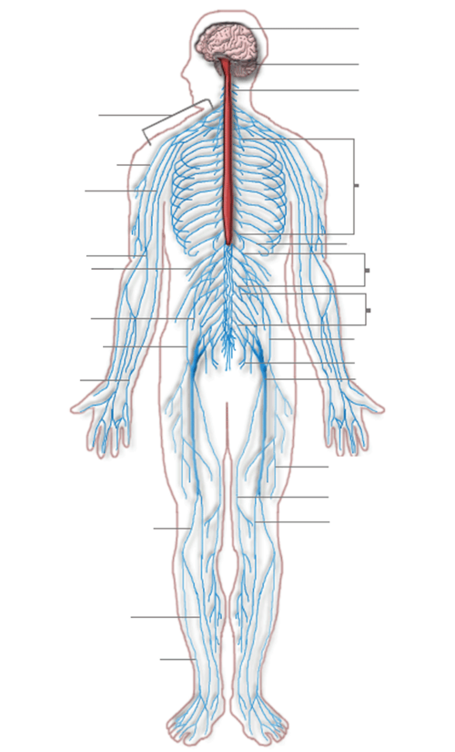 Figure 8.1. Central and Peripheral Nervous Systems[/caption]
Nervous Tissue
Nervous tissue in the CNS and PNS contains two basic types of cells: neurons and neuroglia (also known as glial cells). [pb_glossary id="634"]Neurons[/pb_glossary] are the communicating cells of the nervous system. They are excitable and can generate and conduct electrical signals. [pb_glossary id="635"]Neuroglia[/pb_glossary] include a variety of cells that support the neurons in their communication activities.Neurons
Neurons are the functional cells of the nervous system. They carry electrical signals that communicate sensory information, facilitate thought processes within the brain, and produce movements in response to sensory stimuli. The same neurons last throughout a lifetime, as they don't readily undergo mitosis. If they are lost due to damage or disease, they are not replaced, which may lead to permanent deficits or disability.Anatomy of a Neuron
The main part of a neuron is the cell body, also known as the soma (soma = “body”). The [pb_glossary id="1873"]cell body[/pb_glossary] contains the nucleus and most of the major organelles. Neurons are specialized cells because they have many extensions of their cell membranes that are generally referred to as processes. Neurons usually have one [pb_glossary id="1874"]axon[/pb_glossary] - a fiber that emerges from the cell body and sends an electrical nerve impulse to target cells. That single axon can have many branches, allowing it to communicate with many target cells. The other processes on the neuron are [pb_glossary id="1876"]dendrites[/pb_glossary], which receive information from other neurons at synapses. A [pb_glossary id="1875"]synapse[/pb_glossary] is where two neurons (or a neuron and muscle) meet. Although they are close together, there is a small gap of space between them. Information flows through a neuron from the dendrites, across the cell body, and down the axon. This gives the neuron a polarity—meaning that information flows in one direction. See Figure 8.2[footnote]“1206_The_Neuron” by OpenStax is licensed under CC BY 4.0 ↵ - “Neuron-section” by Panzer VI-II is licensed under CC BY-SA 4.0 ↵
- “1202_White_and_Gray_Matter” by OpenStax is licensed under CC BY 4.0 ↵
- “1203_Concept_of_NucleusN” by OpenStax is licensed under CC BY 4.0 ↵
- “1204_Optic_Nerve_vs_Optic_Tract” by OpenStax is licensed under CC BY 4.0 ↵
- Betts, J. G., Young, K. A., Wise, J. A., Johnson, E., Poe, B., Kruse, D. H., Korol, O., Johnson, J. E., Womble, M., & DeSaix, P. (2022). Anatomy and physiology 2e. OpenStax. https://openstax.org/books/anatomy-and-physiology-2e/pages/1-introduction ↵
- “1207_Neuron_Shape_Classification” by OpenStax is licensed under CC BY 4.0 ↵
- “1208_Other_Types_of_Neurons” by OpenStax is licensed under CC BY 4.0 ↵
- “1209_Glial_Cells_of_the_CNS-02” by OpenStax is licensed under CC BY 4.0 ↵
- “1210_Glial_Cells_of_the_PNS” by OpenStax is licensed under CC BY 4.0 ↵
- “1211_Myelinated_Neuron” by OpenStax is licensed under CC BY 4.0 ↵
- “1305_CerebrumN” by OpenStax is licensed under CC BY 4.0 ↵
- “Slide1oo” by Anatomist90 is licensed under CC BY-SA 3.0 ↵
- “1306_Lobes_of_Cerebral_CortexN” by OpenStax is licensed under CC BY 4.0 ↵
- “1605_Brocas_and_Wernickes_Areas-02.jpg” by OpenStax College is licensed under CC BY 3.0 ↵
- “1307_Brodmann_Areas” by OpenStax is licensed under CC BY 4.0 ↵
- “1511_The_Limbic_Lobe” by OpenStax College is licensed under CC BY 3.0 ↵
- “1310_Diencephalon” by OpenStax is licensed under CC BY 4.0 ↵
- “1311_Brain_Stem” by OpenStax is licensed under CC BY 4.0 ↵
- “1426_Corticospinal_Pathway” by OpenStax College is licensed under CC BY 3.0 ↵
- “1312_CerebellumN” by OpenStax is licensed under CC BY 4.0 ↵
- “1313_Spinal_Cord_Cross_Section” by OpenStax is licensed under CC BY 4.0 ↵
- “Spinal Cord” by Julia Brown for Open RN is licensed under CC BY-NC 4.0 ↵
- Geneedinc. (2008, August 25). Spinal cord anatomy and innervation [Video]. YouTube. All rights reserved. https://www.youtube.com/watch?v=LwuV5JbgCNk ↵
- “1316_Meningeal_LayersN” by OpenStax is licensed under CC BY 4.0 ↵
- Medical Centric. (2023, January 16). Lumbar puncture: Everything you need to know [Video]. YouTube. All rights reserved. https://www.youtube.com/watch?v=RpzqlmzVLp8 ↵
- “1317_CFS_Circulation” by OpenStax is licensed under CC BY 4.0 ↵
- “Dorsal Root Ganglion” by Julia Brown for Open RN is licensed under CC BY-NC 4.0 ↵
- “1319_Nerve_StructureN” by OpenStax College is licensed under CC BY 3.0 ↵
- Ernstmeyer, K., & Christman, E. (Eds.). (2023). Nursing skills 2e. Open RN. https://wtcs.pressbooks.pub/nursingskills/ ↵
- "Brain_human_normal_inferior_view_with_labels_en-2” by Patrick J. Lynch, medical illustrator is licensed under CC BY 2.5 ↵
- “shutterstock_1008694237.jpg” by VectorMine is used under paid license from Shutterstock.com ↵
- “1321_Spinal_Nerve_Plexuses” by OpenStax is licensed under CC BY 4.0 ↵
- “1205_Somatic_Autonomic_Enteric_StructuresN” by OpenStax is licensed under CC BY 4.0 ↵
Includes the brain and spinal cord.
The part of the nervous system outside the brain and spinal cord. It includes all the nerves that connect the CNS to the rest of the body, transmitting sensory and motor information.
A lipid-rich, insulating substance made from neuroglia within some axons.
A gap within the myelin covering an axon.
Ends of axons.
The tip of a neuron’s axon that releases chemical signals (neurotransmitters) to communicate with another cell across a synapse.
Regions within the nervous system structures with many cell bodies and dendrites.
The regions with many myelinated axons.
The control center of a eukaryotic cell, which houses the genetic material (DNA) and is involved in regulating cell activities like growth and reproduction.
In the PNS, a collection of neuron cell bodies in the PNS.
A bundle of axons, or fibers, found in the CNS.
A bundle of axons in the PNS.
These neurons carry sensory information to the spinal cord or brain.
Neurons found in the PNS that carry information to other motor neurons, muscles, or glands; also known as efferent neurons.
Connect sensory neurons to motor neurons and are responsible for interpreting sensory information, storing information, and for decision-making. Most neurons in the central nervous system are interneurons; also referred to as association neurons.
Cells that have only one process emerging from the cell. True unipolar cells are only found in invertebrate animals.
Have two processes, which extend from each end of the cell body, opposite to each other. One is the axon, and one is the dendrite.
Any neurons that are not unipolar or bipolar. They have one axon and two or more dendrites. Most neurons in the human body are multipolar.
A type of neuroglia of the central nervous system.
A physical barrier that restricts what can cross from the blood into the brain.
The neuroglia that insulate axons with myelin in the central nervous system.
A type of macrophage but are smaller than most other neuroglia. When macrophages encounter diseased or damaged cells, they ingest and digest those cells or the pathogens that cause disease.
A neuroglial cell that filters blood to make cerebrospinal fluid.
The fluid that circulates through and around the brain and spinal cord.
Hollow chambers or cavities within organs, such as the four pumping chambers of the heart or the fluid-filled spaces within the brain where cerebrospinal fluid circulates (singular = ventricle).
A specialized structure in the ventricles where ependymal cells come in contact with blood vessels and filter and absorb components of the blood to produce cerebrospinal fluid.
Provide support to neurons, performing similar functions in the PNS as astrocytes do in the CNS—except for establishing the BBB.
Insulates axons with myelin.
Composed of myelin that surrounds an axon and helps increase the speed of electrical signals along the axon.
Process of the central nervous system that requires the analysis of sensory information and development of thoughts, emotions, and conscious experiences in the brain.
The iconic gray structure of the human brain that makes up most of the mass of the brain; responsible for higher neurological functions such as memory, emotion, and consciousness.
The wrinkled outer layer of the brain responsible for higher-level functions like thinking, decision-making, and sensory processing.
The large groove between the two sides of the cerebrum that separates the cerebrum into two distinct halves.
The right and left halves (hemispheres) of the cerebrum.
White matter deep within the cerebrum that provides the major pathway for communication between the two hemispheres of the cerebrum.
A ridge on a wrinkle of the cerebral cortex.
A groove or depression found on body surfaces, such as the fat-filled grooves on the heart that contain blood vessels or the shallow grooves in the brain's surface that create its folded appearance (plural = sulci).
One on each side of the brain, are the regions of the cerebral cortex deep to the temporal bones.
Separates the temporal lobe from the other regions of the brain.
Cerebral cortex regions located near the top and back of the head that process sensory information such as touch, temperature, and spatial awareness.
Regions of the cerebral cortex deep to the frontal bone, which is also superior to the lateral sulcus.
Separates the parietal lobes and the frontal lobes of the brain.
The region of the cerebral cortex at the back of the brain primarily responsible for processing visual information.
A groove that separates the parietal lobe from the occipital lobe, playing a role in dividing regions involved in sensory processing and visual information.
Higher mental functions, such as orientation and memory, language and speech, sensorium, and judgment and abstract reasoning, occurring across regions of the cerebral cortex. If a particular area of the brain becomes injured or diseased, specific cognitive abilities are affected.
A person's awareness of themselves and their surroundings, including knowledge of their name, location, purpose, and the date. This cognitive function is managed by the prefrontal cortex and can be affected by various medical conditions.
Refers to how the brain stores and remembers information. It is a function of the temporal lobe.
Responsible for controlling movements of the structures of speech production.
A condition where speech production is impaired, resulting in non-fluent, broken, or halting speech. Grammar may be affected, but comprehension often remains intact.
Responsible for speech comprehension.
A condition in which speech production is impaired, causing difficulty in forming fluent and coherent speech. While grammar may be disrupted, comprehension generally remains unaffected.
The brain's interpretation of sensory stimuli, primarily managed by the cerebral cortex, which includes areas for processing touch, vision, hearing, and taste, allowing for conscious sensory perception.
The ability to understand concepts and make decisions based on logic or experience.
A system of classification developed in the early 1900s by Korbinian Brodmann to describe the anatomical distinctions within the cortex.
The region of the parietal lobe located just behind the central sulcus, responsible for processing somatosensory information, such as touch, temperature, and pain. It corresponds to Brodmann's areas 1, 2, and 3.
The region of the frontal lobe located just in front of the central sulcus, containing the primary motor cortex. This area is responsible for voluntary control over skeletal muscles, with its cells acting as upper motor neurons that signal the spinal cord to initiate muscle movement.
Association Areas (Integrative Areas): Areas of the brain’s cortex found in the spaces between the primary areas for sensory and motor functions and integrate multisensory information.
Responsible for planning and coordinating movements, along with preventing or suppressing unwanted movements and regulating muscle tone.
Contains nuclei that are important in learning and memory. Alzheimer’s disease is associated with a loss of neurons in this area.
A region of the cerebral cortex that is part of the limbic system, which plays a key role in regulating emotions, memory, and behavior.
A collection of structures involved in emotion, memory, and behavior.
Involved in long-term memory formation and emotional responses.
A brain structure involved in long-term memory formation and emotional responses.
Includes structures that are involved in emotional responses, as well as structures that contribute to memory; has strong connections with the hypothalamus and influences the state of its activity on the basis of emotional state.
Controlled by the sympathetic division of the autonomic nervous system.
Relays information between the cerebral cortex and the brain stem, spinal cord and nerves.
A major region of the diencephalon; the major regulator of homeostasis and also controls temperature, appetite, thirst, and sleep.
A network of gray matter located throughout the brainstem, also known as the reticular activating system. It plays a crucial role in regulating sleep, wakefulness, general brain activity, and attention.
A region of the midbrain that contains four nuclei, called the colliculi (meaning "little hill" in Latin), which are divided into the superior and inferior colliculi. The superior colliculi are involved in visual processing, while the inferior colliculi are involved in auditory processing.
Inferior pair of colliculi of the midbrain’s tectum; neurons within the inferior colliculus project to the thalamus, which then sends auditory information to the cerebrum for the conscious perception of sound.
The upper pair of nuclei in the tectum, responsible for integrating sensory information from visual, auditory, and somatosensory inputs.
A structure in the midbrain that helps regulate automatic skeletal muscle movements, such as arm swinging during walking.
The main connection between the cerebellum and the brain stem.
The most inferior of the brain stem before it transitions into the spinal cord.
The two large bulges of white matter on the anterior surface of the medulla oblongata, formed by descending motor tracts. These tracts are involved in transmitting motor signals from the brain to the spinal cord.
The crossing point of motor tracts at the medullary-spinal border, where the tracts from one side of the brain switch to the opposite side of the body. This explains why each hemisphere of the brain controls movement on the opposite side of the body.
Heart rate, force of the heart’s contraction, and blood pressure are controlled by the medulla oblongata.
An area of the brain attached to the brain stem; accounts for 10 percent of the brain’s mass and is covered in folds and grooves like the cerebrum and looks like a miniature version of that part of the brain.
The anterior midline of the spinal cord.
A shallow groove running along the back (posterior) midline of the spinal cord, helping to divide it into two symmetrical halves.
Most common type of sensory ganglion that have sensory endings in the periphery, such as the skin, and extend into the CNS through the dorsal nerve root.
The branch of a spinal nerve that carries motor signals from the spinal cord to muscles and glands. It exits the spinal cord from the anterior (front) side.
A long bundle of nerves that emerges from the end of the spinal cord and resembles a horse’s tail.
The part of the gray matter in the spinal cord that receives sensory information from the body.
Area of gray matter within the spinal cord that sends out motor signals to the skeletal muscles.
Area of gray matter found in the thoracic, upper lumbar, and sacral regions; contains cell bodies of motor neurons of the autonomic nervous system.
Bundles of white matter in the back (posterior) part of the spinal cord that carry sensory information—such as touch, pressure, vibration, and proprioception—to the brain.
Area of white matter in the spinal column between the two anterior horns of gray matter.
The white matter on either side of the spinal cord, between the posterior horn and the anterior horn.
Nervous system fibers within the spinal cord columns that carry sensory information up to the brain.
Nervous system fibers within the spinal cord columns that carry motor commands down from the brain.
The medical term for disruption of blood supply to the brain, commonly called a stroke.
Three layers of connective tissue membranes—dura mater, arachnoid mater, and pia mater—that surround and protect the brain and spinal cord.
The outermost layer of the meninges.
The space between the dura and the vertebrae.
The narrow space between the dura mater and the arachnoid mater, containing a small amount of fluid. It can accumulate blood in cases of trauma, leading to a subdural hematoma.
A membrane of thin fibrous tissue that forms a loose sac around the CNS and is found in the middle layer of the meninges.
A thin, filamentous mesh deep to the arachnoid.
The space between the arachnoid mater and pia mater that is filled with cerebrospinal fluid (CSF). It helps cushion the brain and spinal cord and allows for the circulation of CSF, which provides nutrients and removes waste.
A thin fibrous membrane that follows the bumps and grooves of the gyri and sulci.
A medical procedure in which a needle is inserted into the subarachnoid space of the lumbar region of the spinal cord to collect cerebrospinal fluid (CSF) for diagnostic purposes or to administer medications, such as anesthesia.
Two ventricles within each cerebral hemisphere of the brain.
A narrow, fluid-filled cavity located in the center of the brain, between the left and right thalamus. It is part of the ventricular system and connects to the lateral ventricles and the fourth ventricle via the cerebral aqueduct. The third ventricle helps circulate cerebrospinal fluid (CSF) throughout the brain.
Area of the brain where the third ventricle opens into and then passes through the midbrain.
The space between the cerebellum and the pons and upper medulla oblongata; narrows into the central canal of the spinal cord.
Most common type of sensory ganglion that have sensory endings in the periphery, such as the skin, and extend into the CNS through the dorsal nerve root.
A layer of fibrous connective tissue that surrounds the outer surface of a nerve.
Groups of axons bundled within a nerve.
A protective layer of fibrous connective tissue that surrounds each fascicle (bundle of nerve fibers).
Loose connective tissue that surrounds individual axons.
Twelve pairs of nerves that emerge directly from the brain, primarily responsible for sensory and motor functions of the head and neck.
The first cranial nerve responsible for the sense of smell.
The second cranial nerve responsible for vision.
Responsible for controlling most of the eye's movements, including pupil constriction and eyelid elevation.
The smallest cranial nerve, it controls the superior oblique muscle, which helps in downward and inward eye movement.
The largest cranial nerve, it has three branches: ophthalmic (sensory), maxillary (sensory), and mandibular (both sensory and motor). It provides sensation to the face and controls muscles for chewing.
Responsible for controlling the lateral rectus muscle, which moves the eye outward (abduction).
Controls the muscles of facial expression, taste sensations on the anterior two-thirds of the tongue and also plays a role in saliva and tear production.
Composed of two parts: the vestibular nerve, which controls balance, and the cochlear nerve, which controls hearing.
Responsible for taste on the posterior one-third of the tongue, sensations from the pharynx and tonsils, and helps with swallowing and saliva production.
The longest cranial nerve, it controls parasympathetic functions like heart rate, digestion, and respiratory rate.
Controls the sternocleidomastoid and trapezius muscles, enabling movements of the head, neck, and shoulders.
Controls the muscles of the tongue, which are important for speech, swallowing, and food manipulation in the mouth.
31 pairs of mixed nerves that emerge from the spinal cord, responsible for transmitting sensory and motor information between the body and the central nervous system.
Axons from different spinal nerves that come together and form networks of nerve fibers.
Composed of axons from spinal nerves C1 through C5 and branches into nerves in the posterior neck and head, as well as the phrenic nerve.
A nerve originating from the cervical plexus (C3-C5) that controls the diaphragm, essential for breathing.
Spinal nerves C4 through T1 reorganize through this plexus to give rise to the nerves of the arms.
Arises from all the lumbar spinal nerves and gives rise to nerves innervating the pelvic region and the anterior leg.
One of the major nerves from the lumbar plexus.
A network of nerves formed by the ventral rami of L4-S4 spinal nerves. It supplies motor and sensory functions to the pelvis, buttocks, legs, and feet, including the sciatic nerve.
The largest nerve in the body, arising from the sacral plexus (L4-S3). It provides motor and sensory functions to the lower leg, foot, and parts of the thigh.
A painful condition resulting from inflammation or compression of the sciatic nerve causing widespread pain that radiates from the lower back down the thigh and into the leg.
Responsible for our conscious perception of the environment (senses) and voluntary motor responses to that perception by the contraction of skeletal muscles, such as moving your leg or raising your hand.
A quick predictable automatic response to a stimulus and occurs without a conscious decision by the brain to perform it.
Responsible for involuntary control of the body to maintain homeostasis.
A division of the nervous system that controls the smooth muscle and glandular tissue in the digestive system.

