6.7 Skeletal System Disorders
Skeletal System Disorders[1]
Arthritis
Arthritis is a general term related to inflammation of a joint. This common disorder often results in significant joint pain, along with swelling, stiffness, and reduced joint mobility. There are more than 100 different forms of arthritis. Arthritis may arise from aging, damage to the articular cartilage, autoimmune diseases, bacterial or viral infections, or unknown (probably genetic) causes. The most common type of arthritis is osteoarthritis, caused by wear and tear on the joint due to aging. Read more about this condition below in the “Osteoarthritis” subsection. A second type of arthritis is rheumatoid arthritis, caused by an autoimmune disease. Read more about this condition below in the “Rheumatoid Arthritis” subsection. Gout is a third common type of arthritis, caused by the deposit of uric acid crystals in a joint. Read more about this condition in the “Gout” subsection below.
See Figure 6.55[2] for an illustration comparing a normal joint with osteoarthritis and rheumatoid arthritis.

Bursitis
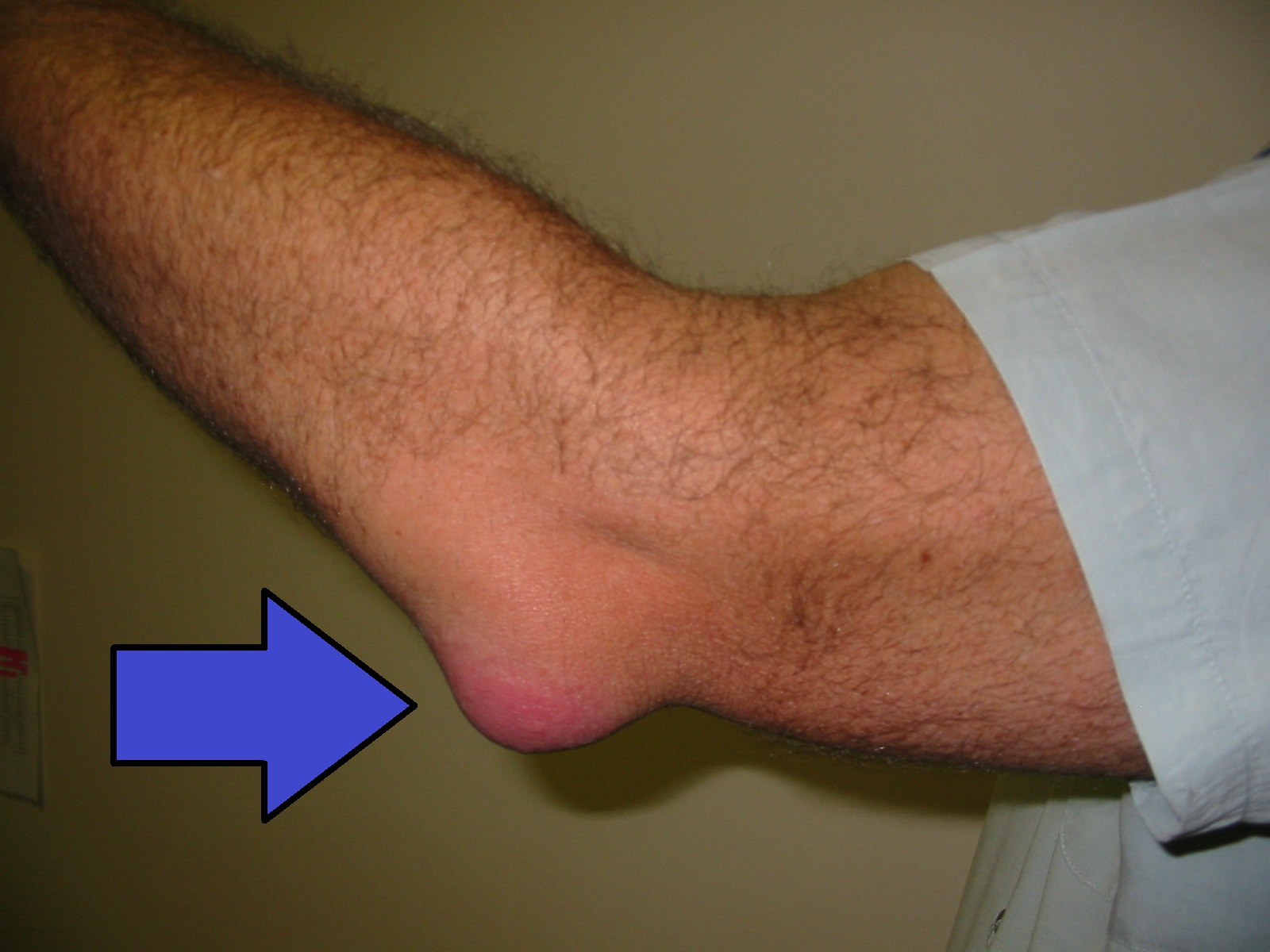
Bursitis is the inflammation of a bursa near a joint. It can cause pain, swelling, tenderness, and joint stiffness. Bursitis is commonly associated with the bursae in the shoulder, hip, knee, or elbow joints. Bursitis can be either acute (lasting only a few days) or chronic. It can arise from muscle overuse, trauma, excessive or prolonged pressure on the skin, rheumatoid arthritis, gout, or infection of the joint. Repeated acute episodes of bursitis can result in a chronic condition. Treatments for the disorder include antibiotics if the bursitis is caused by an infection or anti-inflammatory agents, such as nonsteroidal anti-inflammatory drugs (NSAIDs) or corticosteroids, if the bursitis is due to trauma or overuse. Chronic bursitis may require that fluid be drained, but additional surgery is usually not required. See Figure 6.56[3] for an image of bursitis.
Dislocation
A dislocation (or luxation) is an injury that forces the bones in a joint out of position. The cause is usually a fall, a car accident, or an injury during contact sports. Dislocation mostly involves the body’s larger joints. The most common site of dislocation is the shoulder. For young children, the elbow is a common site. Smaller joints, such as the thumbs and fingers, also can be dislocated if bent the wrong way with force. Dislocation may cause sudden and severe pain and swelling. It requires prompt medical attention to move the bones back in place.[4]
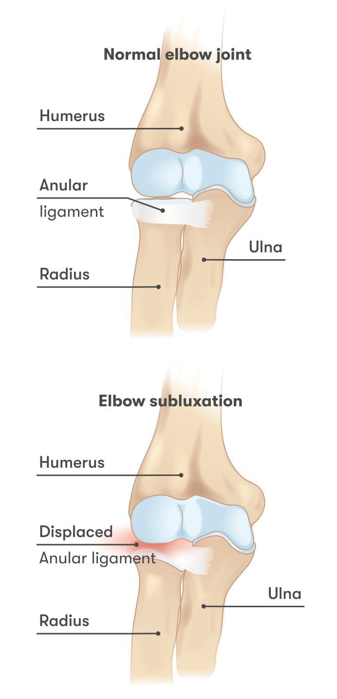
Subluxation is a partial dislocation of a bone. See Figure 6.57[5] for an illustration of subluxation in the elbow.
Gout
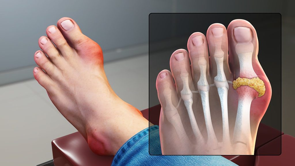
Gout is a form of arthritis caused when uric acid crystals (a waste product from nucleic acid breakdown) are deposited in a body joint. Usually, only one or a few joints are affected, such as the big toe, knee, or ankle. The attack may only last a few days but may return to the same or a different joint. Gout occurs when the body makes too much uric acid, or the kidneys do not remove it properly. A diet with excessive fructose (a simple sugar) has been shown to increase the chances of developing gout in some people. See Figure 6.58[6] for an illustration of gout.
Herniated Disc
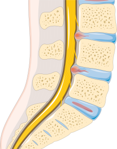
A herniated disc is a condition in which an intervertebral disc protrudes beyond the normal limits of the vertebrae and puts pressure on a spinal nerve causing pain. The most common sites for disc herniation are the L4/L5 or L5/S1 intervertebral discs, which can cause sciatica. A herniated disc is also known as a ruptured or slipped disc. View supplementary videos in the following box to learn more about herniated discs and their treatment. See Figure 6.59[7] for an illustration of a herniated disc.
View this supplementary YouTube video[8] to learn more about a herniated disc: Herniated Disc – Patient Education.
View this supplementary YouTube video[9] about one potential treatment for a herniated disc, removing the damaged portion of the disc: Lumbar Microdiscectomy – Spine Center Northern Nevada, Northern California – Spine Surgery.
Multiple Myeloma
Multiple myeloma is a cancer of the white blood cells that cause them to overgrow and crowd out normal cells in the bone marrow that make red blood cells, platelets, and other white blood cells. Advanced symptoms of multiple myeloma often include severe bone pain, especially in the back or ribs and multiple fractures.[10]
Osteoarthritis
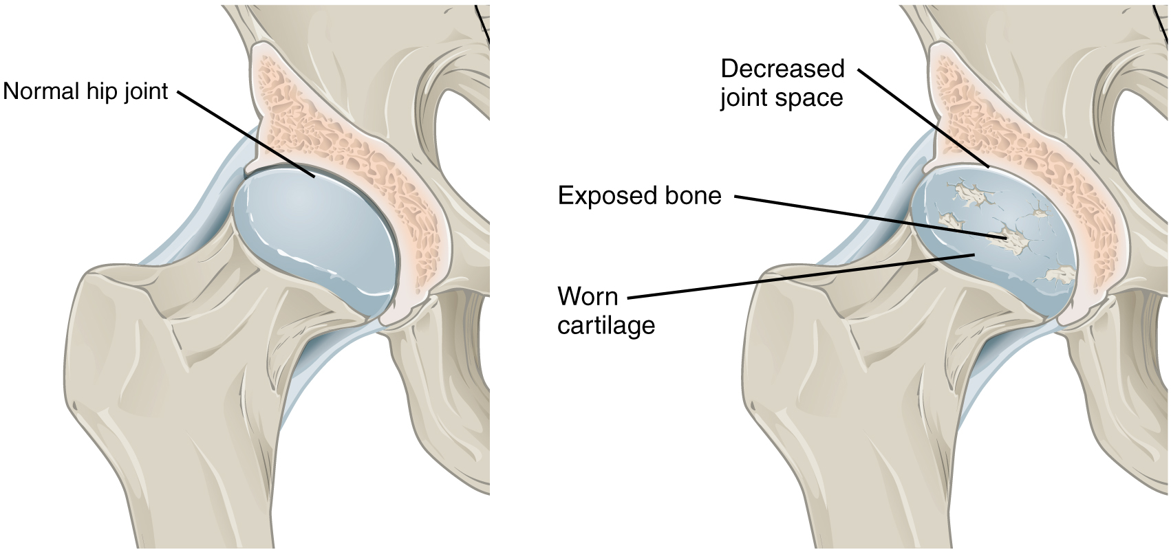
The most common type of arthritis is osteoarthritis, which is associated with aging and “wear and tear” of the articular cartilage. Risk factors that may lead to osteoarthritis later in life include injury to a joint; jobs that involve physical labor; sports with running, twisting, or throwing actions; and being overweight. These factors put stress on the articular cartilage that covers the surfaces of bones at synovial joints, causing the cartilage to gradually become thinner. As the articular cartilage layer wears down, more pressure is placed on the bones. The joint responds by increasing production of the lubricating synovial fluid, but this can lead to swelling of the joint cavity, causing pain and joint stiffness as the articular capsule is stretched. The bone tissue underlying the damaged articular cartilage also responds by thickening, producing irregularities and causing the articulating surfaces of the bones to become rough or bumpy. Joint movement then results in pain and inflammation. In its early stages, symptoms of osteoarthritis may be reduced by mild activity that “warms up” the joint, but the symptoms may worsen following exercise. In individuals with more advanced osteoarthritis, the affected joints can become more painful and, therefore, are difficult to use effectively, resulting in decreased mobility. There is no cure for osteoarthritis, but several treatments can help alleviate the pain. Treatments may include lifestyle changes, such as weight loss and low-impact exercise, and over-the-counter or prescription medications that help to alleviate the pain and inflammation. For severe cases, joint replacement surgery (arthroplasty) may be required. See Figure 6.60[11] for an illustration comparing a normal hip joint to one with osteoarthritis.
Osteomalacia and Rickets
Osteomalacia is softening of the bones due to a lack of mineralization with calcium and phosphate and most often caused by a lack of vitamin D. In children, osteomalacia is termed rickets.[12] See Figure 6.61[13] for an image of a child with rickets before and after treatment.

Osteomyelitis
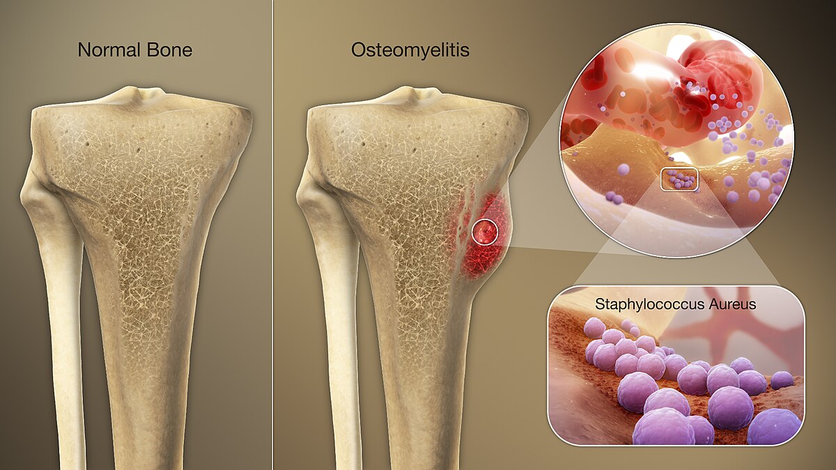
Osteomyelitis is an infection in a bone. It can affect one or more parts of a bone. Infections can reach a bone through the bloodstream or from nearby infected tissue. Infections also can begin in the bone if an injury opens the bone to germs. People who smoke and people with chronic health conditions, such as diabetes or kidney failure, are at higher risk of getting osteomyelitis. People who have diabetes with foot ulcers may get osteomyelitis in the bones of their feet.[14] See Figure 6.62[15] for an illustration of osteomyelitis.
Osteonecrosis
Osteonecrosis, also known as avascular necrosis, is the death of bone tissue due to a lack of blood supply. It can lead to tiny breaks in the bone and cause the bone to collapse. The process usually takes months to years. Osteonecrosis is often associated with long-term use of high-dose steroid medications and too much alcohol. Anyone can be affected, but the condition is most common in people between the ages of 30 and 50.[16]
Osteoporosis
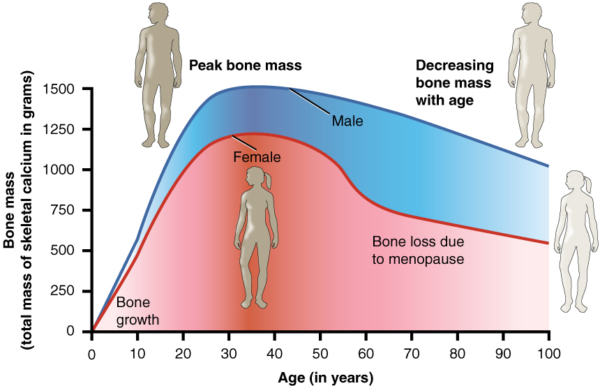
Osteopenia is loss of bone mass that may lead to osteoporosis. Osteoporosis is a disease characterized by a decrease in bone mass, a common occurrence as the body ages. See Figure 6.63[17] for an illustration of loss of bone mass in males and females due to age. Females lose bone mass more quickly than males starting around age 50 due to the loss of estrogen, a hormone that promotes osteoblastic (bone building) activity. Family history is another risk factor for developing osteoporosis.
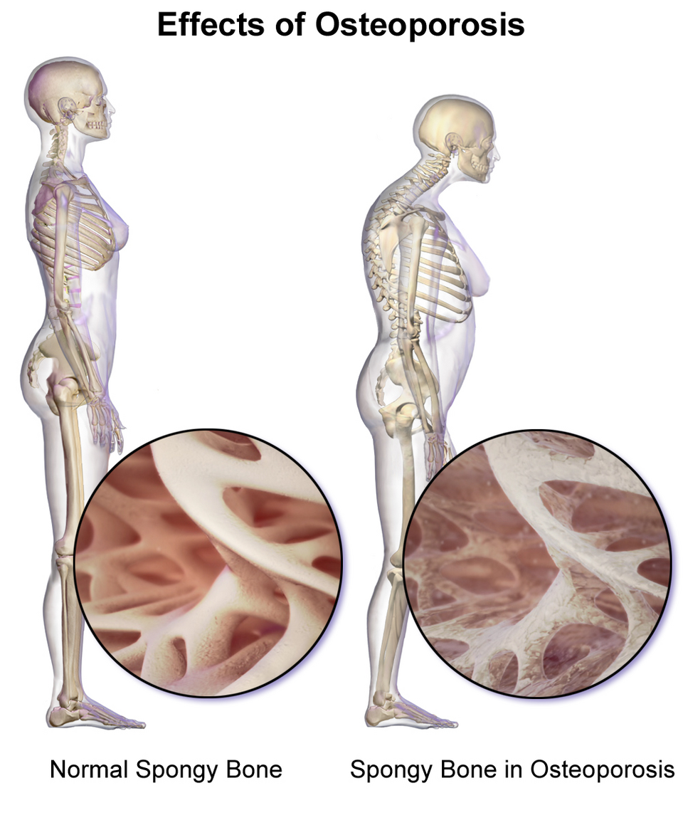
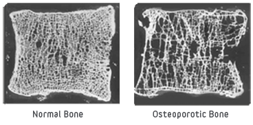
While osteoporosis can involve any bone, it most commonly affects the proximal ends of the femur, vertebrae, and wrist. As a result, these bones may not provide enough support for everyday functions, and something as simple as a sneeze can cause a vertebral fracture. Osteoporosis can result in kyphosis, an excessive outward curvature of the spine in the upper back because of compression and collapse of the vertebrae. See Figure 6.64[18] for an illustration of the effects of osteoporosis and Figure 6.65[19] for a microscopic image of normal bone tissue and osteoporotic bone.
The best treatment for osteoporosis is prevention, which includes a diet with adequate intake of calcium and vitamin D and daily physical activity that includes weight-bearing exercise. Medications are also prescribed for treating osteoporosis and preventing further loss of bone mass.
Osteosarcoma
Osteosarcoma is a cancer that begins in the cells that form bones. Osteosarcoma tends to happen most often in teenagers and young adults. Osteosarcoma can start in any bone, but it’s most often found in the long bones of the legs and sometimes the arms. Very rarely, it happens in soft tissue outside the bone. Advances in the treatment of osteosarcoma have improved the outlook for this cancer.[20]
Paget’s Disease
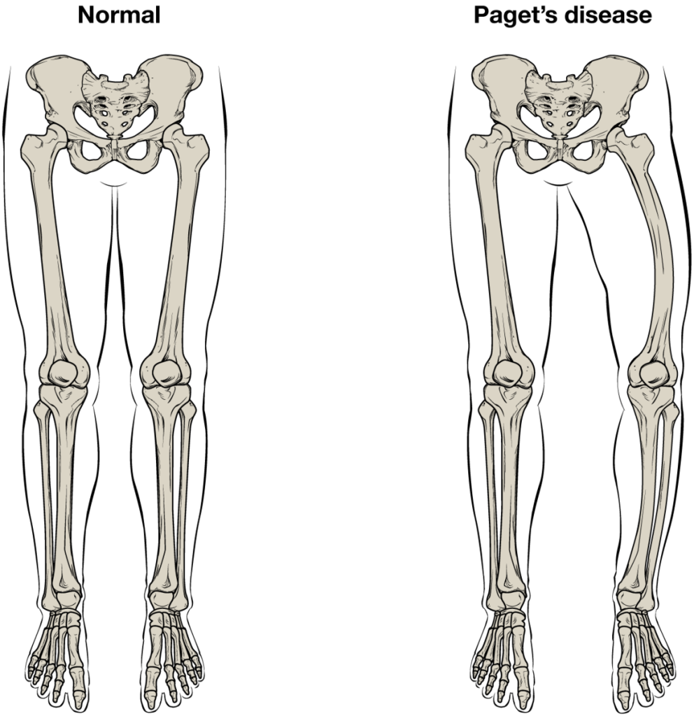
Paget's disease of bone interferes with the body’s normal recycling process, in which new bone tissue gradually replaces old bone tissue. Over time, bones can become fragile and misshapen. The pelvis, skull, spine, and legs are most commonly affected. The risk of Paget’s disease of bone increases with age and if family members have the disorder. Complications can include broken bones, hearing loss, and pinched nerves in your spine.[21] See Figure 6.66[22] for an illustration of Paget’s disease.
Patellofemoral Syndrome
Patellofemoral syndrome, also known as “Runner’s Knee,” is the most common overuse injury among runners. It is most frequent in adolescents and young adults and is more common in females. It often results from excessive running, particularly downhill, but may also occur in athletes who do a lot of knee bending, such as jumpers, skiers, cyclists, weight lifters, and soccer players. It is felt as a dull, aching pain around the front of the knee and deep to the patella. The pain may be felt when walking or running, going up or down stairs, kneeling or squatting, or after sitting with the knee bent for an extended period. Patellofemoral syndrome may be initiated by a variety of causes, including individual variations of the patella, a direct blow to the patella, flat feet, or improper shoes that cause excessive turning in or out of the feet or leg.
Rheumatoid Arthritis
Rheumatoid arthritis is an autoimmune disease where the immune system attacks the joints. As a result, the joint capsule and synovial membrane become inflamed. As the disease progresses, the articular cartilage is severely damaged or destroyed, resulting in joint deformation, loss of movement, and severe disability. The most commonly involved joints are the hands, feet, and cervical spine, with corresponding joints on both sides of the body usually affected, though not always to the same extent. Rheumatoid arthritis is also associated with lung fibrosis, vasculitis (inflammation of blood vessels), coronary heart disease, and premature mortality. With no known cure, treatments are aimed at alleviating symptoms. Exercise, anti-inflammatory and pain medications, disease-modifying anti-rheumatic drugs, biologic medications that suppress an overactive immune system, or surgery are used to treat rheumatoid arthritis. See Figure 6.67[23] for an image of rheumatoid arthritis in a person’s hands.

Sciatica
Sciatica is a painful condition resulting from inflammation or compression of the sciatic nerve, causing widespread pain that radiates from the lower back down the thigh and into the leg. The most common causes of sciatica include herniated discs, spinal stenosis, and conditions affecting the lower back or pelvis that compress the nerve.
Scoliosis, Kyphosis, and Lordosis
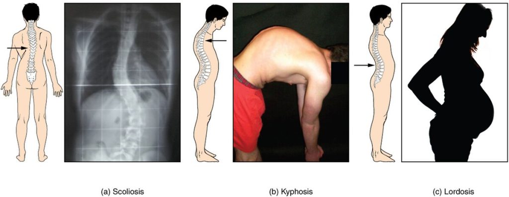
Scoliosis, kyphosis, and lordosis are abnormal curvatures of the spine. Scoliosis is an abnormal lateral curvature of the spine accompanied by twisting of the vertebral column. Other curves of the vertebral column may also develop to help keep the head positioned over the feet. Scoliosis is the most common vertebral abnormality among girls. The cause is usually unknown, but it may result from weakness of the back muscles, defects such as different growth rates in the right and left sides of the vertebral column, or differences in the length of the lower limbs. Scoliosis tends to get worse during adolescent growth spurts. Although most individuals do not require treatment, a back brace may be recommended for growing children. In extreme cases, surgery may be required.
Kyphosis is a condition sometimes referred to as humpback or hunchback. It is due to an excessive posterior curvature of the upper thoracic region of the vertebral column.
Lordosis, or swayback, is an excessive anterior curvature of the lumbar region and is most commonly associated with obesity or late pregnancy. The extra body weight in the abdominal region causes a shift in the line of gravity that carries the weight of the body. This causes an anterior tilt of the pelvis and a pronounced enhancement of the lumbar curve.
See Figure 6.68[24] for an illustration of abnormal curvatures of the spine, including scoliosis, kyphosis, and lordosis.
Spondylosis
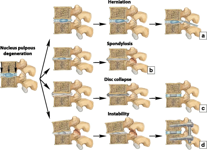
Spondylosis is a painful condition of the spine caused by the breakdown of the intervertebral discs (cartilage found between the vertebrae). See Figure 6.69[25] that compares herniation, spondylosis, disc collapse, and instability.
Sprain
A sprain is the stretching or tearing of ligaments in a joint. Commonly sprained joints include the ankle, wrist, knee, and thumb.
- Ernstmeyer, K., & Christman, E. (Eds.). (2024). Medical terminology 2e. Open RN | WisTech Open. https://wtcs.pressbooks.pub/medterm/ ↵
- “Osteoarthritis_and_rheumatoid_arthritis_-_Normal_joint_Osteoarthr_--_Smart-Servier” by Laboratoires Servier is licensed under CC BY-SA 3.0 ↵
- “Bursitis_Elbow_WCArrow” by en:User:NJC123 is licensed under CC0, Public Domain ↵
- Mayo Clinic. (2024). Dislocation: First aid. https://www.mayoclinic.org/first-aid/first-aid-dislocation/basics/art-20056693 ↵
- “Elbow_subluxation_2” by InjuryMap is licensed under CC BY-SA 4.0 ↵
- “Gout_Signs_and_Symptoms” by www.scientificanimations.com is licensed under CC BY-SA 4.0 ↵
- “Disc_herniation_--_Smart-Servier” by Smart Servier is licensed under CC BY-SA 3.0 ↵
- NuVasiveInc. (2017, September 12). Herniated disc - Patient education [Video]. YouTube. All rights reserved. https://www.youtube.com/watch?v=lZm4j6Ls128 ↵
- Swift Institute. (2014, September 23). Lumbar microdiscectomy - Spine Center Northern Nevada, Northern California - Spine surgery [Video]. YouTube. All rights reserved. https://youtu.be/i5xZrmoamsA?feature=shared ↵
- Centers for Disease Control and Prevention. (2025). Myeloma basics. https://www.cdc.gov/myeloma/about/index.html ↵
- “e805ac89b71c1ed264f89d79472b1ed9d7d0834e” by OpenStax College is licensed under CC BY 4.0. Access for free at https://openstax.org/books/anatomy-and-physiology-2e/pages/9-4-synovial-joints#fig-ch09_04_04 ↵
- Mayo Clinic. (2021). Rickets. https://www.mayoclinic.org/diseases-conditions/rickets/symptoms-causes/syc-20351943 ↵
- “Before_and_after_photographs_for_therapy_for_rickets_Wellcome_L0074524” by unknown author, Wellcome Images is licensed under CC BY 4.0 ↵
- Mayo Clinic. (2024). Osteomyelitis. https://www.mayoclinic.org/diseases-conditions/osteomyelitis/symptoms-causes/syc-20375913 ↵
- “3D_Medical_Animation_Staphylococcus_Aureus” by https://www.scientificanimations.com is licensed under CC BY-SA 4.0 ↵
- Mayo Clinic. (2022). Avascular necrosis (osteonecrosis). https://www.mayoclinic.org/diseases-conditions/avascular-necrosis/symptoms-causes/syc-20369859 ↵
- “615_Age_and_Bone_Mass” by OpenStax College is licensed under CC BY 3.0 ↵
- “Osteoporosis_02” by BruceBlaus is licensed under CC BY-SA 4.0 ↵
- “Bone_Comparison_of_Healthy_and_Osteoporotic_Vertibrae” by Turner Biomechanics Laboratory is licensed under CC0, Public Domain. ↵
- Mayo Clinic. (2023). Osteosarcoma. https://www.mayoclinic.org/diseases-conditions/osteosarcoma/symptoms-causes/syc-20351052 ↵
- Mayo Clinic. (2023). Paget's disease of bone. https://www.mayoclinic.org/diseases-conditions/pagets-disease-of-bone/symptoms-causes/syc-20350811 ↵
- “610_Feature_Pagets_Disease” by OpenStax College is licensed under CC BY 3.0 ↵
- “Swan_neck_deformity_in_a_65_year_old_Rheumatoid_Arthritis_patient-_2014-05-27_01-49” by User:Phoenix119 is licensed under CC BY-SA 3.0 ↵
- “717_Abnormal_Curves_of_Vertebral_Column” by OpenStax College is licensed under CC BY 3.0 ↵
- “Surgical_treatment_options_for_degenerative_changes” by Irina Nefedova is licensed under CC BY 4.0 ↵
A general term related to inflammation of a joint.
The inflammation of a bursa near a joint.
An injury that forces the bones in a joint out of position.
A partial dislocation of a bone.
A form of arthritis caused when uric acid crystals (a waste product from nucleic acid breakdown) are deposited in a body joint.
A condition in which an intervertebral disc protrudes beyond the normal limits of the vertebrae and puts pressure on a spinal nerve causing pain.
A cancer of the white blood cells that cause them to overgrow and crowd out normal cells in the bone marrow that make red blood cells, platelets, and other white blood cells.
Most common type of arthritis, which is associated with aging and “wear and tear” of the articular cartilage.
Softening of the bones due to a lack of mineralization with calcium and phosphate, most often caused by a lack of vitamin D.
Osteomalacia in children.
Infection in a bone.
The death of bone tissue due to a lack of blood supply.
Loss of bone mass that may lead to osteoporosis.
A disease characterized by a decrease in bone mass, a common occurrence as the body ages.
A cancer that begins in the cells that form bones.
A chronic condition where the body's natural process of bone remodeling is disrupted, leading to bones becoming larger, weaker, and more brittle than normal.
Also known as runner's knee or anterior knee pain, is a condition characterized by pain around the kneecap (patella) and front of the knee.
An autoimmune disease where the immune system attacks the joints.
A painful condition resulting from inflammation or compression of the sciatic nerve causing widespread pain that radiates from the lower back down the thigh and into the leg.
An abnormal lateral curvature of the spine accompanied by twisting of the vertebral column.
a condition sometimes Referred to as humpback or hunchback. It is due to an excessive posterior curvature of the upper thoracic region of the vertebral column.
An excessive anterior curvature of the lumbar region and is most commonly associated with obesity or late pregnancy. Also known as swayback.
A painful condition of the spine caused by the breakdown of the intervertebral discs (cartilage found between the vertebrae).
The stretching or tearing of ligaments in a joint.

